1. Introduction
The pancreas, a vital organ in both the digestive and endocrine systems, plays a critical role in maintaining overall bodily function. Pancreatitis, a common pancreatic ailment, is recognized by clinicians as a complex abdominal condition requiring precise diagnosis and management. Categorization of pancreatitis relies on clinical, morphological, and histological assessments. Acute pancreatitis (AP), frequently triggered by gallstone migration and excessive alcohol consumption, stands out as the most prevalent form. Accurate diagnosis and severity assessment of AP are paramount for determining effective treatment strategies and predicting disease progression, thereby preventing severe complications, organ dysfunction, or failure. This review synthesizes current recommendations and guidelines for managing patients with acute pancreatitis, with a focus on diagnostic criteria.
2. Diagnostic Criteria for Acute Pancreatitis
Establishing a diagnosis of acute pancreatitis requires fulfilling at least two of the following established criteria: characteristic abdominal pain, elevated serum enzyme levels, and radiological evidence. These criteria are crucial for initiating appropriate management and differentiating AP from other abdominal conditions.
2.1. Clinical Presentation: Abdominal Pain
Abdominal pain is a hallmark symptom of acute pancreatitis. It is typically described as persistent, severe epigastric pain that often radiates to the back and has an acute onset. This pain is usually the primary reason patients seek medical attention and is a critical factor in prompting further diagnostic evaluation for pancreatitis. The nature and location of the pain, while suggestive, must be corroborated with other diagnostic criteria for a definitive diagnosis.
2.2. Laboratory Tests: Elevated Serum Enzyme Levels
Elevated serum levels of pancreatic enzymes, specifically lipase or amylase, are a cornerstone in the diagnosis of acute pancreatitis. For diagnostic purposes, the activity of serum lipase or amylase must be at least three times greater than the upper limit of normal. Lipase is generally considered a more sensitive and specific marker for AP compared to amylase.
2.2.1. Serum Lipase
Serum lipase is a pancreatic enzyme that hydrolyzes triglycerides. In acute pancreatitis, damage to pancreatic cells leads to the release of lipase into the bloodstream, causing a significant elevation in serum levels. Lipase levels typically rise within 4-8 hours of symptom onset, peak at 24 hours, and remain elevated for up to 8-14 days. Its prolonged elevation compared to amylase makes it particularly useful for diagnosing patients who present to medical facilities a few days after symptom onset. Serum lipase is also noted to have a higher sensitivity in cases of alcohol-induced pancreatitis.
2.2.2. Serum Amylase
Serum amylase is another enzyme used in the diagnosis of acute pancreatitis, although it is less specific than lipase. Amylase is produced not only by the pancreas but also by salivary glands and other tissues, which can lead to elevated levels in conditions other than pancreatitis. Serum amylase levels usually increase within 6-24 hours of symptom onset, peak at 48 hours, and return to normal within 5-7 days. Despite its lower specificity, serum amylase is still a valuable diagnostic marker, especially when considered alongside clinical symptoms and other diagnostic findings. However, serum lipase is generally preferred due to its higher specificity and prolonged elevation in AP.
2.3. Radiological Imaging
Radiological imaging plays a crucial role when clinical presentation and enzyme levels are not definitive or when assessing the severity and complications of acute pancreatitis. Ultrasonography (US), computed tomography (CT), and magnetic resonance imaging (MRI) are the primary imaging modalities used to visualize the pancreas and identify characteristic signs of pancreatitis.
2.3.1. Ultrasonography (US)
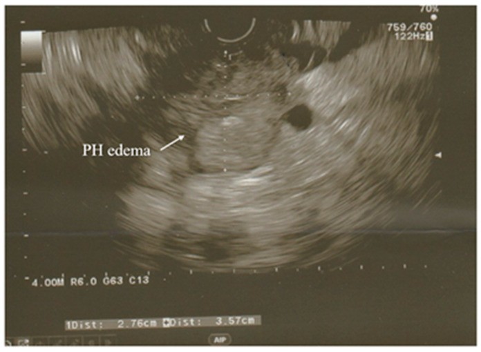
Ultrasonography is often the initial imaging test performed in suspected acute pancreatitis cases due to its accessibility, lack of radiation, and cost-effectiveness. US is particularly useful for identifying gallstones, a major etiological factor in AP. It can visualize the gallbladder and common bile duct (CBD) to detect biliary stones.
Image alt text: Ultrasound image showing acute pancreatitis with hypoechoic enlarged pancreatic head (PH). EUS examination detail highlighting pancreatic head enlargement.
However, US has limitations in visualizing the pancreas itself due to bowel gas interference. Its sensitivity in detecting gallstones can also decrease if stones are small or located in specific areas of the gallbladder. Despite these limitations, US remains a valuable first-line imaging tool, especially for detecting biliary causes of pancreatitis.
2.3.2. Computed Tomography (CT)
Computed tomography is a more detailed imaging modality often used to assess the extent of pancreatic inflammation, necrosis, and complications. CT scans provide cross-sectional images of the pancreas and surrounding tissues, allowing for a comprehensive evaluation of pancreatic morphology and peripancreatic changes.
Image alt text: Early phase acute pancreatitis CT scan showing fat stranding (FS) and enlarged lymph node. Arterial phase CT highlighting pancreatic head (PH) inflammation.
CT is particularly useful in identifying local complications such as fluid collections, pseudocysts, and pancreatic necrosis. It is typically performed with intravenous contrast to better delineate pancreatic perfusion and necrosis. CT severity indices, like the Balthazar CTSI and modified CTSI, are used to grade the severity of pancreatitis based on CT findings. However, CT is not routinely recommended early in the disease course unless there is diagnostic uncertainty or suspicion of complications, as pancreatic necrosis may not be evident in the first 48-72 hours.
2.3.3. Magnetic Resonance Imaging (MRI) and Magnetic Resonance Cholangiopancreatography (MRCP)
Magnetic resonance imaging, especially when combined with magnetic resonance cholangiopancreatography (MRCP), offers excellent soft tissue contrast without ionizing radiation. MRI is valuable for visualizing the pancreatic parenchyma, peripancreatic fluid collections, and biliary tree. MRCP specifically provides detailed images of the pancreatic and bile ducts, aiding in the detection of ductal abnormalities, strictures, and stones.
Image alt text: EUS image of a partially calcified gallstone in the distal common bile duct with surrounding edematous pancreatic parenchyma. Endoscopic ultrasound view of CBD stone in pancreatitis case.
MRI is particularly advantageous in patients with contraindications to CT contrast, such as renal insufficiency or contrast allergy. It is also superior to CT in characterizing fluid collections and detecting subtle pancreatic necrosis, especially in the early stages of AP. MRCP is often used as a non-invasive alternative to diagnostic ERCP for evaluating biliary obstruction.
2.3.4. Endoscopic Ultrasonography (EUS)
Endoscopic ultrasonography combines endoscopy with ultrasound to provide high-resolution images of the pancreas and biliary system from within the gastrointestinal tract. EUS offers superior image quality compared to transabdominal US due to its closer proximity to the pancreas and biliary ducts, overcoming limitations posed by bowel gas.
Image alt text: CT arterial phase showing inflammation of pancreatic head (PH) with surrounding fluid and enlarged lymph nodes. CT scan detail of inflamed pancreatic head in acute pancreatitis.
EUS is highly sensitive for detecting small gallstones and microlithiasis, which may be missed by other imaging modalities. It is also valuable in evaluating idiopathic acute pancreatitis and recurrent acute pancreatitis to identify occult biliary or pancreatic pathology. While EUS is minimally invasive, it is not typically the first-line imaging test for AP diagnosis but is often used for further investigation when the etiology remains unclear or for evaluating complications.
3. Classification and Severity Criteria
The Revised Atlanta Classification (2012) is the widely accepted system for classifying acute pancreatitis and defining its severity. This classification categorizes AP into two morphological subtypes: interstitial edematous pancreatitis and necrotizing pancreatitis. Furthermore, it stratifies AP severity into mild, moderately severe, and severe, based on the presence and duration of organ failure and local or systemic complications.
3.1. Severity Grading Based on Atlanta Classification
- Mild Acute Pancreatitis: Characterized by the absence of organ failure, local complications, or systemic complications. It usually resolves within the first week.
- Moderately Severe Acute Pancreatitis: Defined by transient organ failure (resolving within 48 hours) and/or local or systemic complications without persistent organ failure. It may also include exacerbation of co-morbid conditions.
- Severe Acute Pancreatitis: Characterized by persistent organ failure lasting more than 48 hours. This category carries the highest risk of mortality and requires intensive care management.
3.2. Organ Failure Criteria
Organ failure is a critical determinant of severity in acute pancreatitis. The Revised Atlanta Classification defines organ failure based on the modified Marshall score, assessing respiratory, cardiovascular, and renal systems.
- Respiratory Failure: PaO2/FiO2 ratio ≤ 300.
- Cardiovascular Failure: Systolic blood pressure < 90 mmHg or need for vasopressors to maintain adequate blood pressure.
- Renal Failure: Serum creatinine ≥ 170 μmol/L (≥ 2 mg/dL) or urine output < 0.5 mL/kg/h for > 6 hours despite fluid resuscitation.
Persistent organ failure, lasting beyond 48 hours, indicates severe acute pancreatitis and is associated with increased morbidity and mortality.
4. Etiology of Acute Pancreatitis
Determining the etiology of acute pancreatitis is essential for guiding treatment and preventing recurrence. The most common causes are gallstones and alcohol abuse, accounting for approximately 70-80% of cases. Other causes include hypertriglyceridemia, medications, post-ERCP pancreatitis, and autoimmune pancreatitis.
4.1. Gallstone Pancreatitis
Gallstones are the leading cause of acute pancreatitis. Migration of gallstones into the common bile duct can obstruct the pancreatic duct, leading to pancreatic enzyme activation and inflammation. Ultrasonography is crucial for detecting gallstones in patients presenting with acute pancreatitis.
4.2. Alcohol-Induced Pancreatitis
Excessive alcohol consumption is another major cause of AP. Chronic alcohol abuse can damage pancreatic cells and predispose individuals to pancreatitis. A detailed alcohol history is important in diagnosing alcohol-induced pancreatitis.
4.3. Hypertriglyceridemia
Elevated serum triglyceride levels (> 1000 mg/dL) can cause acute pancreatitis. Hypertriglyceridemia-induced pancreatitis requires specific management, including lipid-lowering therapies.
4.4. Drug-Induced Pancreatitis
Certain medications are associated with drug-induced pancreatitis. Identifying potential causative drugs is important, especially in patients without other obvious etiologies.
4.5. Post-ERCP Pancreatitis
Endoscopic retrograde cholangiopancreatography (ERCP) can, in some cases, induce pancreatitis. Post-ERCP pancreatitis is a recognized complication of this procedure.
4.6. Autoimmune Pancreatitis (AIP)
Autoimmune pancreatitis is a distinct form of pancreatitis characterized by autoimmune mechanisms. It is classified into Type 1 and Type 2 AIP, with different clinical and pathological features.
4.7. Idiopathic Pancreatitis
In a subset of patients, the etiology of acute pancreatitis remains undetermined after initial investigations. These cases are classified as idiopathic acute pancreatitis. Further investigations, including EUS and MRCP, may be necessary to identify occult causes in idiopathic cases, especially in recurrent pancreatitis.
5. Severity Assessment Tools and Prognostic Criteria
Beyond the Revised Atlanta Classification, several scoring systems and biomarkers are used to assess the severity and predict the prognosis of acute pancreatitis. These tools aid in risk stratification and guiding management decisions.
5.1. Clinical Scoring Systems
- BISAP (Bedside Index of Severity in Acute Pancreatitis) Score: A simple scoring system assessed within the first 24 hours of admission, utilizing variables like blood urea nitrogen, impaired mental status, SIRS, age, and pleural effusion.
- APACHE II (Acute Physiology and Chronic Health Evaluation II) Score: A more complex scoring system evaluating multiple physiological variables over the first 24 hours, commonly used in intensive care settings to predict mortality risk.
- Ranson’s Criteria: An older, more extensive scoring system that assesses severity based on parameters at admission and after 48 hours. It is less frequently used now due to its complexity and the need for a 48-hour assessment period.
- Harmless Acute Pancreatitis Score (HAPS): Designed to identify patients with a very low risk of severe AP early in admission, facilitating early discharge for low-risk patients.
5.2. Biomarkers
- C-Reactive Protein (CRP): Serum CRP levels rise in response to inflammation and are often used to predict the severity of AP. Levels > 150 mg/L at 48 hours are suggestive of severe disease.
- Procalcitonin (PCT): PCT levels can be helpful in detecting infected necrosis in patients with AP.
- Blood Urea Nitrogen (BUN): Elevated BUN levels on admission or rising BUN are associated with increased severity and mortality.
- Hematocrit: Elevated hematocrit (> 44%) or rising hematocrit can indicate hemoconcentration and is associated with pancreatic necrosis.
5.3. Systemic Inflammatory Response Syndrome (SIRS)
The presence of SIRS at admission and persistent SIRS beyond 48 hours are useful indicators of severity. SIRS criteria include body temperature, heart rate, respiratory rate, and white blood cell count. Persistent SIRS is associated with a higher risk of organ failure and mortality.
6. Management Based on Diagnostic Criteria and Severity
Management of acute pancreatitis is primarily supportive, focusing on fluid resuscitation, pain control, nutritional support, and monitoring for complications. The intensity of management is guided by the severity of pancreatitis.
6.1. Supportive Care
- Fluid Resuscitation: Aggressive intravenous fluid resuscitation with isotonic crystalloids is crucial in the early management of AP. Ringer’s lactate may be preferred over normal saline. Fluid administration should be guided by hemodynamic status and markers of volume status like hematocrit and BUN.
- Pain Management: Effective pain control is essential. A multimodal analgesic approach, including opioids, non-steroidal anti-inflammatory drugs (NSAIDs) (if not contraindicated), and acetaminophen, is often used. Patient-controlled analgesia (PCA) can be beneficial.
- Nutritional Support: Early enteral nutrition is recommended for patients with severe AP. Oral feeding can be initiated early in mild AP as tolerated. Nasogastric or nasojejunal feeding can be used for enteral nutrition. Total parenteral nutrition (TPN) should be avoided unless enteral nutrition is not feasible.
- Monitoring and ICU Care: Patients with severe AP or organ failure require intensive monitoring, often in an intensive care unit (ICU). Vital signs, organ function, and laboratory parameters should be closely monitored.
6.2. Interventional and Surgical Management
- ERCP: In gallstone pancreatitis with cholangitis or CBD obstruction, early ERCP with sphincterotomy may be indicated to remove obstructing stones.
- Drainage of Necrotic Collections: In infected necrotizing pancreatitis, drainage of necrotic collections is necessary. Percutaneous catheter drainage is often the initial approach, followed by endoscopic or surgical necrosectomy if needed. Intervention is generally delayed until walled-off necrosis has formed (typically > 4 weeks).
- Cholecystectomy: For gallstone pancreatitis, cholecystectomy is recommended, usually during the same hospitalization for mild cases or after resolution of acute inflammation in severe cases, to prevent recurrence.
- Surgical Necrosectomy: Surgical necrosectomy may be necessary in cases of persistent infected necrosis or when minimally invasive approaches are unsuccessful. Open surgical approaches are generally reserved for specific indications or when other methods fail.
7. Prevention of Recurrent Pancreatitis
Preventing recurrent pancreatitis involves identifying and addressing the underlying etiology.
- Gallstone Pancreatitis: Cholecystectomy prevents recurrence in gallstone-induced AP.
- Alcohol-Induced Pancreatitis: Alcohol abstinence is crucial for preventing recurrence.
- Hypertriglyceridemia: Management of hypertriglyceridemia with diet and lipid-lowering medications is necessary.
- Idiopathic Pancreatitis: Thorough investigation for underlying causes, including EUS and MRCP, is important. Genetic testing may be considered in recurrent idiopathic pancreatitis.
8. Conclusion
Accurate diagnosis of acute pancreatitis relies on a combination of clinical criteria, including characteristic abdominal pain, elevated serum pancreatic enzyme levels (primarily lipase), and supportive radiological imaging findings. The Revised Atlanta Classification provides a framework for defining AP severity based on organ failure and complications, guiding management strategies. Early recognition of diagnostic criteria and severity assessment is crucial for optimizing patient outcomes, preventing complications, and reducing mortality in acute pancreatitis. This comprehensive understanding of diagnostic criteria and management principles is essential for healthcare professionals involved in the care of patients with acute pancreatitis.
Author Contributions
J.W.—project development, data collection and management, data analysis, and manuscript writing; N.Z.—data analysis and manuscript editing; M.P.—data analysis and manuscript editing; R.S.T.—data analysis and manuscript editing; J.D.-R.—data analysis and manuscript editing; Ł.O.—data collection, data analysis, and manuscript editing. All authors have read and agreed to the published version of the manuscript.
Institutional Review Board Statement
Not applicable.
Informed Consent Statement
Not applicable.
Data Availability Statement
Please contact authors for data requests (Łukasz Olewnik—email address: [email protected]).
Conflicts of Interest
The authors declare that they have no competing interests.
Funding Statement
The authors have no financial or personal relationship with any third party whose interests could be positively or negatively influenced by the article’s content. This research did not receive any specific grant from funding agencies in the public, commercial, or not-for-profit sectors.
Footnotes
Publisher’s Note: MDPI stays neutral with regard to jurisdictional claims in published maps and institutional affiliations.
References
[References]
Associated Data
Data Availability Statement
Please contact authors for data requests (Łukasz Olewnik—email address: [email protected]).