Pancreatitis, characterized by the inflammation of the pancreas, arises when pancreatic enzymes initiate autodigestion, the process of digesting the pancreas’s own tissues. The severity of pancreatitis can vary widely, ranging from mild discomfort to life-threatening conditions, and it manifests in both acute and chronic forms. Acute pancreatitis typically presents with intense abdominal pain and tenderness, accompanied by abdominal distention, nausea, vomiting, and fever. Chronic pancreatitis may mirror these symptoms or, in some instances, remain asymptomatic, posing diagnostic challenges.
Nurses play a pivotal role in the care of patients with pancreatitis, primarily in inpatient settings. The management of pain stemming from pancreatic inflammation and the administration of intravenous fluids are critical components of care, often necessitating hospitalization. Furthermore, patient education on crucial lifestyle modifications, such as abstaining from alcohol and smoking and adopting specific dietary changes, is an essential aspect of nursing responsibilities.
Nursing Process in Pancreatitis Management
The nursing process is fundamental in managing pancreatitis, guiding nurses through assessment, diagnosis, planning, implementation, and evaluation of patient care. This structured approach ensures comprehensive and individualized care for patients with pancreatitis.
Nursing Assessment for Pancreatitis
The initial step in providing effective nursing care is a thorough nursing assessment. This involves gathering comprehensive data encompassing physical, psychosocial, emotional, and diagnostic aspects. A detailed assessment allows for the identification of specific nursing diagnoses and the formulation of tailored care plans.
Review of Health History
A detailed health history is crucial in identifying potential causes and risk factors for pancreatitis, as well as understanding the patient’s symptom presentation.
1. Identifying General Symptoms of Pancreatitis: Acute pancreatitis often emerges as the pancreas attempts to recover from an injury, triggering a cascade of symptoms. Key indicators of acute pancreatitis include:
- Nausea and vomiting, often persistent and severe.
- Rapid heartbeat (tachycardia), reflecting the body’s stress response.
- Sudden and severe epigastric abdominal pain, a hallmark symptom.
- Diarrhea, resulting from digestive enzyme disruption.
2. Recognizing Signs of Deteriorating Pancreas in Chronic Pancreatitis: Chronic pancreatitis, characterized by progressive pancreatic damage, presents with symptoms that indicate long-term dysfunction. These signs include:
- Bloating and discomfort post-eating, due to impaired digestion.
- Unintentional weight loss, a consequence of malabsorption.
- Loss of appetite, reflecting digestive system distress.
3. In-depth Investigation of Abdominal Pain: Abdominal pain is the cardinal symptom of pancreatitis, requiring careful evaluation. Pancreatitis-related pain is often described as:
- Moderate to severe dull abdominal pain, developing abruptly.
- Radiation to the back or below the left shoulder blade.
- In acute pancreatitis, pain is typically more intense and penetrating.
- Abdominal tenderness upon palpation.
- In chronic pancreatitis, pain may be intermittent but rarely disappears entirely, commonly exacerbated after meals.
4. Determining Modifiable Risk Factors: Several lifestyle and dietary factors can significantly elevate the risk of pancreatitis. Identifying and addressing these modifiable factors is essential for prevention and management:
- Obesity: Increases pancreatitis risk due to elevated insulin levels and dysregulation of fat metabolism.
- Diet: Diets high in processed and red meats, and saturated fats, contribute to elevated triglyceride levels, increasing acute pancreatitis susceptibility.
- Smoking: A significant risk factor for chronic pancreatitis, causing pancreatic damage and functional impairment.
- Alcohol consumption: Excessive and chronic alcohol intake is the most prevalent cause of pancreatitis.
5. Collecting Family History: A family history of chronic pancreatitis increases an individual’s risk, particularly when combined with other risk factors. Genetic predispositions can play a role in pancreatic vulnerability.
6. Reviewing Medical History for Underlying Conditions: A comprehensive medical history review is vital to identify pre-existing conditions that can trigger pancreatitis. These include:
- Gallstones, a common cause of biliary pancreatitis.
- Viral infections, which can sometimes induce pancreatic inflammation.
- Autoimmune diseases, potentially leading to autoimmune pancreatitis.
- Inherited gene mutations, predisposing to hereditary pancreatitis.
- Cystic fibrosis, associated with pancreatic dysfunction.
- High blood triglyceride levels (hypertriglyceridemia), a metabolic risk factor.
- High blood calcium levels (hypercalcemia), another metabolic trigger.
- Restricted blood supply (ischemia) to the pancreas.
- Cancer of the pancreas or biliary tract.
- Traumatic injury to the pancreas.
- Surgical procedures that inadvertently damage the pancreas.
- Conditions affecting pancreatic blood flow (e.g., ischemia, vasculitis).
- Medications with potential pancreatic side effects.
Alt text: A nurse assesses a patient experiencing severe abdominal pain, a key symptom of pancreatitis, by using a pain scale and palpating the abdomen to identify the location and intensity of the discomfort.
Physical Assessment
A thorough physical examination is essential to identify objective signs and symptoms of pancreatitis, differentiating between acute and chronic presentations.
1. Comprehensive Physical Examination: Assess for the following signs and symptoms indicative of acute or chronic pancreatitis:
- General: Fever, restlessness, indicating systemic inflammation and discomfort.
- CNS: Decreased mentation, potentially due to severe illness or electrolyte imbalances.
- HEENT: Yellowish eyes (jaundice), suggesting biliary obstruction or liver involvement.
- Respiratory: Tachypnea, basilar rales upon auscultation, reflecting potential pulmonary complications.
- Cardiovascular: Tachycardia, hypotension, indicating hemodynamic instability.
- Gastrointestinal: Abdominal tenderness, guarding, distention, hematemesis, melena, clay-colored stool, steatorrhea, epigastric pain radiating to the back, all pointing to gastrointestinal dysfunction.
- Genitourinary: Dark urine, possibly due to bilirubinuria.
- Integumentary: Jaundice, pruritus, pale skin, diaphoresis, reflecting systemic effects and potential complications.
2. Auscultation of Bowel Sounds: Diminished or absent bowel sounds are expected in cases of ileus, a common complication of acute pancreatitis, indicating reduced gastrointestinal motility.
3. Checking for Pancreatic Necrosis or Hemorrhaging: Specific signs can indicate severe complications like pancreatic necrosis or retroperitoneal bleeding:
- Cullen sign: Bluish discoloration around the umbilicus, suggestive of intraperitoneal bleeding.
- Grey-Turner sign: Ecchymosis along the flanks, indicating retroperitoneal hemorrhage, often seen with Cullen’s sign.
- Fox’s sign: Bruising over the inguinal ligament, also indicating retroperitoneal bleeding.
Alt text: Visual representation of Cullen’s sign, a bluish discoloration around the umbilicus, and Grey-Turner’s sign, bruising along the flanks, both clinical indicators of severe pancreatitis-related internal bleeding.
Diagnostic Procedures
Diagnostic procedures are essential to confirm the diagnosis of pancreatitis, determine its severity, identify underlying causes, and guide treatment strategies.
1. Blood Sample Analysis: Blood tests are crucial for evaluating various parameters:
- White blood cell count, elevated in inflammation and infection.
- Kidney function tests, assessing for renal complications.
- Liver enzyme levels, indicating potential biliary involvement.
- Pancreatic enzyme levels (amylase, lipase), key markers of pancreatic inflammation.
2. Blood Glucose Monitoring: Blood glucose tests assess pancreatic endocrine function. Elevated glucose levels in pancreatitis suggest impaired insulin production. Glucose levels are typically elevated in pancreatitis due to pancreatic cell damage.
3. Stool Sample Examination: Stool tests provide insights into pancreatic exocrine function:
- Stool elastase test, assessing digestive enzyme adequacy.
- Fecal fat analysis, detecting fat malabsorption (steatorrhea), indicated by excess fat in stool.
4. Review of Imaging Scan Findings: Imaging techniques play a vital role in visualizing the pancreas and surrounding structures:
- Computed Tomography (CT) scan of the abdomen: Visualizes gallstones, pancreatic inflammation extent, and is indicated for severe acute pancreatitis.
- Ultrasound of the abdomen: Recommended as the initial test for pancreatic inflammation and gallstones due to its non-invasiveness and accessibility.
- Endoscopic Ultrasound (EUS): Primarily used to detect inflammation and obstructions in pancreatic or bile ducts, offering detailed visualization.
- Magnetic Resonance Imaging (MRI) scan: Shows abnormalities of the gallbladder, pancreas, and ducts, providing comprehensive soft tissue imaging.
- Endoscopic Retrograde Cholangiopancreatography (ERCP): Not a first-line diagnostic test due to risks of infection and perforation. Used therapeutically to remove gallstones or address ductal issues.
- Magnetic Resonance Cholangiopancreatography (MRCP): Non-invasive and safer alternative to ERCP for visualizing biliary and pancreatic ducts.
5. Prognosis and Level of Care Determination: Assessing the severity of acute pancreatitis is crucial for determining prognosis and appropriate care level. Severity scoring systems help guide decisions:
- Persistent SIRS (systemic inflammatory response syndrome), Glasgow Coma Scale >3, APACHE score >8, or a Ranson score >3 indicate severe pancreatitis and need for Intensive Care Unit (ICU) admission.
Nursing Interventions for Pancreatitis
Effective nursing interventions are critical for patient recovery and management of pancreatitis. These interventions focus on supportive care, managing the underlying condition, and preventing recurrence.
Initiate Supportive Care
Supportive care is the cornerstone of pancreatitis management, aiming to stabilize the patient and alleviate symptoms.
1. Fluid Resuscitation: Rapid intravenous (IV) hydration in the first 24 hours is crucial. Aggressive fluid resuscitation, especially early in acute pancreatitis, is vital to maintain hemodynamic stability and pancreatic perfusion. Patients should be kept NPO (nothing per oral) if nausea, vomiting, or abdominal pain is present to reduce pancreatic stimulation.
2. Adherence to Fluid Resuscitation Guidelines: The American College of Gastroenterology guidelines recommend Ringer’s lactate (LR) as the preferred IV crystalloid for fluid resuscitation in acute pancreatitis. LR has been shown to reduce systemic inflammation compared to normal saline.
3. Avoiding Overhydration: While aggressive hydration is necessary, overhydration can paradoxically increase the risk of sepsis and mortality. Close monitoring of fluid balance is essential. Insertion of a urinary catheter is recommended for accurate monitoring of intake and output.
4. Judicious Use of Antibiotics: Antibiotics are not routinely indicated for non-infected pancreatitis. They should be reserved for cases with confirmed or suspected infection, such as infected necrosis.
5. Consideration of Enteral Feeding: Once abdominal pain subsides and the patient’s condition stabilizes, enteral feeding should be considered. Enteral nutrition, preferably via a nasogastric (NG) tube, is preferred over parenteral nutrition as it helps maintain gut integrity and reduce infectious complications.
6. Anticipating Total Parenteral Nutrition (TPN): Total parenteral nutrition (TPN) may be necessary for patients who cannot tolerate enteral feeding. TPN solutions should include adequate fat emulsions to prevent essential fatty acid deficiency, while minimizing potential metabolic complications.
7. Pain Management: Intense pain is a hallmark of pancreatitis. Effective pain management is crucial for patient comfort and reducing physiological stress. Opioids and patient-controlled analgesia (PCA) are often necessary for adequate pain control. Regular pain assessment and adjustment of analgesia are important.
Alt text: A nurse carefully monitors the IV fluid administration for a patient with pancreatitis, highlighting the importance of fluid resuscitation in managing the condition and ensuring hemodynamic stability.
Management of Pancreatitis
Managing pancreatitis involves addressing the underlying cause and implementing specific interventions to resolve the acute episode and prevent complications.
1. Treating the Underlying Condition: Identifying and treating the root cause of pancreatitis is paramount. Once the acute phase is controlled, focus shifts to addressing the underlying etiology. Specific interventions vary depending on the cause, such as:
- Necrotizing pancreatitis: May require surgical or endoscopic debridement.
- Gallstone pancreatitis: Requires gallstone removal, often cholecystectomy.
- Alcohol-induced pancreatitis: Mandates alcohol cessation and addiction treatment.
2. Preparation for Surgical or Invasive Procedures: Surgical or minimally invasive procedures may be necessary to remove the cause of pancreatitis or manage complications:
- Endoscopic Retrograde Cholangiopancreatography (ERCP): Used to remove blockages like gallstones in the bile duct. Urgent ERCP within 24-72 hours is indicated for acute pancreatitis with cholangitis.
- Cholecystectomy: Surgical removal of the gallbladder, indicated for gallstone pancreatitis to prevent recurrence.
- Pancreaticojejunostomy: A surgical procedure to relieve blockages in the pancreatic ducts and reduce chronic pain.
- Stenting: Placement of stents to open narrowed pancreatic ducts, facilitating drainage of pancreatic secretions.
In severe chronic pancreatitis, partial or complete removal of the pancreas may be considered. Total pancreatectomy is avoided when possible due to the pancreas’s vital role in insulin and digestive enzyme production, leading to diabetes and malabsorption.
3. Encouraging Alcohol Cessation: Alcohol is a major risk factor for pancreatitis. For patients with alcohol-induced pancreatitis, complete abstinence from alcohol is crucial. Referral to alcohol addiction treatment programs is highly recommended. Continued alcohol consumption will worsen pancreatitis and can be life-threatening.
4. Discontinuing Offending Medications: Certain medications can induce pancreatitis. Reviewing the patient’s medication list and discontinuing any potentially causative drugs is essential. Collaboration with the healthcare provider and pharmacist is needed to find alternative medications if necessary.
Prevention of Recurrent Pancreatitis
Preventing recurrence is a key aspect of long-term management, especially for chronic pancreatitis. Lifestyle modifications and ongoing medical management are crucial.
1. Promoting Lifestyle Changes: Lifestyle modifications are essential to prevent recurrent pancreatitis, especially chronic forms:
- Smoking cessation is critical as smoking exacerbates pancreatic damage.
- Complete abstinence from alcohol is vital for patients with alcohol-induced pancreatitis and beneficial for all pancreatitis patients.
These changes significantly impact pancreatic health and reduce the risk of complications.
2. Dietary Modifications: Low-Fat Diet: Dietary management is crucial. Patients should adhere to a low-fat diet, rich in fruits, vegetables, and whole grains. A low-fat diet reduces pancreatic workload. Adequate hydration, drinking plenty of water daily, is also important to maintain pancreatic function and prevent exacerbations.
3. Nutritional Supplements: Exocrine Pancreatic Insufficiency (EPI) is a common long-term complication of chronic pancreatitis, leading to malabsorption. Nutritional supplements are often necessary:
- Pancreatic enzyme replacement therapy (PERT) to aid digestion.
- Vitamin and mineral supplementation to address deficiencies.
4. Diabetes Prevention: Diabetes mellitus is a common sequela of pancreatitis, particularly after acute episodes. Preventing severe pancreatitis and managing risk factors can reduce the risk of developing diabetes. The exact pathogenesis of diabetes post-pancreatitis is complex and multifactorial.
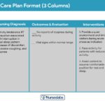
Pancreatitis Nursing Care Plans
Nursing care plans are essential tools to guide individualized patient care, prioritize nursing diagnoses, and outline interventions to achieve both short-term and long-term goals. Here are examples of nursing care plans for common nursing diagnoses related to pancreatitis.
Acute Pain related to Pancreatitis
Pain associated with pancreatitis is a primary concern, stemming from pancreatic inflammation and biliary duct obstruction.
Nursing Diagnosis: Acute Pain related to inflammation of the pancreas and obstruction of biliary ducts.
Related Factors:
- Gallstones causing obstruction.
- Inflammation of pancreatic tissue.
- Obstructed and damaged biliary ducts.
- Autodigestion of the pancreas releasing inflammatory toxins.
As Evidenced By:
- Verbal reports of abdominal pain, using pain scales.
- Guarding behavior and protective body language.
- Facial grimacing and expressions of distress.
- Agitation and restlessness due to pain.
- Changes in vital signs (increased heart rate, blood pressure).
Expected Outcomes:
- Patient will verbalize a reduction in pain intensity or achieve pain relief through pharmacologic and non-pharmacologic interventions within a specified timeframe.
- Patient will actively participate in the prescribed treatment plan to maintain pain relief and comfort.
Nursing Assessments:
1. Pain Assessment: Regularly assess pain characteristics, including location, intensity (using a 0-10 scale), quality, onset, duration, aggravating, and relieving factors. For non-verbal patients, utilize non-verbal pain assessment tools.
2. Abdominal Tenderness Assessment: Palpate the abdomen gently to identify areas of tenderness, guarding, and rebound tenderness. Note the location and extent of abdominal pain to understand the severity and location of inflammation.
3. Monitoring Vital Signs and Nonverbal Pain Cues: Observe for nonverbal indicators of pain such as sweating, restlessness, grimacing, and changes in vital signs (heart rate, blood pressure, respiratory rate). Correlate these with patient’s verbal reports to evaluate pain management effectiveness.
Nursing Interventions:
1. Routine Pain Medication Administration: Administer prescribed pain medications, including analgesics (opioids, NSAIDs) and antispasmodics, routinely and proactively, based on pain assessment and physician orders. Smaller, more frequent doses may be preferable to large boluses to maintain consistent pain control while minimizing side effects.
2. Maintaining NPO Status and Gastric Suctioning: Maintain NPO status to reduce pancreatic stimulation and enzyme secretion. Insert and maintain a nasogastric (NG) tube for gastric decompression to alleviate abdominal distension and pain, and to allow the pancreas to rest. Administer IV fluids for hydration and electrolyte balance.
3. Non-Pharmacologic Pain Management: Implement alternative pain relief strategies, including distraction techniques (TV, music, games), relaxation techniques (massage, guided imagery, deep breathing exercises), and thermal therapies (hot/cold packs) to complement pharmacologic interventions.
4. Promoting Comfort Positioning: Assist the patient to assume positions of comfort, such as the side-lying position with knees flexed, to reduce abdominal pressure and muscle tension. Elevate the head of the bed to improve respiratory effort and reduce abdominal discomfort.
Deficient Knowledge related to Pancreatitis
Patients often lack understanding of pancreatitis, its triggers, and preventive measures.
Nursing Diagnosis: Deficient Knowledge related to lack of information regarding pancreatitis, its causes, management, and prevention of recurrence.
Related Factors:
- Lack of prior exposure to information about pancreatitis.
- Information misinterpretation or lack of recall.
- Cognitive limitations affecting learning.
- Lack of perceived need to learn or disinterest.
- Limited access to reliable educational resources.
As Evidenced By:
- Inability to accurately recall or explain instructions regarding diet, medications, or lifestyle modifications.
- Verbalization of lack of understanding about pancreatitis and its management.
- Expressed disinterest in learning or denial of the need for education.
- Worsening of pancreatitis symptoms or recurrence due to lack of self-management.
Expected Outcomes:
- Patient will verbalize understanding of pancreatitis, its causes, symptoms, treatment, and potential complications prior to discharge.
- Patient will demonstrate adherence to the prescribed medication regimen and dietary recommendations by discharge and during follow-up.
- Patient will verbalize understanding of required diagnostic tests, follow-up appointments, and emergency signs and symptoms necessitating prompt medical attention.
Nursing Assessments:
1. Assessing Willingness to Learn: Evaluate the patient’s readiness and willingness to learn about pancreatitis and self-management. Identify barriers to learning, such as pain, anxiety, or lack of motivation.
2. Identifying Learning Styles: Assess the patient’s preferred learning style (visual, auditory, kinesthetic) and health literacy level to tailor educational strategies effectively. Determine the most effective methods for information delivery (verbal, written, visual aids).
3. Evaluating Current Knowledge: Assess the patient’s existing knowledge base about pancreatitis, treatment plan, and lifestyle modifications. Use open-ended questions and teach-back methods to identify knowledge gaps and misconceptions.
Nursing Interventions:
1. Utilizing Diverse Teaching Techniques: Employ a variety of teaching methods to cater to different learning styles. Provide verbal explanations, written materials (pamphlets, discharge instructions), visual aids (diagrams, videos), and demonstrations. Reinforce teaching through repetition and clarification.
2. Creating a Conducive Learning Environment: Ensure a comfortable, quiet, and private learning environment, free from distractions. Manage pain and anxiety to enhance the patient’s ability to focus and learn. Schedule teaching sessions when the patient is most receptive.
3. Providing Supportive Resources: Offer resources for ongoing support and information, such as support groups, online resources, and community services. For alcohol-induced pancreatitis, provide information and referrals to alcohol abuse treatment programs, social workers, or case managers.
4. Emphasizing Follow-Up Importance: Educate the patient on the crucial role of follow-up appointments, lab work, and adherence to prescribed therapies in managing pancreatitis and preventing recurrence. Provide clear instructions on symptoms to monitor and when to seek immediate medical attention.
Imbalanced Nutrition: Less Than Body Requirements related to Pancreatitis
Pancreatitis often leads to nutritional imbalances due to impaired digestion and reduced appetite.
Nursing Diagnosis: Imbalanced Nutrition: Less Than Body Requirements related to impaired digestion, vomiting, and reduced oral intake secondary to pancreatitis.
Related Factors:
- Persistent vomiting and nausea.
- Impaired digestion and malabsorption due to pancreatic enzyme deficiency.
- Anorexia and loss of appetite.
- NPO status or dietary restrictions during acute phases.
As Evidenced By:
- Reports of insufficient food intake and anorexia.
- Unintentional weight loss and muscle wasting.
- Aversion to food and reduced interest in eating.
- Decreased energy levels and fatigue.
- Reduced muscle tone and weakness.
- Laboratory findings indicating nutritional deficits (e.g., hypoalbuminemia).
Expected Outcomes:
- Patient will maintain stable weight or minimize weight loss (not exceeding 5 pounds) during hospitalization.
- Patient will verbalize understanding of dietary recommendations and meal planning strategies for pancreatitis management prior to discharge.
- Patient will demonstrate improvement or maintenance of nutritional laboratory values within acceptable limits.
Nursing Assessments:
1. Comprehensive Nutritional Assessment: Conduct a detailed assessment of the patient’s current nutritional status, including dietary history, food preferences, eating patterns, weight history, and any recent weight changes. Identify factors affecting nutritional intake, such as nausea, vomiting, pain, and anorexia.
2. Monitoring for Hyperglycemia: Regularly monitor blood glucose levels to detect hyperglycemia, a common complication of pancreatitis due to impaired insulin production. Assess for signs and symptoms of hyperglycemia (polyuria, polydipsia, polyphagia).
3. Laboratory Data Monitoring: Monitor relevant laboratory values, including serum amylase and lipase (to assess pancreatic enzyme levels), electrolytes, prealbumin, albumin, and glucose levels, to evaluate nutritional status and pancreatic function.
Nursing Interventions:
1. Providing Nutritional Support and Education: Collaborate with a registered dietitian to provide individualized nutritional counseling and meal planning. Educate the patient on dietary modifications, emphasizing a low-fat, high-protein diet, rich in fruits, vegetables, and whole grains. Advise avoidance of alcohol, greasy, and fried foods.
2. Optimizing Oral Hygiene: Promote good oral hygiene before and after meals to stimulate appetite and enhance taste. Provide mouth care to alleviate dry mucous membranes and unpleasant tastes associated with nausea and vomiting.
3. Administering Antiemetics: Administer prescribed antiemetics as needed, especially before mealtimes, to reduce nausea and vomiting and improve oral intake. Monitor the effectiveness of antiemetics and report persistent nausea.
4. Nutritional Supplements: Provide nutritional supplements, such as oral liquid supplements or vitamins and minerals, as prescribed, to address nutritional deficits and meet increased metabolic demands. For patients with chronic pancreatitis and EPI, ensure pancreatic enzyme replacement therapy (PERT) is administered with meals and snacks to improve digestion and absorption.
Ineffective Breathing Pattern related to Pancreatitis
Pancreatitis can lead to respiratory compromise due to abdominal distension and pain.
Nursing Diagnosis: Ineffective Breathing Pattern related to abdominal distension, pain, and increased intra-abdominal pressure secondary to pancreatitis.
Related Factors:
- Abdominal distension secondary to ileus and inflammation.
- Abdominal discomfort and pain restricting deep breathing.
- Increased intra-abdominal pressure impairing diaphragmatic excursion.
- Acid-base imbalances affecting respiratory drive.
- Fatigue and weakness.
- Anxiety exacerbating breathing difficulties.
As Evidenced By:
- Altered chest excursion and shallow breathing.
- Tachypnea (increased respiratory rate).
- Cyanosis (late sign of hypoxia).
- Hypoxemia (decreased blood oxygen levels).
- Hypoxia (decreased tissue oxygenation).
- Hyperventilation or hypoventilation.
- Decreased SpO2 (oxygen saturation).
- Altered Arterial Blood Gases (ABGs) indicating respiratory imbalance.
Expected Outcomes:
- Patient will exhibit a normal respiratory rate and breathing pattern, without signs of respiratory distress, throughout hospitalization.
- Patient will report the ability to breathe comfortably and effectively manage shortness of breath.
Nursing Assessments:
1. Comprehensive Respiratory Status Assessment: Monitor respiratory rate, rhythm, depth, and effort. Auscultate lung sounds for adventitious sounds (rales, wheezes). Assess for signs of respiratory distress (dyspnea, nasal flaring, use of accessory muscles).
2. Assessing Respiration Pattern in Relation to Symptoms: Evaluate the relationship between breathing pattern, abdominal pain, and distension. Note if shortness of breath worsens after meals or with specific positions.
3. Monitoring ABGs and Oxygen Saturation: Regularly monitor oxygen saturation (SpO2) via pulse oximetry and review arterial blood gas (ABG) results to assess for hypoxemia, hypercapnia, or acid-base imbalances.
Nursing Interventions:
1. Promoting Comfort Positioning: Encourage and assist the patient to assume positions that ease breathing, such as the semi-Fowler’s or high-Fowler’s position to improve diaphragmatic excursion. The fetal position with knees flexed towards the abdomen may also reduce pain and improve breathing comfort.
2. Encouraging Controlled Breathing Exercises: Teach and encourage controlled breathing techniques, such as deep breathing and pursed-lip breathing, to improve ventilation and oxygenation. Guide the patient in slow, deep breaths to maximize lung expansion and reduce tachypnea.
3. Providing Supplemental Oxygenation: Administer supplemental oxygen as prescribed to maintain adequate oxygen saturation and alleviate hypoxemia. Monitor oxygen therapy effectiveness and adjust flow rate as needed based on SpO2 and ABG results.
4. Monitoring for Respiratory Failure Signs: Closely monitor for signs of impending respiratory failure, such as increasing dyspnea, cyanosis, altered mental status, and worsening ABG values. Be prepared to escalate care and notify the physician promptly if respiratory distress worsens.
Ineffective Tissue Perfusion related to Pancreatitis
Pancreatitis can lead to impaired tissue perfusion due to inflammation and complications.
Nursing Diagnosis: Ineffective Tissue Perfusion related to inflammatory processes, potential hypovolemia, and pancreatic tissue necrosis secondary to pancreatitis.
Related Factors:
- Disease process of pancreatitis and systemic inflammation.
- Inflammatory mediators affecting vascular permeability and blood flow.
- Obstruction in pancreatic ducts or gallbladder impairing perfusion.
- Potential for blood supply loss due to necrosis or hemorrhage.
- Dehydration and hypovolemia.
- Pancreatic tissue death or necrosis affecting local perfusion.
As Evidenced By:
- Altered level of consciousness (LOC) indicating cerebral hypoperfusion.
- Oliguria (decreased urine output) reflecting renal hypoperfusion.
- Persistent vomiting leading to dehydration and reduced perfusion.
- Fever indicating systemic inflammation and potential infection.
- Jaundice suggesting biliary obstruction and liver dysfunction.
- Pallor, diaphoresis, and decreased capillary refill time indicating peripheral hypoperfusion.
- Elevated liver enzymes, kidney function tests (BUN, creatinine), and pancreatic enzymes in laboratory results.
Expected Outcomes:
- Patient will maintain stable vital signs, adequate urine output, and normal level of consciousness, indicating effective tissue perfusion.
- Patient will not exhibit worsening abdominal pain, jaundice, or further elevations in liver enzymes, WBC, BUN, or creatinine, reflecting stable perfusion status.
- Patient will remain free from signs of perfusion complications, including infection, peritonitis, and pancreatic tissue necrosis throughout hospitalization.
Nursing Assessments:
1. Pain History and Characteristics Assessment: Assess pain characteristics, including location, intensity, duration, and relieving factors. Worsening pain unrelieved by analgesia may indicate complications like peritonitis or worsening necrosis.
2. Past Medical History Review: Review past medical history for pre-existing conditions (peptic ulcer disease, vascular disorders, renal disease, hyperparathyroidism, hyperlipidemia) or prior abdominal surgeries/procedures (cholecystectomy, ERCP) that may complicate perfusion status and pancreatitis management.
3. Laboratory Test Results Review: Monitor trends in laboratory values, including amylase, lipase, liver enzymes (AST, ALT, bilirubin), kidney function tests (BUN, creatinine), and complete blood count (CBC). Persistent elevation or worsening trends may signal perfusion problems, duct obstruction, or necrosis.
Nursing Interventions:
1. Maintaining NPO Status and Bowel Rest: Keep the patient NPO if exhibiting abdominal pain, nausea, or vomiting to reduce pancreatic enzyme secretion and allow the pancreas and gastrointestinal system to rest, improving local perfusion and reducing inflammation.
2. Monitoring for Organ Failure Signs: Closely monitor for signs of Systemic Inflammatory Response Syndrome (SIRS), including temperature, heart rate, respiratory rate, and WBC count, as SIRS can progress to organ dysfunction and ineffective tissue perfusion. Assess for signs of multi-organ failure (renal, respiratory, cardiovascular).
3. Intravenous Fluid Replacement Administration: Administer intravenous fluids, such as Lactated Ringer’s solution, as prescribed to correct dehydration, maintain circulating volume, and support tissue perfusion. Monitor fluid balance closely, including intake and output, and adjust IV fluid rates as needed.
4. Electrolyte Balance Maintenance: Monitor serum electrolytes, particularly calcium and magnesium, as hypocalcemia and hypomagnesemia can occur in pancreatitis and affect cardiovascular function and tissue perfusion. Replace electrolytes as necessary to prevent cardiac arrhythmias and optimize perfusion.