Abdominal pain, a common complaint, refers to discomfort felt anywhere between the chest and groin. This pain can manifest in various ways – acute or chronic, with differing levels of severity and characteristics. Understanding the nuances of abdominal pain is crucial for effective nursing care.
Abdominal pain can be categorized based on its characteristics:
- Cramp-like pain: Often associated with gas, bloating, and may precede diarrhea.
- Colicky pain: Described as sharp, abrupt spasms, frequently linked to gallstones or kidney stones.
- Localized pain: Confined to a specific area, suggesting issues with organs such as the gallbladder, stomach, or appendix.
- Generalized pain: Diffuse pain across a larger abdominal area, often nonspecific, potentially indicating indigestion, gas, or, in severe cases, blockage.
This article provides a comprehensive guide for nurses on assessing, diagnosing, and creating a nursing care plan for patients experiencing abdominal pain.
Nursing Process in Abdominal Pain Management
Nurses play a pivotal role in the diagnosis and management of abdominal pain. A thorough nursing history and physical assessment are essential first steps. This involves gathering information on the patient’s diet, medical and surgical history, and conducting a detailed pain assessment. Nurses prepare patients for necessary diagnostic tests and review the results in collaboration with the healthcare team.
The management strategy for abdominal pain is determined by its underlying cause. This may include addressing fluid and electrolyte imbalances, providing pain relief, and, in severe cases, surgical interventions.
Nursing Assessment for Abdominal Pain
The nursing assessment is the cornerstone of care. It involves collecting subjective and objective data to understand the patient’s condition comprehensively.
Review of Health History: Subjective Data Collection
1. Comprehensive Pain Assessment: A detailed examination of the pain’s characteristics is paramount. This includes onset, progression, migration, nature, intensity, location, and triggers.
2. PQRST Pain Assessment: Utilize the PQRST method as a structured approach to evaluate and document abdominal pain effectively.
- P = Provocation/Palliation: What triggers or alleviates the pain?
- Q = Quality/Quantity: How would you describe the pain? (e.g., sharp, dull, cramping)
- R = Region/Radiation: Where is the pain located? Does it spread?
- S = Severity: On a scale of 0-10, how intense is the pain?
- T = Timing/Treatment: When did the pain start? What treatments have you tried?
3. Onset of Pain: Determine whether the pain onset was sudden, rapid, or gradual.
-
Sudden-onset pain: The patient can pinpoint the exact moment the pain began, often associated with a specific activity. Potential causes include:
- Colonic diverticulum
- Gastric or duodenal ulcer
- Ectopic pregnancy rupture
- Mesenteric infarction
- Ruptured aortic aneurysm
- Embolism of an abdominal artery
-
Rapid-onset pain: Pain starts mildly and progressively worsens. The patient can usually recall the time of onset, though less precisely than sudden-onset pain. Associated conditions include:
- Cholecystitis
- Pancreatitis
- Intestinal obstruction
- Diverticulitis
- Appendicitis
- Ureteral stone
- Penetrating gastric or duodenal ulcer
-
Progressive-onset pain: Pain gradually intensifies over hours or days. The patient may have a vague recollection of the pain’s initial start. This type of pain can be linked to:
- Cancer
- Chronic inflammatory processes
- Large bowel obstruction
4. Pain Shifting or Radiation: Investigate if the pain moves or spreads to other areas. Pain shifting from the initial site to another abdominal location can indicate acute appendicitis, where somatic right lower quadrant pain may be replaced by visceral epigastric pain due to an inflamed peritoneum.
5. Pain Characteristics: Encourage the patient to describe the pain in their own words. Is it constant or intermittent? Cramping, dull, sharp, or aching? Understanding the pain quality is vital for identifying the underlying pathology.
6. Pain Intensity: Assess pain intensity using a standardized pain scale (0-10) or other appropriate pain assessment tools. Remember that pain perception is subjective and varies among individuals.
7. Pain Location: Ask the patient to pinpoint the pain location. The site of abdominal pain can offer clues to the affected organ.
- Visceral pain: Originating from the stretching of smooth muscle, visceral pain is typically localized in the epigastric, mid-abdominal, or lower abdominal midline areas. It may be less precisely localized.
- Somatic pain: Somatic pain is more precisely located and is aggravated by pressure on the abdominal wall, palpation, or deep inspiration.
8. Associated Symptoms: Identify accompanying symptoms, which are crucial for accurate diagnosis. Key symptoms to inquire about include:
- Chills
- Fever
- Urinary frequency
- Hematuria
- Jaundice
- Abdominal distension
- Diarrhea
- Constipation
- Obstipation
- Tarry/bloody stools
- Nausea and vomiting
9. Medical and Surgical History: Review the patient’s past and present medical and surgical conditions. This history provides context and can reveal potential causes or complications related to abdominal pain.
10. Treatments and Medications: Explore current treatments and medications, as abdominal pain can be a side effect. Common medications that may induce abdominal discomfort include:
-
Antibiotics
-
Antidiarrheals
-
Aspirin
-
Ibuprofen
-
Iron supplements
-
Laxatives
-
Naproxen
Treatments like chemotherapy and radiation can also contribute to abdominal pain.
11. Family History: Inquire about family history, particularly of cancers like colon cancer or gastrointestinal disorders such as colonic polyps and inflammatory bowel diseases (Crohn’s disease, ulcerative colitis). Genetic predispositions can increase risk.
12. Social History: Social factors are relevant to abdominal health. Assess alcohol consumption, tobacco usage, drug use, food access, and living arrangements, as these can influence diagnosis and management.
13. Dietary Habits: Evaluate dietary and food choices. Food and fluid intake are directly related to GI motility and metabolism. Certain foods can trigger food poisoning, gas, and bloating, leading to abdominal pain.
14. Bowel Movements and Practices: Discuss bowel movement frequency, consistency, color, and any abnormalities. Diarrhea, constipation, or obstipation are significant factors in abdominal pain.
15. Aggravating and Alleviating Factors: Determine what worsens or improves the pain. This includes positions, activities, medications, or foods that aggravate the pain and any treatments that provide relief.
Physical Assessment: Objective Data Collection
1. IAPP Sequence: Follow the correct order for abdominal examination: Inspection, Auscultation, Percussion, and Palpation (IAPP). Perform the assessment with the patient supine. Auscultate before percussion and palpation to avoid altering bowel sounds. Palpate last to minimize patient discomfort and gather more data before potentially exacerbating pain.
2. Inspection: Begin by visually examining the abdomen. Note any abdominal distention, abnormal masses, or medical devices like feeding tubes, drains, or catheters that could be sources of pain or infection.
3. Auscultation of Bowel Sounds: Position the stethoscope diaphragm on the right side of the umbilicus to listen for bowel sounds. Auscultate for at least two minutes to assess the rate and character. Normal bowel sounds are typically low-pitched, bubbling, and occur at a rate of 2 to 5 per minute. Absent bowel sounds may indicate paralytic ileus, while hyperactive sounds (borborygmi) are often present in small intestine obstruction.
4. Percussion: Percuss the abdomen to assess underlying structures. Tympany, a high-pitched, drum-like sound, is expected over air-filled structures like the stomach. Dullness may indicate organomegaly or an underlying mass. Percussion is also used to estimate liver size.
5. Palpation: Perform both light and deep palpation.
- Light Palpation: Start in the region furthest from the pain, progressing through all nine abdominal regions superficially. If no pain is reported, begin anywhere.
- Deep Palpation: Use one or two hands for deeper pressure. Apply pressure steadily and firmly, avoiding rapid pressing, which can trap gas and cause false-positive pain. Observe for tenderness and guarding during palpation.
Alt text: A visual guide to PQRST pain assessment for abdominal pain, outlining Provocation/Palliation, Quality/Quantity, Region/Radiation, Severity, and Timing/Treatment with example questions for each category.
Diagnostic Procedures for Abdominal Pain
1. Laboratory and Diagnostic Tests: The specific tests are guided by the suspected cause, symptoms, and patient history. Common tests include:
- Stool tests
- Urine tests
- Pregnancy tests (for females)
- Blood tests (CBC, electrolytes, liver enzymes, amylase, lipase)
- Barium swallow and enema
- Ultrasound
- Plain radiography of the abdomen (KUB X-ray)
- CT scan (with or without contrast)
- MRI
- Colonoscopy
- Sigmoidoscopy
- Endoscopy
Nursing Interventions for Abdominal Pain
Effective nursing interventions are crucial for patient recovery and comfort.
1. Non-Pharmacological Interventions: These are vital for managing abdominal pain, offering cost-effective ways to reduce analgesic drug dosages, minimize side effects, and decrease drug dependence. Nonpharmacologic options include:
- Heating pads
- Positioning (knee-chest, side-lying)
- Distraction techniques
2. Pharmacological Management: Administer medications as prescribed based on the pain source and associated symptoms.
- Proton pump inhibitors or antacids for acid reduction
- Antispasmodics for irritable bowel syndrome spasms
- Loperamide for diarrhea control
- Bismuth-containing products for nausea, indigestion, and diarrhea
- Stool softeners and laxatives for constipation
- Antiemetics for nausea and vomiting
- Simethicone for gas relief
- Opioid analgesics for severe pain
3. Bowel Rest: Resting the digestive system by limiting oral intake. This can range from complete NPO to clear liquids, gradually advancing to bland foods and a normal diet as tolerated. Bowel rest aids in recovery from infection, disease, trauma, or injury.
4. Nasogastric Tube Insertion: Common for bowel obstruction to decompress the stomach.
5. Hydration Management: Ensure adequate hydration to prevent constipation and electrolyte imbalances. Dehydration can worsen abdominal pain and related symptoms.
6. Warm Fluids: Encourage warm fluid intake to stimulate the digestive system and aid elimination by promoting intestinal contractions.
7. Natural Remedies: Consider natural remedies like peppermint, chamomile, and ginger to alleviate GI upset and nausea.
8. Trigger Avoidance: Advise patients to limit gastric irritants such as alcohol, coffee, caffeinated tea, and spicy foods.
9. BRAT Diet: Recommend the BRAT diet (bananas, rice, applesauce, toast) for vomiting, diarrhea, and GI upset. These bland, low-fiber foods are gentle on the stomach and help firm stools.
10. Ambulation: Promote ambulation to increase blood flow, aid healing (especially post-surgery), promote peristalsis, and improve abdominal muscle tone.
11. Treat Underlying Cause: Address the root cause of abdominal pain, which can range from mild conditions like IBS and gastroenteritis to severe conditions requiring medical or surgical intervention. Serious causes include appendicitis, cholecystitis, pancreatitis, peritonitis, ruptured spleen, hernias, endometriosis, cancer, bowel obstruction, gallstones, kidney stones, pelvic inflammatory disease, and IBD.
12. Patient Education on Pain Management: Educate patients about effective pain management, including proper medication use and avoidance of pain triggers.
Alt text: A nurse educating a patient about managing abdominal pain, highlighting the importance of communication and personalized care strategies.
Nursing Care Plans for Abdominal Pain
Nursing care plans prioritize assessments and interventions to achieve short and long-term care goals. Here are examples of nursing diagnoses and associated care plans for abdominal pain.
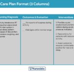
Acute Pain Care Plan
Nursing Diagnosis: Acute Pain
Related to: Disease processes, inflammatory processes, infection, pathological processes
As evidenced by: Reports of pain, appetite changes, altered physiological parameters, diaphoresis, distraction behavior, expressive behavior, facial grimacing/crying, guarding behavior, positioning to ease pain, protective behavior
Expected Outcomes:
- Patient will report abdominal pain of 2/10 or less by discharge.
- Patient will report relief from associated symptoms like nausea, cramping, and gas by discharge.
Assessments:
- Comprehensive Pain Assessment: Identify pain location, intensity, frequency, and characteristics to determine the underlying cause and treatment effectiveness.
- Review Diagnostic Studies: Assess results from ultrasounds, abdominal x-rays, and CT scans to aid in diagnosis.
Interventions:
- Administer Medications: Provide analgesics, sedatives, and medications for gas, nausea, constipation, or diarrhea as ordered.
- Position for Comfort: Assist patient to a comfortable position, such as knee-chest or side-lying, or elevate the head of the bed.
- Nasogastric Tube Insertion: Insert NG tube as needed for conditions like bowel obstruction.
- Prepare for Surgical Intervention: Assist and prepare the patient for surgery if indicated by the underlying cause.
Dysfunctional Gastrointestinal Motility Care Plan
Nursing Diagnosis: Dysfunctional Gastrointestinal Motility
Related to: Food intolerance, ingestion of contaminated materials, malnutrition, disease processes, anxiety, stressors
As evidenced by: Abdominal cramping, abdominal pain, absence of flatus, altered bowel sounds, diarrhea, constipation, nausea, vomiting, distended abdomen
Expected Outcomes:
- Patient will exhibit normal bowel sounds and remain free of abdominal pain and distention.
Assessments:
- Assess Abdominal Symptoms: Evaluate abdominal pain, nausea, vomiting, indigestion, symptom duration, and precipitating factors.
- Assess Dietary Habits: Thoroughly review daily food and liquid intake to identify potential causes.
- Assess Bowel Habits: Assess bowel movement frequency, consistency, color, and odor to aid in diagnosis.
Interventions:
- Administer Medications: Provide antidiarrheals, antibiotics, antacids, proton-pump inhibitors, or other medications as prescribed.
- Encourage Ambulation: Promote ambulation and exercise to increase GI motility and relieve symptoms.
- Dietary Education: Provide tailored dietary education, such as fiber supplements for constipation or elimination diets for diarrhea triggers.
- Obtain Stool Sample: Collect stool samples for analysis to identify infections or other abnormalities.
Imbalanced Nutrition: Less Than Body Requirements Care Plan
Nursing Diagnosis: Imbalanced Nutrition: Less Than Body Requirements
Related to: Abdominal pain, food aversion, pathological processes, inflammatory processes, loss of appetite, nausea and vomiting
As evidenced by: Body weight below ideal range, constipation, diarrhea, food intake less than RDA, hypoglycemia, abnormal bowel sounds, poor appetite
Expected Outcomes:
- Patient will progressively gain weight towards the desired goal.
- Patient will be free of signs of malnutrition.
- Patient will consume adequate caloric intake without discomfort.
Assessments:
- Nutritional Screening: Conduct a comprehensive nutritional screening, including physical findings, labs, diet history, weight/BMI, and food access.
- Assess Laboratory Values: Monitor prealbumin, albumin, C-reactive protein, and WBC count for indicators of inflammation and nutritional status.
- Identify Barriers to Eating: Assess for factors like nausea, pain, and socioeconomic barriers affecting nutritional intake.
Interventions:
- Promote Conducive Eating Environment: Minimize environmental stimulants to improve appetite and intake.
- Monitor Weight and Muscle Mass: Track weight gain and muscle mass to assess nutritional progress.
- Promote Oral Hygiene: Ensure good oral health to enhance appetite and functional ability to eat.
- Dietary Modifications: Advise avoiding high-fiber, raw, and spicy foods that can exacerbate pain.
- Small Frequent Bland Feedings: Offer small, frequent meals with bland foods like plain rice, oatmeal, toast, crackers, and clear soup.
- Refer to Dietitian: Refer to a dietitian or nutritionist for personalized meal planning.
Ineffective Tissue Perfusion Care Plan
Nursing Diagnosis: Ineffective Tissue Perfusion
Related to: Abdominal pain, inflammatory process, disease process
As evidenced by: Hypoactive or absent bowel sounds, bloating, abdominal rigidity, constipation, abdominal pain, nausea and vomiting, malnutrition, weight loss, fatigue
Expected Outcomes:
- Patient will remain free from nausea, vomiting, or abdominal discomfort.
- Patient will verbalize improved comfort and show no signs of tissue perfusion complications.
Assessments:
- Abdominal Assessment (IAPP): Assess, inspect, palpate, and auscultate the abdomen to identify perfusion issues like peritonitis and bowel obstruction.
- Assess Lab Results: Review liver enzymes, kidney function, and occult blood tests to determine underlying causes.
- Assess Diagnostic Imaging: Evaluate ultrasound results to visualize abdominal organs and identify causes of pain.
Interventions:
- NPO Status: Maintain NPO status until diagnosis to prevent pain aggravation and complications.
- Fluid Management: Monitor intake and output and administer fluid replacement for blood and fluid loss.
- Gradual Diet Progression: Encourage slow dietary progression from clear liquids to bland foods after NPO.
- Document Bowel Movements: Accurately document bowel movements to monitor for obstruction or bleeding.
- Rest After Meals: Encourage rest after meals to maximize blood flow for digestion.
Risk for Deficient Fluid Volume Care Plan
Nursing Diagnosis: Risk for Deficient Fluid Volume
Related to: Fluid loss through vomiting or diarrhea, aversion to food, decreased fluid intake, disease processes
As evidenced by: (Risk diagnosis – no actual evidence, interventions are preventative)
Expected Outcomes:
- Patient will maintain adequate hydration and fluid balance.
- Patient will consume at least 500 mL of fluid per day.
Assessments:
- Intake and Output: Monitor and document all fluid intake and output.
- Assess for Dehydration: Check skin turgor and mucous membranes for signs of dehydration.
- Monitor Lab Values: Review hematocrit, electrolytes, urinalysis, BUN, and creatinine levels.
Interventions:
- Administer IV Fluids: Provide IV fluids and electrolytes as prescribed to maintain hydration.
- Encourage Fluid Intake: Offer various fluids like jello, popsicles, soups, fruits, and Pedialyte.
- Parenteral/Enteral Nutrition: Provide nutrition and fluids via parenteral or enteral routes if NPO status is prolonged.
References
(Keep the original references if they were present in the source article. If not, consider adding relevant, credible sources.)