Acute cholangitis is a critical clinical syndrome arising from bacterial infection of the biliary tree, most frequently triggered by biliary obstruction. Prompt diagnosis and treatment are paramount to prevent severe complications and mortality. While the classic presentation of Charcot’s triad (fever, jaundice, and right upper quadrant pain) is suggestive, it lacks sensitivity. Therefore, a thorough understanding of the differential diagnosis is essential for accurate and timely management. This article provides an in-depth exploration of the differential diagnosis of acute cholangitis, aiming to enhance diagnostic accuracy and improve patient outcomes.
ETIOLOGY and Risk Factors
Understanding the underlying causes of biliary obstruction is crucial in considering acute cholangitis and its differential diagnosis. Choledocholithiasis, or gallstones in the common bile duct, is the most prevalent cause. However, other etiologies must be considered, including:
- Benign and Malignant Biliary Strictures: These can result from primary sclerosing cholangitis, IgG4-related cholangitis, or cholangiocarcinoma, as well as postsurgical strictures.
- Pancreatic and Periampullary Tumors: Malignancies in these areas can compress or obstruct the distal common bile duct.
- Biliary Stent Obstruction: Biofilm formation, sludge deposition, and reflux can lead to stent occlusion and subsequent cholangitis.
- Mirizzi Syndrome: Extrinsic compression of the common hepatic duct by a gallstone impacted in the cystic duct.
- Parasitic Infestations: Ascaris lumbricoides and liver flukes like Clonorchis sinensis can obstruct the biliary system and introduce infection.
- AIDS Cholangiopathy: Biliary strictures and papillary stenosis in the context of HIV infection.
- Choledochocele and Narrow-Caliber Bile Ducts: Congenital anomalies predisposing to obstruction and cholangitis.
- Post-ERCP Cholangitis: A recognized complication following endoscopic retrograde cholangiopancreatography.
Certain risk factors increase susceptibility to gallstone formation and, consequently, acute cholangitis. These include a high-fat diet, sedentary lifestyle, obesity, rapid weight loss, and heavy alcohol consumption, which can contribute to liver cirrhosis and gallstone development.
EPIDEMIOLOGY
The prevalence of gallstones, the primary risk factor for acute cholangitis, varies across ethnicities. While gallstones are common in White populations (10%-15%) and Native Americans (60%-70%), they are less prevalent in Asians and African Americans. A significant proportion of patients hospitalized for gallstone disease (6%-9%) develop acute cholangitis. The condition affects males and females equally, typically presenting in individuals aged 50 to 60 years.
PATHOPHYSIOLOGY
Biliary obstruction is the cornerstone of acute cholangitis pathogenesis. While bacteria may be present in bile even under normal conditions, obstruction leads to bile stasis, increased intraductal pressure, and impaired local defense mechanisms. Normally, bile salts possess bacteriostatic properties, and the biliary epithelium secretes IgA and mucus to prevent bacterial adhesion. Kupffer cells and tight junctions between cholangiocytes limit bacterial translocation. Normal bile flow effectively flushes bacteria into the duodenum.
Obstruction disrupts these protective mechanisms. Elevated intraductal pressure exceeding 25 cm H2O can trigger cholangiovenous and cholangiolymphatic reflux, leading to bacteremia and endotoxemia. Systemic release of inflammatory mediators like TNF, IL-1, and IL-6 further contributes to hemodynamic instability.
The most common bacterial pathogens in acute cholangitis are Gram-negative enteric organisms, including Escherichia coli, Klebsiella species, Enterococcus species, and Enterobacter species. Anaerobic bacteria and parasites can also be causative agents, particularly in specific patient populations.
CLINICAL PRESENTATION and Diagnostic Criteria
The clinical spectrum of acute cholangitis ranges from mild to severe. The classic Charcot’s triad, while highly specific (95.9%), exhibits low sensitivity (26.4%). Reynolds’ pentad, which includes Charcot’s triad plus hypotension and altered mental status, indicates severe cholangitis.
To improve diagnostic accuracy, the Tokyo Guidelines (TG) were developed. TG07 criteria, established in 2006, and subsequently refined in TG13 (2012), provide more sensitive diagnostic frameworks. TG13 criteria categorize diagnostic items into:
(A) Systemic Inflammation:
- Fever (temperature > 38°C) and/or chills.
- Laboratory evidence of inflammatory response (WBC > 10,000/cmm, CRP ≥ 1 mg/dL).
(B) Cholestasis:
- Jaundice (total bilirubin ≥ 2 mg/dL).
- Laboratory evidence of cholestasis (Alkaline phosphatase, GGT, AST, ALT > 1.5 × upper limit of normal).
(C) Imaging:
- Biliary dilatation.
- Evidence of etiology (stone, stricture, stent, etc.).
A suspected diagnosis requires one item from A plus one item from either B or C. A definite diagnosis requires one item from A, one from B, and one from C. TG13 criteria significantly improved sensitivity (91.8%) compared to Charcot’s triad and TG07, while maintaining reasonable specificity (77.7%).
Physical examination findings may include fever, tachycardia, hypotension, jaundice, right upper quadrant tenderness, and altered mental status.
Severity Grading
The severity of acute cholangitis is classified into three grades based on response to initial medical management and organ dysfunction:
- Grade I (Mild): Patients respond to initial antibiotics and lack organ dysfunction or criteria for moderate cholangitis.
- Grade II (Moderate): Patients do not respond to initial antibiotics but lack organ dysfunction. At least two of the following conditions are present: WBC > 12,000/cmm or < 4,000/cmm, temperature > 39°C or < 36°C, age > 75 years, total bilirubin ≥ 5 mg/dL, or hypoalbuminemia.
- Grade III (Severe): Patients fail to respond to initial medical treatment and exhibit organ dysfunction in at least one of the following systems: cardiovascular (hypotension requiring dopamine ≥ 5 μg/kg/min or norepinephrine), neurological (altered consciousness), respiratory (PaO2/FiO2 ratio < 300), renal (oliguria or creatinine > 2.0 mg/dL), hepatic (PT-INR > 1.5), or hematologic (platelet count < 100,000/cmm).
Acute suppurative cholangitis (ASC), characterized by pus in the bile duct, is sometimes diagnosed during ERCP. However, ASC does not always correlate with severe acute cholangitis.
ACUTE CHOLANGITIS DIFFERENTIAL DIAGNOSIS
The differential diagnosis of acute cholangitis is broad and includes several conditions that can mimic its clinical presentation. A systematic approach, incorporating clinical findings, laboratory data, and imaging, is crucial for accurate differentiation. Key differential diagnoses include:
-
Acute Cholecystitis: Inflammation of the gallbladder, often due to gallstones. While sharing right upper quadrant pain and fever with acute cholangitis, jaundice is less common in uncomplicated cholecystitis. Imaging, particularly ultrasound, is essential to differentiate. Ultrasound in cholecystitis typically reveals gallbladder wall thickening, pericholecystic fluid, and Murphy’s sign. In contrast, cholangitis imaging focuses on biliary duct dilatation.
-
Acute Hepatitis: Viral or drug-induced hepatitis can present with jaundice, fever, and abdominal discomfort. However, hepatitis typically lacks the marked right upper quadrant tenderness and biliary dilatation seen in cholangitis. Liver function tests are abnormal in both conditions, but the pattern may differ. Viral hepatitis serology is crucial in differentiation.
-
Liver Abscess: Pyogenic or amebic liver abscess can cause fever, right upper quadrant pain, and leukocytosis. Jaundice may be present if the abscess obstructs bile ducts. Imaging, particularly CT or MRI, is critical to identify the abscess. Blood cultures and aspiration of the abscess can identify the causative organism.
-
Cirrhosis of the Liver: Decompensated cirrhosis can present with jaundice, ascites, and encephalopathy, mimicking severe cholangitis. However, cirrhosis is a chronic condition with characteristic stigmata of chronic liver disease, such as spider angiomata, palmar erythema, and splenomegaly. While infection can trigger decompensation in cirrhosis, biliary dilatation is typically absent unless there is superimposed cholangitis.
-
Septic Shock of Other Origin: Sepsis from any source, such as pneumonia or urinary tract infection, can present with fever, hypotension, and altered mental status, resembling severe cholangitis (Reynolds’ pentad). However, in non-biliary sepsis, jaundice and right upper quadrant pain are usually absent or less prominent. Thorough clinical examination and laboratory investigations are needed to identify the primary source of infection.
-
Right-Sided Diverticulitis: Inflammation of the colonic diverticula in the right colon can cause right lower quadrant or flank pain, fever, and leukocytosis, potentially mimicking cholangitis, especially if the pain radiates to the right upper quadrant. However, jaundice is absent, and bowel symptoms may be more prominent. CT scan of the abdomen and pelvis can differentiate diverticulitis from cholangitis.
-
Right-Sided Pyelonephritis: Kidney infection can present with flank pain, fever, and leukocytosis, mimicking cholangitis, especially if pain radiates to the upper abdomen. However, urinary symptoms are typically present in pyelonephritis, and jaundice is absent. Urinalysis and urine culture are diagnostic.
-
Pancreatitis: Acute pancreatitis, especially biliary pancreatitis, can present with abdominal pain, elevated amylase and lipase, and potentially jaundice if the common bile duct is obstructed. While imaging might show biliary dilatation in biliary pancreatitis, the clinical focus in primary pancreatitis is typically centered around epigastric pain and pancreatic enzyme elevation, rather than cholestasis markers to the same degree as cholangitis.
-
Hepatic Flexure Colitis: Inflammation of the hepatic flexure of the colon can cause right upper quadrant pain and mimic some aspects of cholangitis, but typically lacks jaundice and significant cholestasis. Colonoscopy can help differentiate.
-
Subphrenic Abscess: An abscess below the diaphragm can cause fever and right upper quadrant pain, potentially mimicking cholangitis. Imaging, especially CT scan, is crucial for diagnosis.
INVESTIGATIONS
Diagnostic workup for suspected acute cholangitis and its differential diagnosis includes:
-
Laboratory Tests:
- Complete blood count (CBC) to assess for leukocytosis.
- Erythrocyte sedimentation rate (ESR) or C-reactive protein (CRP) to evaluate inflammation.
- Complete metabolic panel, including liver function tests (bilirubin, alkaline phosphatase, GGT, AST, ALT) to assess cholestasis and liver injury.
- Prothrombin time (PT) and INR to assess coagulation.
- Blood cultures to identify bacteremia. Bile culture, if drainage is performed, can also be informative.
-
Imaging Studies:
- Abdominal Ultrasound: Initial imaging modality to assess for gallstones, biliary dilatation, and gallbladder abnormalities (cholecystitis). Less sensitive for distal common bile duct stones.
- Computed Tomography (CT): More sensitive than ultrasound for detecting common bile duct stones and assessing for other intra-abdominal pathology (abscess, diverticulitis, pancreatitis). CT without contrast is superior for stone detection.
- Magnetic Resonance Cholangiopancreatography (MRCP): Highly sensitive for detecting choledocholithiasis and biliary strictures. Excellent for visualizing the biliary tree and identifying the level and cause of obstruction.
- Endoscopic Ultrasound (EUS): Most sensitive imaging modality for choledocholithiasis, particularly small stones. Can also assess for periampullary tumors and guide fine-needle aspiration if needed.
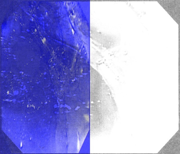
Alt text: Purulent discharge from the ampulla of Vater during ERCP, indicative of acute suppurative cholangitis.
MANAGEMENT
Management of acute cholangitis requires a multi-faceted approach:
-
Resuscitation: Intravenous fluids to address dehydration and hypotension.
-
Antibiotics: Empiric broad-spectrum antibiotics should be initiated promptly to cover Gram-negative and anaerobic bacteria. Common choices include piperacillin-tazobactam, ceftriaxone plus metronidazole, or ciprofloxacin plus metronidazole in penicillin-allergic patients. Antibiotic selection should be guided by local resistance patterns and adjusted based on blood and bile culture results.
-
Biliary Drainage: Essential to relieve biliary obstruction and decompress the infected biliary system.
- Endoscopic Retrograde Cholangiopancreatography (ERCP): The preferred method for biliary drainage. Sphincterotomy and stone extraction are performed for choledocholithiasis. Stent placement may be necessary for strictures or when stone extraction is not feasible.
- Endoscopic Nasobiliary Drainage (ENBD): Placement of a nasobiliary catheter for drainage. Less comfortable for patients but allows for repeat cholangiography and effective drainage of purulent bile.
- Percutaneous Transhepatic Biliary Drainage (PTBD): Reserved for cases where ERCP is unsuccessful or contraindicated. Involves percutaneous puncture and drainage of the biliary system.
- EUS-Guided Biliary Drainage: Emerging technique for biliary drainage when ERCP fails, particularly in altered anatomy.
- Surgical Drainage: Rarely needed in the modern era, reserved for failed endoscopic and percutaneous approaches.
Alt text: Endoscopic drainage of pus from the bile duct following biliary stent insertion during ERCP, demonstrating successful biliary decompression for acute cholangitis.
Timing of Biliary Drainage
- Grade I (Mild): Drainage within 24-48 hours.
- Grade II (Moderate): Early drainage, ideally within 24 hours of admission.
- Grade III (Severe): Urgent drainage as soon as possible.
Following resolution of acute cholangitis due to gallstones, laparoscopic cholecystectomy is recommended to prevent recurrence.
Alt text: Endoscopic biliary sphincterotomy, a key step in ERCP for acute cholangitis management, widening the bile duct opening to facilitate stone removal.
Alt text: Endoscopic retrieval of a biliary stone using a basket after sphincterotomy, a common procedure to relieve obstruction in acute cholangitis.
Alt text: Fluoroscopic image showing lithotripsy basket-assisted extraction of a large bile duct stone during ERCP, a technique used when standard methods fail in acute cholangitis.
Alt text: Endoscopic nasobiliary drainage catheter in situ, providing external biliary drainage in a patient with acute cholangitis after ERCP.
PROGNOSIS
The prognosis of acute cholangitis is significantly improved with timely biliary drainage and antibiotic therapy. Mortality rates have decreased substantially since the advent of ERCP. However, delayed drainage and severe cholangitis still carry a higher risk. Poor prognostic factors include advanced age, high fever, leukocytosis, hyperbilirubinemia, hypoalbuminemia, pre-existing comorbidities, and elevated pre-drainage serum creatinine.
CONCLUSION
Acute cholangitis is a potentially life-threatening condition requiring prompt recognition and management. While Charcot’s triad is suggestive, a comprehensive understanding of the differential diagnosis is crucial for accurate and timely intervention. The Tokyo Guidelines provide a robust framework for diagnosis and severity assessment. A systematic approach incorporating clinical evaluation, laboratory investigations, and imaging, coupled with prompt antibiotic therapy and biliary drainage, is essential to optimize patient outcomes and minimize mortality. Differentiating acute cholangitis from its mimics, such as acute cholecystitis, hepatitis, and liver abscess, is paramount to ensure appropriate and targeted treatment strategies are implemented.