Headache stands as a ubiquitous human experience, frequently encountered in emergency departments (EDs). While the majority of headaches are benign, a subset signifies serious, potentially life-threatening underlying conditions. For automotive repair experts at xentrydiagnosis.store, understanding diagnostic differentiation is crucial; similarly, for medical professionals, accurately distinguishing between benign and dangerous headaches is paramount to avert severe outcomes. This article provides an in-depth guide to the differential diagnosis of acute headaches, focusing on critical aspects for emergency evaluation and management.
Understanding Headache Etiology for Differential Diagnosis
Headaches are broadly categorized into primary and secondary types. Primary headaches, such as migraines, tension headaches, and cluster headaches, occur independently and are not caused by an underlying medical condition. Secondary headaches, conversely, arise as symptoms of other medical issues, some of which are life-threatening. Accurate differential diagnosis hinges on recognizing the subtle yet critical differences between these categories, especially in the acute setting.
The International Classification of Headache Disorders (ICHD-III) provides a standardized framework for headache classification, essential for systematic differential diagnosis:
- Primary Headaches: Migraine, tension-type headache, cluster headache, and other trigeminal autonomic cephalalgias.
- Secondary Headaches: Attributed to underlying conditions like head trauma, vascular disorders, intracranial pressure changes, infections, substance abuse, or other systemic illnesses.
- Cranial Neuralgias and Facial Pain: Including trigeminal neuralgia and other pain syndromes affecting the cranial nerves.
For emergency clinicians, a strong familiarity with conditions that manifest as secondary headaches is crucial. These conditions, demanding prompt recognition and intervention, include:
- Vascular Disorders: Subarachnoid hemorrhage, intracranial hemorrhage, stroke, transient ischemic attack (TIA), carotid or vertebral artery dissection, cerebral venous sinus thrombosis.
- Intracranial Pressure Issues: Idiopathic intracranial hypertension, hydrocephalus, space-occupying lesions (tumors, abscesses).
- Infections: Meningitis, encephalitis, sinusitis.
- Systemic Illnesses: Hypertensive emergencies, giant cell arteritis, acute glaucoma, carbon monoxide poisoning, medication overuse headache, toxin exposure or withdrawal.
Epidemiology of Headaches: Context for Acute Presentations
Headaches are incredibly common, with a lifetime prevalence estimated at 96% in the United States. While not all headaches require emergency attention, their high prevalence means EDs frequently encounter patients presenting with acute headache. Understanding the epidemiology provides context for assessing risk:
- Prevalence Peaks: Headaches are most prevalent in individuals aged 25 to 40, but concerning secondary headaches can occur at any age.
- Gender Differences: Women experience migraines and severe headaches more frequently than men, but both genders are susceptible to dangerous secondary headaches.
- Emergency Department Visits: Headaches account for approximately 3% of all ED visits, emphasizing the need for efficient and accurate differential diagnosis in this setting.
Pathophysiology: Differentiating Pain Mechanisms
While brain tissue itself lacks pain receptors, headaches arise from nociceptor activation in surrounding structures. These include blood vessels, meninges, muscles of the head and neck, facial structures, and cranial nerves. Understanding the potential pain sources aids in differential diagnosis:
- Primary Headaches: Pathophysiology is complex and not fully understood but involves neuronal and vascular mechanisms. It’s crucial to recognize that primary headaches are diagnoses of exclusion, made after ruling out secondary causes.
- Secondary Headaches: Pathophysiology directly relates to the underlying condition. For example, subarachnoid hemorrhage causes sudden, severe headache due to blood irritating meninges and increasing intracranial pressure. Meningitis involves inflammation of the meninges, leading to headache, neck stiffness, and other systemic symptoms.
History and Physical Examination: Cornerstones of Acute Headache Differential Diagnosis
A detailed history and thorough physical exam are the most critical tools in differentiating acute headaches. For automotive diagnostics, expert technicians rely on systematic troubleshooting; similarly, clinicians use a structured approach to headache evaluation. The goal is to identify “red flags” suggesting serious secondary headaches, guiding further investigation and management.
Key Historical Features for Differential Diagnosis:
-
Detailed Headache Characterization:
- Location: Unilateral, bilateral, localized, generalized?
- Onset: Sudden (thunderclap), gradual? Time of onset?
- Provoking/Palliating Factors: What triggers or relieves the headache?
- Progression: Worsening, improving, constant?
- Quality: Throbbing, sharp, dull, pressure-like?
- Severity: Pain scale rating (1-10).
- Radiation: Does the pain spread to other areas?
- Prior Headaches: History of similar headaches? Change in headache pattern?
-
Crucial Questions to Rule Out Secondary Headaches:
- “Worst headache of my life?” (Subarachnoid hemorrhage)
- Sudden, maximal onset? (Subarachnoid hemorrhage, cervical artery dissection)
- Fever, stiff neck? (Meningitis, encephalitis)
- Neurological symptoms: Weakness, numbness, speech difficulty, vision changes, confusion? (Stroke, TIA, space-occupying lesion, dissection)
- Trauma history? (Subdural hematoma, post-concussion headache, cervical artery dissection)
- Systemic symptoms: Fever, weight loss, malaise? (Infection, malignancy, giant cell arteritis)
- Recent travel, sick contacts, immunocompromised state? (Infection)
- Medications, substance use? (Medication overuse headache, withdrawal)
- Pregnancy, postpartum period? (Preeclampsia/eclampsia, cerebral venous sinus thrombosis)
- Eye pain, vision changes? (Acute angle-closure glaucoma)
- Jaw claudication, scalp tenderness (elderly)? (Giant cell arteritis)
-
SNOOP Mnemonic: Red Flags for Secondary Headaches: This is an invaluable tool for rapid risk stratification:
- Systemic symptoms, secondary risk factors (fever, cancer, HIV, pregnancy, hypertension).
- Neurologic symptoms or abnormal signs (confusion, weakness, seizures, vision changes, papilledema).
- Onset sudden (“thunderclap”) or new onset, especially in older individuals (>50 years).
- Other associated conditions or features: trauma, toxins, Valsalva maneuver/exertion induced, awakens from sleep, positional headache.
- Prior headache history: progressive change in pattern, or significant difference from typical headaches.
Physical Examination Findings Guiding Differential Diagnosis:
- General Appearance: Signs of systemic illness, distress level.
- Vital Signs: Blood pressure (hypertensive emergency), heart rate, temperature (infection).
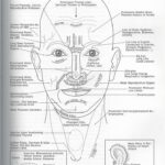
- HEENT Exam:
- Fundoscopic exam: Papilledema (increased intracranial pressure).
- Conjunctival injection, fixed dilated pupil: Acute angle-closure glaucoma.
- Temporal artery tenderness: Giant cell arteritis.
- Sinus tenderness: Sinusitis.
- Ear exam: Otitis media/externa.
- Neurological Exam:
- Mental status: Alertness, orientation, cognition.
- Cranial nerves: Visual fields, pupillary reflexes, extraocular movements, facial droop, bulbar function.
- Motor and sensory function: Weakness, numbness, ataxia.
- Reflexes: Asymmetry, hyperreflexia.
- Meningeal signs: Nuchal rigidity (meningitis), Kernig’s and Brudzinski’s signs (less sensitive in adults).
Table
Table 1. Characteristics of migraine, tension-type, and cluster headache syndromes.
This table, directly from the original article, is crucial for differentiating common primary headaches. Understanding these features is essential after excluding secondary causes in the differential diagnosis.
Evaluation: Diagnostic Testing for Acute Headache
If red flags are present, or the history and physical suggest a secondary headache, further investigation is warranted. Similar to using advanced diagnostic tools in automotive repair, clinicians employ specific tests to confirm or exclude serious conditions.
Laboratory Studies:
- Routine labs: Generally not helpful for primary headache diagnosis.
- Pregnancy test: In women of childbearing age with headache and hypertension.
- Serum glucose: In patients with altered mental status or focal neurological deficits.
- Carboxyhemoglobin level: Suspected carbon monoxide poisoning.
- Erythrocyte sedimentation rate (ESR) and C-reactive protein (CRP): Suspected giant cell arteritis. Elevated ESR/CRP supports the diagnosis, but normal values do not rule it out.
- Coagulation studies and D-dimer: Suspected cerebral venous thrombosis. D-dimer can help rule out CVT in low-risk patients, but MRV/CTV is often needed in higher-risk cases.
- Lumbar puncture (LP): Considered if meningitis, encephalitis, or subarachnoid hemorrhage (if CT is negative and suspicion remains high) are suspected. Also diagnostic for idiopathic intracranial hypertension (opening pressure). Crucially, CT scan should precede LP to rule out mass effect and prevent herniation.
Radiographic Imaging:
- Non-contrast head CT: Initial imaging modality of choice for acute headache with red flags, especially to rule out intracranial hemorrhage, hydrocephalus, and mass lesions.
- CT angiography (CTA) or MR angiography (MRA): For suspected vascular pathologies like subarachnoid hemorrhage (especially if CT is negative but suspicion is high), arterial dissections, or cerebral venous sinus thrombosis.
- MRI and MR venography (MRV): Highly sensitive for cerebral venous sinus thrombosis and posterior fossa lesions.
- Imaging Indications (Based on ACEP guidelines and clinical judgment):
- New neurological deficits.
- Sudden onset, severe headache (“thunderclap headache”).
- New-onset headache in HIV-positive patients.
- New headache in patients >50 years old.
- Presence of SNOOP red flags.
- Worsening headache despite treatment.
- History of cancer.
- Immunocompromised state.
Differential Diagnosis of Acute Headache: Key Conditions to Consider
The differential diagnosis for acute headache is extensive. Focusing on acute presentations, particularly in the emergency setting, the most critical differential diagnoses include:
- Subarachnoid Hemorrhage (SAH): “Thunderclap headache,” sudden onset, maximal intensity at onset, often with neck stiffness, vomiting, and altered consciousness. CT angiography is essential for diagnosis.
- Meningitis/Encephalitis: Fever, nuchal rigidity, altered mental status, photophobia, headache. Lumbar puncture is crucial for CSF analysis.
- Cerebral Venous Sinus Thrombosis (CVST): Headache (often worsening, “thunderclap” in some cases), papilledema, focal neurological deficits, seizures. MRV or CTV is diagnostic. Risk factors include hypercoagulable states, pregnancy, and infection.
- Cervical Artery Dissection (CAD): Headache (often unilateral), neck pain, Horner’s syndrome, stroke or TIA symptoms (younger patients). MRA or CTA of the neck and head is used for diagnosis.
- Stroke/Transient Ischemic Attack (TIA): Sudden onset neurological deficits (weakness, speech difficulty, vision loss), headache may be present but not always the primary symptom. CT or MRI of the brain is essential.
- Hypertensive Emergency: Severe headache, markedly elevated blood pressure, end-organ damage (encephalopathy, retinopathy, nephropathy). Immediate blood pressure management is crucial.
- Idiopathic Intracranial Hypertension (IIH): Headache (often daily, worse with straining/lying down), papilledema, vision changes, pulsatile tinnitus. Lumbar puncture (elevated opening pressure) and MRI (to exclude other causes) are diagnostic.
- Giant Cell Arteritis (GCA): New onset headache (often temporal, unilateral), age >50, scalp tenderness, jaw claudication, vision loss. Elevated ESR/CRP, temporal artery biopsy for definitive diagnosis.
- Acute Angle-Closure Glaucoma: Sudden onset eye pain, headache, vision changes (halos around lights), nausea/vomiting. Ophthalmologic exam (increased intraocular pressure) is diagnostic.
- Carbon Monoxide Poisoning: Headache, dizziness, nausea, vomiting, confusion, history of exposure to CO (e.g., faulty heating, car exhaust). Carboxyhemoglobin level is diagnostic.
- Space-Occupying Lesions (Tumors, Abscesses): Progressive headache, focal neurological deficits, seizures. MRI or CT of the brain is diagnostic.
Other conditions to consider in the broader differential include sinusitis, temporomandibular joint (TMJ) disorders, dental problems, medication overuse headache, and post-concussion syndrome.
Treatment and Management of Acute Headache in the Emergency Department
Emergency management of acute headache focuses on two primary goals:
- Identify and treat life-threatening secondary headaches: This is the priority. Specific treatment depends on the underlying diagnosis (e.g., neurosurgical intervention for SAH, antibiotics for meningitis, antihypertensives for hypertensive emergency).
- Provide symptomatic relief for benign headaches: For primary headaches or benign secondary headaches after serious causes are ruled out, treatment aims to reduce pain and associated symptoms.
Symptomatic Treatment Options for Benign Headaches in the ED:
- Non-steroidal anti-inflammatory drugs (NSAIDs): Ketorolac, ibuprofen, naproxen. Effective for mild to moderate headaches.
- Acetaminophen: For mild to moderate headaches, often used in combination with other agents.
- Triptans: Sumatriptan, rizatriptan, etc. Effective for migraines, but contraindicated in certain vascular conditions.
- Antiemetics: Prochlorperazine, metoclopramide. Help with nausea and vomiting, and some (like prochlorperazine) have inherent analgesic properties for headache.
- Dopamine antagonists (Neuroleptics): Prochlorperazine, metoclopramide. Effective for migraine and tension-type headaches in the ED setting.
- Corticosteroids (Dexamethasone): May reduce migraine recurrence in the 72 hours post-ED discharge. Not for routine use, but can be considered in specific cases.
- Sphenopalatine Ganglion Block: Intranasal lidocaine or bupivacaine may provide rapid relief for various headache types, a promising newer technique.
- Oxygen: High-flow oxygen is first-line treatment for cluster headaches.
Medications to Avoid in Routine Headache Management:
- Opioids: Generally avoided for primary headaches in the ED due to limited efficacy, risk of dependence, and potential to worsen chronic headache problems (medication overuse headache). Reserved for very specific situations when other options fail and secondary causes are ruled out.
Table
Table 2. Acute therapy for migraine.
This table summarizes acute migraine treatments, providing a quick reference for ED management after ruling out secondary headaches.
Table
Table 3. Short-term preventive therapy for episodic cluster therapy.
This table is relevant for managing cluster headaches in the acute setting, highlighting short-term preventative strategies.
Prognosis and Complications
- Primary Headaches: Prognosis is generally good in terms of long-term health, but chronic or recurrent headaches can significantly impact quality of life. Proper management and preventative strategies are crucial.
- Secondary Headaches: Prognosis varies widely depending on the underlying condition. Early diagnosis and treatment are critical to minimize morbidity and mortality associated with serious secondary headaches.
- Complications: Complications of primary headaches are mainly related to disability and reduced productivity. A major complication of headache treatment is medication overuse headache. Complications of secondary headaches are those of the underlying condition, ranging from neurological deficits to death. Delayed diagnosis of serious secondary headaches can have devastating consequences.
Deterrence and Patient Education
Patient education is crucial for both primary and secondary headaches:
- Primary Headaches: Educate patients about the benign nature of their headaches, triggers to avoid, lifestyle modifications (sleep, caffeine), and appropriate use of acute and preventative medications. Emphasize follow-up with a primary care physician or neurologist for ongoing management.
- Secondary Headaches: Educate patients about the underlying cause of their headache, treatment plan, potential complications, and red flags that should prompt them to seek immediate medical attention.
Enhancing Healthcare Team Outcomes
Effective management of acute headaches, especially the differential diagnosis and management of serious secondary causes, requires a collaborative interprofessional team. This includes emergency physicians, nurses, neurologists, radiologists, pharmacists, and potentially neurosurgeons and critical care specialists. Clear communication, coordinated care pathways, and shared decision-making are essential to optimize patient outcomes and minimize errors in diagnosis and treatment.
Conclusion
Differentiating between benign and dangerous causes of acute headache is a critical skill in emergency medicine. A thorough history, meticulous physical examination utilizing tools like the SNOOP mnemonic, and judicious use of diagnostic testing are essential for accurate differential diagnosis. By focusing on red flags and systematically considering serious secondary headache etiologies, clinicians can ensure timely and appropriate management, improving outcomes for patients presenting with this common yet potentially life-threatening complaint. This systematic approach mirrors the diagnostic rigor demanded in fields like automotive repair, where accurate and timely diagnosis is equally critical.