Angina bullosa haemorrhagica (ABH) is a benign oral condition characterized by the sudden appearance of blood-filled blisters on the oral mucosa. While ABH is generally harmless and resolves spontaneously, it is crucial for clinicians to accurately diagnose it and differentiate it from other more serious blistering conditions. This article provides a comprehensive overview of the Angina Bullosa Haemorrhagica Differential Diagnosis, aiding in accurate identification and patient management.
Understanding Angina Bullosa Haemorrhagica
Angina bullosa haemorrhagica, first described by Badham in 1967, is a benign condition presenting with single or multiple vesicles or bullae filled with blood, arising on the oral mucosa. These lesions are typically painless and often rupture spontaneously within 24-48 hours, leaving behind superficial erosions that heal within a week without scarring. The soft palate is the most commonly affected site, but ABH can occur anywhere in the oropharynx, including the lips, buccal mucosa, tongue, and even extending to the esophagus.
While the exact etiology of ABH remains unclear, several predisposing factors have been identified. Trauma, even minor, such as from dental procedures, hot or spicy foods, or forceful coughing, is frequently cited as a trigger. Systemic conditions like diabetes mellitus, hypertension, and chronic renal failure have also been associated with an increased incidence of ABH. Inhaled corticosteroids are another recognized cause. However, in many cases, no specific trigger can be identified.
Clinical Presentation of ABH
Patients with ABH typically present with a sudden onset of one or more blood-filled blisters in the oral cavity. As exemplified in a case of a 48-year-old male with hypertension and diabetes, ABH can manifest as an asymptomatic, single hemorrhagic bulla. Upon examination, a non-tender, blood-filled bulla, approximately 1.5–2 cm in diameter, may be observed on the inner aspect of the lower lip, as shown in Figure 1.
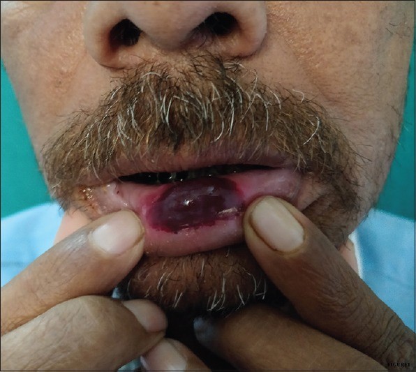 Hemorrhagic bulla on the inner lower lip in Angina Bullosa Haemorrhagica: A clinical presentation.
These bullae are fragile and tend to rupture quickly, often within a day or two, leading to a superficial ulcer. The rapid onset and spontaneous healing without scarring are characteristic features of ABH. Hematological and coagulation studies are typically normal in patients with ABH, further supporting the diagnosis and helping to rule out other conditions.
Angina Bullosa Haemorrhagica Differential Diagnosis
The primary importance of understanding ABH lies in its differential diagnosis. Several other vesiculobullous conditions can mimic ABH, necessitating careful clinical evaluation and, in some cases, further investigations to reach an accurate diagnosis. The key conditions in the angina bullosa haemorrhagica differential diagnosis include:
1. Thrombocytopenia
Thrombocytopenia, a condition characterized by a low platelet count, can lead to spontaneous bleeding and bruising, including the formation of blood blisters in the oral mucosa. While hemorrhagic bullae can be seen in thrombocytopenia, these are usually associated with other signs of bleeding disorders, such as petechiae, purpura, and easy bruising in other parts of the body. Hematological investigations, specifically a platelet count, are crucial to differentiate thrombocytopenia from ABH. In thrombocytopenia, the platelet count will be significantly reduced, whereas it is normal in ABH.
2. Pemphigus and Bullous Pemphigoid
Pemphigus vulgaris and bullous pemphigoid are autoimmune blistering diseases that can affect the oral mucosa. Pemphigus vulgaris typically presents with fragile vesicles and bullae that easily rupture, leading to painful erosions. Oral involvement is common and often precedes skin lesions. Bullous pemphigoid, on the other hand, is characterized by tense bullae that are less prone to rupture. While oral involvement can occur, it is less frequent and less severe than in pemphigus vulgaris.
Distinguishing pemphigus and bullous pemphigoid from ABH involves considering the clinical presentation and utilizing diagnostic tools. Pemphigus and bullous pemphigoid lesions are typically more persistent and may not heal spontaneously within a week as seen in ABH. Furthermore, these conditions often involve other mucosal sites and the skin. Biopsy and immunofluorescence studies are essential for definitive diagnosis, revealing characteristic histopathological and immunopathological features of pemphigus and bullous pemphigoid, which are absent in ABH.
3. Bullous Lichen Planus
Bullous lichen planus is a variant of lichen planus that presents with bullae in addition to the typical features of lichen planus, such as reticular white striae (Wickham’s striae) and papules. Oral lichen planus commonly affects the buccal mucosa, tongue, and gingiva. While bullae can occur, they are less frequent than the characteristic lichen planus lesions.
The presence of typical lichen planus lesions, along with bullae, is a key differentiating factor from ABH. Biopsy can help confirm the diagnosis of bullous lichen planus, showing characteristic lichenoid tissue reaction and, in some cases, subepithelial bullae.
4. Dermatitis Herpetiformis
Dermatitis herpetiformis is a chronic blistering skin condition associated with celiac disease. While primarily affecting the skin, oral lesions can occur, although they are less common. Oral manifestations may include small vesicles or erosions.
Dermatitis herpetiformis is less likely to present with prominent, isolated blood-filled bullae like ABH in the oral cavity. Skin lesions are typically pruritic and symmetrically distributed, often on extensor surfaces. Diagnosis is confirmed by skin biopsy with direct immunofluorescence showing IgA deposits in the dermal papillae, and serological testing for celiac disease antibodies.
5. Epidermolysis Bullosa
Epidermolysis bullosa (EB) is a group of genetic disorders characterized by skin and mucous membrane fragility, leading to blister formation with minor trauma. There are various types of EB, ranging in severity. Oral involvement is common in many forms of EB, with blister formation and erosions occurring in response to minimal friction.
Epidermolysis bullosa is typically evident from early childhood or infancy, with a history of easy blistering since birth. The presence of skin blistering, often in multiple locations and triggered by minor trauma, along with a family history, helps differentiate EB from ABH. Genetic testing and skin biopsy with immunohistochemistry are used to diagnose and classify different types of epidermolysis bullosa.
6. Oral Amyloidosis
Oral amyloidosis, although rare, can present with various oral lesions, including vesicles and bullae. Amyloidosis involves the deposition of abnormal amyloid protein in tissues and organs. In the oral cavity, amyloid deposition can lead to macroglossia, mucosal plaques, and, less commonly, bullae.
Oral amyloidosis is a less common consideration in the differential diagnosis of ABH. Other clinical features of amyloidosis, such as macroglossia, systemic involvement, and purpuric papules, may be present. Biopsy with Congo red staining and immunohistochemistry is essential to confirm the diagnosis of amyloidosis and identify amyloid deposits in the tissue.
Diagnostic Approach for ABH
The diagnosis of angina bullosa haemorrhagica is primarily clinical, based on the characteristic presentation of sudden onset, painless blood-filled blisters in the oral cavity that rupture spontaneously and heal quickly without scarring. The absence of systemic symptoms and normal hematological and coagulation parameters further support the diagnosis.
However, in cases where the clinical presentation is atypical, or if there is suspicion of other blistering conditions, further investigations may be warranted. These may include:
- Hematological investigations: To rule out thrombocytopenia and other bleeding disorders.
- Biopsy: To exclude other vesiculobullous conditions such as pemphigus, bullous pemphigoid, and bullous lichen planus. Histopathology of ABH typically shows subepithelial bulla with hemorrhage, without specific inflammatory features.
- Immunofluorescence studies: To rule out autoimmune blistering diseases like pemphigus and bullous pemphigoid. Immunofluorescence is negative in ABH.
It is crucial to emphasize that in typical cases of ABH, these investigations are not necessary. The clinical diagnosis is usually sufficient, preventing unnecessary anxiety and invasive procedures for the patient.
Management and Prognosis
Angina bullosa haemorrhagica is a benign, self-limiting condition. No specific treatment is required, as the lesions heal spontaneously within a week. Reassurance and patient education about the benign nature of the condition are essential. Patients should be advised to avoid potential triggers, if identifiable, such as sharp or spicy foods, and to practice gentle oral hygiene.
The prognosis for ABH is excellent. Recurrences can occur, but they are also self-limiting and do not lead to any long-term complications. The primary goal of diagnosis is to differentiate ABH from more serious vesiculobullous diseases and to provide reassurance to the patient.
Conclusion
Angina bullosa haemorrhagica, while sometimes alarming due to its dramatic presentation of blood-filled blisters, is a benign oral condition. Understanding the angina bullosa haemorrhagica differential diagnosis is paramount for accurate diagnosis and appropriate patient management. By carefully considering the clinical presentation and, when necessary, utilizing appropriate investigations, clinicians can confidently diagnose ABH and differentiate it from other vesiculobullous disorders, ensuring optimal patient care and avoiding unnecessary interventions.
References
[1] Badham J. Angina bullosa haemorrhagica. Oral Surg Oral Med Oral Pathol. 1967 Dec;24(6):817-9.
[2] Grinspan D, Abad ME, López-de-Silanes C, et al. Angina bullosa hemorrhagica of the mouth: clinical study of 64 cases. Int J Dermatol. 2000 Dec;39(12):905-8.
[3] Sapp JP, Eversole LR, Wysocki GP. Contemporary oral and maxillofacial pathology. 2nd ed. St. Louis: Mosby; 1997.
[4] Femiano F, Scully C. Angina bullosa haemorrhagica. Oral Dis. 1999 Jan;5(1):8-9.
[5] Lauritano D, Cura F, Lodi G, Carinci F, Leonardi R, Nardini G, Lo Muzio L. Angina bullosa haemorrhagica: systematic review. J Eur Acad Dermatol Venereol. 2012 Nov;26(11):1347-51.