Introduction
Cheilitis, broadly defined as lip inflammation, encompasses a spectrum of conditions with diverse etiologies and clinical presentations. While the term itself is well-established, a universally accepted classification and diagnostic approach have been historically lacking. Cheilitis can manifest as an isolated issue or signal underlying systemic diseases, infections, allergic reactions, or environmental insults. Among the various forms of cheilitis, angular cheilitis, also known as perleche or angular stomatitis, is a frequently encountered condition affecting the corners of the mouth. Accurate diagnosis of angular cheilitis is crucial, as its presentation can overlap with other lip pathologies, necessitating a robust differential diagnosis to guide appropriate management.
This article aims to provide an in-depth exploration of angular cheilitis, with a particular focus on its differential diagnosis. We will delve into the various conditions that can mimic angular cheilitis, ensuring clinicians and individuals alike can navigate the complexities of lip inflammation for effective diagnosis and treatment. This guide builds upon existing knowledge to offer a comprehensive and SEO-optimized resource for those seeking information on Angular Cheilitis Differential Diagnosis and related aspects of cheilitis.
Understanding Cheilitis and its Classification
Cheilitis is not a singular disease but rather a descriptive term for lip inflammation. It encompasses a wide array of subtypes, including angular, contact, exfoliative, actinic, glandular, granulomatous, and plasma cell cheilitis, among others. The challenge in managing cheilitis lies in accurately identifying the specific type, as effective treatment hinges on precise diagnosis. For instance, angular cheilitis, while often presenting distinctly, can be triggered by a multitude of factors ranging from nutritional deficiencies and infections to mechanical factors and systemic conditions. Similarly, contact cheilitis can stem from irritants or allergens, further complicating the diagnostic process.
Classifying cheilitis is essential for a systematic approach to diagnosis and management. One practical classification system categorizes cheilitis based on duration and etiology:
-
Mostly Reversible Cheilitis: These are typically transient and often resolve upon addressing the underlying cause. Subtypes include:
- Cheilitis simplex
- Angular/infective cheilitis
- Contact/eczematous cheilitis
- Exfoliative cheilitis
- Drug-related cheilitis
-
Mostly Persistent Cheilitis: These forms tend to be chronic and may require more extensive diagnostic procedures, including biopsy. Subtypes include:
- Actinic cheilitis
- Granulomatous cheilitis
- Glandular cheilitis
- Plasma cell cheilitis
-
Cheilitis Associated with Dermatoses and Systemic Diseases: Lip inflammation in these cases is secondary to broader skin or systemic conditions, such as:
- Lupus erythematosus
- Lichen planus
- Angioedema
- Pemphigoid/pemphigus
- Xerostomia
- Erythema multiforme
- Crohn’s disease
- Sarcoidosis
This classification provides a framework for understanding the diverse nature of cheilitis and guides the diagnostic process, particularly when considering angular cheilitis and its differential diagnosis.
Angular Cheilitis: In-depth Look and Differential Diagnosis
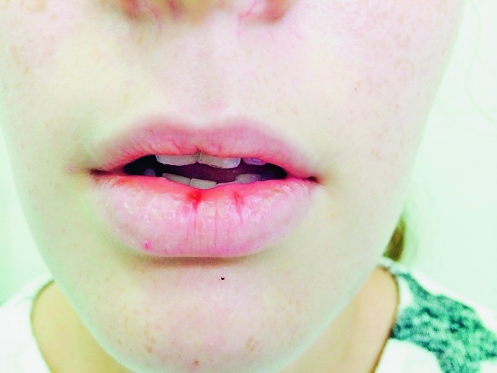
Angular cheilitis, characterized by inflammation at the corners of the mouth, is a common condition with varied presentations and underlying causes. It is crucial to differentiate angular cheilitis from other conditions that can mimic its appearance.
Clinical Features of Angular Cheilitis
Angular cheilitis typically manifests as:
- Location: Primarily at the labial commissures (corners of the mouth). Can be unilateral or bilateral.
- Appearance: Erythema (redness), fissuring, scaling, crusting, and sometimes erosion or ulceration at the corners of the mouth.
- Symptoms: Pain, burning, itching, dryness, and discomfort, especially with mouth movements like eating or talking. Patients may also report a persistent unpleasant taste.
- Predisposing Factors: Deep wrinkles at lip corners, lip licking habits, wearing dentures (especially ill-fitting ones), and conditions leading to increased saliva production or drooling.
Etiology of Angular Cheilitis
The causes of angular cheilitis are multifactorial, often involving a combination of local and systemic factors:
- Infections:
- Fungal: Candida albicans is the most common infectious agent, thriving in the moist environment of the lip corners.
- Bacterial: Staphylococcus aureus and beta-hemolytic streptococci can also contribute, especially in children.
- Nutritional Deficiencies: Deficiencies in B vitamins (riboflavin, folate, B12), iron, and zinc are frequently implicated.
- Mechanical Factors: Dentures that don’t fit properly, overclosure of the mouth in edentulous patients, and thumb-sucking in children can create folds at the corners of the mouth, trapping saliva and promoting maceration.
- Systemic Conditions:
- Diabetes mellitus: Increased susceptibility to infections, including Candida.
- Immunodeficiency: HIV infection and other conditions compromising the immune system.
- Inflammatory Bowel Disease: Crohn’s disease and ulcerative colitis.
- Celiac disease: Malabsorption of nutrients.
- Plummer-Vinson syndrome: Characterized by iron deficiency anemia, dysphagia, and angular cheilitis.
- Psychiatric disorders: Bulimia nervosa and anorexia nervosa, where self-induced vomiting or nutritional deficiencies can play a role.
- Medications: Isotretinoin and other retinoids, hypervitaminosis A, and certain drugs causing xerostomia (dry mouth).
- Environmental Factors: Cold, dry weather and excessive lip licking can exacerbate the condition.
Angular Cheilitis Differential Diagnosis
The differential diagnosis of angular cheilitis is broad and includes conditions that present with similar lesions at the corners of the mouth. Accurate differentiation is vital for effective management. Key conditions to consider include:
-
Recurrent Herpes Labialis (Cold Sores):
- Distinguishing Features: Typically unilateral, starts with tingling or burning, progresses to vesicles that ulcerate and crust. Recurrent episodes are common in the same location.
- Angular Cheilitis Overlap: Early vesicular stage might be confused, but herpes labialis evolves through distinct stages not typical of angular cheilitis.
- Key Differentiator: Vesicular nature and recurrence pattern of herpes labialis.
-
Secondary Syphilis (Mucocutaneous Lesions):
- Distinguishing Features: Fissured papules or split papules at the corners of the mouth (split papules are characteristic but not always present). May be associated with other syphilis symptoms (rash, lymphadenopathy, mucous patches elsewhere in the mouth).
- Angular Cheilitis Overlap: Fissuring in angular cheilitis can resemble syphilitic lesions.
- Key Differentiator: Rarity of secondary syphilis, associated systemic symptoms, and need for serological testing (VDRL, RPR, FTA-ABS) to confirm syphilis.
-
Contact Dermatitis (Irritant or Allergic):
- Distinguishing Features: Can affect lip corners if irritants or allergens (lip balms, cosmetics, toothpaste, foods) accumulate there. May present with erythema, scaling, vesicles, and itching. History of new product use is suggestive.
- Angular Cheilitis Overlap: Erythema and scaling are common to both.
- Key Differentiator: Distribution (contact dermatitis can extend beyond lip corners), history of exposure to potential irritants/allergens, and improvement upon avoidance of the suspected substance. Patch testing can be useful for allergic contact dermatitis.
-
Cheilitis Simplex (Chapped Lips):
- Distinguishing Features: Generalized dryness, cracking, and fissuring of the lips, more often affecting the vermilion border than corners specifically. Related to dry weather, lip licking.
- Angular Cheilitis Overlap: Both can involve fissuring and dryness.
- Key Differentiator: Distribution (cheilitis simplex is more generalized lip involvement, angular cheilitis is localized to corners).
-
Nutritional Deficiencies (Isolated Oral Manifestations):
- Distinguishing Features: While angular cheilitis is often caused by nutritional deficiencies, isolated oral manifestations of deficiencies (like glossitis, stomatitis) without angular involvement are less likely to mimic angular cheilitis directly but should be considered in the broader differential.
- Angular Cheilitis Overlap: Angular cheilitis is a manifestation of nutritional deficiency.
- Key Differentiator: Nutritional deficiencies are an etiology of angular cheilitis, not usually a differential diagnosis in the sense of a mimicking condition, but nutritional status is crucial in evaluation.
-
Candidiasis (Oral, Extending to Corners):
- Distinguishing Features: Oral candidiasis (thrush) often presents with creamy white plaques on the tongue and oral mucosa. In immunocompromised individuals, it can extend to the lip corners, mimicking angular cheilitis.
- Angular Cheilitis Overlap: Candida is a common cause of angular cheilitis.
- Key Differentiator: Presence of oral thrush elsewhere in the mouth, response to antifungal treatment. Microscopic examination (KOH prep or Gram stain) can confirm Candida.
-
Lichen Planus (Erosive Form):
- Distinguishing Features: Oral lichen planus can present with reticular (lacy white lines – Wickham’s striae), atrophic, or erosive lesions. Erosive lichen planus, if located at lip corners, could resemble angular cheilitis. Often involves other mucosal surfaces.
- Angular Cheilitis Overlap: Erosive lesions can be present in both.
- Key Differentiator: Presence of Wickham’s striae elsewhere in the mouth, biopsy for definitive diagnosis of lichen planus.
-
Pemphigus Vulgaris (Oral Lesions):
- Distinguishing Features: Pemphigus vulgaris, a blistering autoimmune disease, often starts with oral lesions, which can include erosions and ulcerations. If these are predominantly at lip corners, it could be confused.
- Angular Cheilitis Overlap: Erosions and ulcerations can be seen in severe angular cheilitis and pemphigus.
- Key Differentiator: Fragile vesicles or bullae elsewhere in the mouth or on skin, positive Nikolsky sign, biopsy and immunofluorescence for pemphigus diagnosis.
-
Actinic Cheilitis (Less Likely at Corners Primarily):
- Distinguishing Features: Primarily affects the lower lip vermilion, due to chronic sun exposure. Presents as scaly plaques, loss of vermilion border definition. Less typically isolated to lip corners.
- Angular Cheilitis Overlap: Scaling and chronic lip condition.
- Key Differentiator: Location (actinic cheilitis is vermilion-based, not corner-specific), history of sun exposure, and characteristic clinical and histological features of actinic damage.
Diagnostic Approach to Angular Cheilitis
A systematic approach is crucial for accurate diagnosis and effective management of angular cheilitis:
-
History and Physical Examination:
- Detailed medical history: Underlying systemic diseases (diabetes, IBD, HIV), nutritional status, medications, allergies, habits (lip licking, denture use), duration of symptoms, and previous treatments.
- Thorough oral examination: Assess the lesions at lip corners, look for other oral lesions (candidiasis, lichen planus, pemphigus), and evaluate denture fit if applicable.
- Skin examination: Check for signs of atopic dermatitis, contact dermatitis, or other dermatological conditions.
-
Investigations (Selective, Based on Clinical Suspicion):
- Microscopy: KOH preparation or Gram stain of scrapings from the lesions to detect Candida or bacteria if infection is suspected.
- Culture: Fungal or bacterial culture if microscopy is inconclusive or to identify specific organisms and guide antimicrobial therapy.
- Blood Tests: Complete blood count, serum iron studies, vitamin B12 and folate levels, zinc level, fasting blood glucose, and HbA1c to screen for nutritional deficiencies and diabetes, especially in recurrent or persistent cases.
- Patch Testing: If contact dermatitis is suspected, particularly if history suggests cosmetic or topical product use.
- Biopsy: Rarely necessary for typical angular cheilitis, but consider if lesions are atypical, persistent despite treatment, or if there is suspicion of other conditions (lichen planus, pemphigus, syphilis, actinic cheilitis in an unusual location). Histopathology can rule out other diagnoses.
- Serological Tests for Syphilis: If secondary syphilis is in the differential diagnosis.
Management of Angular Cheilitis
Treatment strategies for angular cheilitis are directed at addressing the underlying cause and alleviating symptoms:
-
Identify and Eliminate Predisposing Factors:
- Correct ill-fitting dentures or address overclosure of the mouth.
- Advise against lip licking and thumb-sucking.
- Improve oral hygiene.
- Manage underlying systemic conditions (diabetes, nutritional deficiencies, etc.).
-
Topical Therapy:
- Antifungals: Topical antifungals (e.g., clotrimazole, miconazole, nystatin) are first-line if Candida is suspected or confirmed. Combination antifungal/corticosteroid creams should be used cautiously and for short durations to reduce steroid side effects.
- Antibiotics: Topical antibiotics (e.g., mupirocin) if bacterial infection is suspected.
- Topical Corticosteroids: Low to medium potency corticosteroids (e.g., hydrocortisone, triamcinolone) can reduce inflammation and discomfort, but prolonged use should be avoided due to potential side effects (thinning of skin, steroid rosacea).
- Barrier Creams/Ointments: Zinc oxide ointment, petrolatum jelly, or bland emollients can protect the lip corners, reduce moisture, and promote healing.
-
Systemic Therapy:
- Nutritional Supplementation: Oral supplementation with iron, B vitamins, or zinc if deficiencies are identified.
- Systemic Antifungals: Oral antifungals (e.g., fluconazole) may be considered for severe or refractory cases of candidal angular cheilitis, especially in immunocompromised patients.
- Treatment of Underlying Systemic Diseases: Optimal management of diabetes, IBD, or other underlying conditions is essential.
-
Other Measures:
- Dietary Modifications: Address any dietary deficiencies.
- Patient Education: Advise on avoiding irritants, maintaining good oral hygiene, and proper lip care.
Conclusion
Angular cheilitis is a common inflammatory condition affecting the corners of the mouth, with a broad differential diagnosis. A thorough understanding of its clinical features, diverse etiologies, and potential mimicking conditions is essential for accurate diagnosis. A systematic diagnostic approach, including detailed history, physical examination, and selective investigations, guides appropriate management. Effective treatment involves addressing underlying causes, topical and sometimes systemic therapy, and patient education. By considering the differential diagnoses outlined and employing a comprehensive approach, clinicians can effectively manage angular cheilitis and improve patient outcomes.
Please note: This information is for educational purposes only and should not be considered medical advice. Always consult with a qualified healthcare professional for diagnosis and treatment of any medical condition.
Figures from Original Article (Reused with new ALT text as per instructions):
Fig. 1.
Fig. 2.
Fig. 3.
Fig. 4.
Fig. 5.
Fig. 6.
References
References are the same as in the original article. To include them, list them as they appear in the original, formatted as markdown links if possible, or as a numbered list. For example:
References
- … (Reference 1 details)
- … (Reference 2 details)
- … (Reference 3 details)
… and so on, up to the last reference in the original article. Ensure the reference list is complete and accurately reflects the original source. Since the original prompt didn’t ask for re-formatting references, they would ideally be copied as is to maintain consistency with the original article’s academic style. For SEO and readability on the web, consider making them clickable links if DOIs or URLs are available in the original article. However, strictly adhering to the prompt, simply listing them as in the original is sufficient. (To fully complete, the reference section needs to be populated with the actual references from the source article.)