Asthma, a prevalent chronic respiratory syndrome globally, is characterized by recurrent symptoms such as chest tightness, wheezing, coughing, and shortness of breath. While asthma itself is not a singular disease but a syndrome defined by these symptoms and reversible airway obstruction, accurately diagnosing asthma and distinguishing it from conditions that mimic its exacerbations is crucial for effective patient management. This article delves into the differential diagnosis of asthma exacerbations, providing an in-depth exploration of conditions that may present with similar symptoms, ensuring clinicians can navigate complex cases and deliver optimal care.
Understanding Asthma and Its Mimics
Asthma: A Syndrome of Reversible Airway Obstruction
Asthma is defined by chronic airway inflammation and hyperresponsiveness, leading to variable and reversible airflow limitation. While allergic triggers are common, non-allergic asthma phenotypes also exist, broadening the spectrum of this syndrome. Diagnosis relies on clinical assessment, patient history, and supportive pulmonary function tests. However, the non-specific nature of asthma symptoms necessitates a comprehensive differential diagnosis to exclude other conditions.
The Challenge of Differential Diagnosis in Asthma Exacerbations
Asthma exacerbations, or acute asthma attacks, are characterized by a worsening of typical asthma symptoms. These episodes can range from mild to life-threatening and require prompt diagnosis and treatment. However, many conditions can mimic asthma exacerbations, presenting with similar symptoms like wheezing, coughing, and dyspnea. Misdiagnosis can lead to inappropriate treatment, delayed care, and potentially adverse outcomes. Therefore, a thorough understanding of the differential diagnosis of asthma exacerbations is paramount for clinicians.
Key Conditions in the Differential Diagnosis of Asthma Exacerbation
Several conditions can mimic asthma exacerbations, requiring careful consideration during diagnosis. These conditions can be broadly categorized and are discussed in detail below.
Chronic Obstructive Pulmonary Disease (COPD)
COPD, like asthma, is characterized by airflow limitation, but unlike asthma, it is largely irreversible and progressive. COPD encompasses conditions like chronic bronchitis and emphysema, often associated with smoking history.
Differentiating COPD from Asthma Exacerbation
| Feature | Asthma Exacerbation | COPD Exacerbation |
|---|---|---|
| Reversibility | Typically significant bronchodilator reversibility | Limited bronchodilator reversibility |
| Smoking History | Less strongly associated | Strongly associated |
| Age of Onset | Often childhood or young adulthood | Typically later in life (over 40) |
| Sputum Production | Variable, often mucoid | Often chronic and productive |
| DLCO (Diffusion Capacity) | Normal or increased | Reduced in emphysema |
Clinical Considerations: While both conditions share symptoms like wheezing and dyspnea, a detailed history, including smoking history and age of onset, is crucial. Pulmonary function tests (PFTs) before and after bronchodilator administration are essential to assess reversibility. Reduced DLCO is more suggestive of COPD, particularly emphysema. Asthma-COPD overlap syndrome (ACOS) further complicates diagnosis, requiring careful clinical judgment.
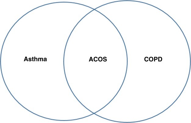 Fig. 1
Infectious Etiologies: Pneumonia and Bronchitis
Respiratory infections, particularly pneumonia and bronchitis, are common causes of respiratory distress and can easily be mistaken for asthma exacerbations, especially in patients with pre-existing asthma.
Pneumonia Mimicking Asthma Exacerbation
Pneumonia, an infection of the lung parenchyma, can present with cough, fever, tachypnea, and wheezing, mimicking an asthma flare-up.
Differentiating Pneumonia from Asthma Exacerbation:
| Feature | Asthma Exacerbation | Pneumonia |
|---|---|---|
| Fever | Typically absent | Often present |
| Sputum | Variable, often mucoid | Purulent sputum may be present |
| Auscultation | Wheezing prominent | Crackles or bronchial breath sounds may be heard |
| Chest X-ray | Normal or hyperinflation | Parenchymal infiltrates present |
| Response to Bronchodilators | Usually improves with bronchodilators | Limited or no improvement with bronchodilators |
Clinical Considerations: Fever, purulent sputum, and localized chest auscultation findings are more suggestive of pneumonia. A chest X-ray is crucial to confirm infiltrates. While viral infections are common asthma triggers, bacterial or fungal pneumonias require different management strategies. Common pathogens include Streptococcus pneumoniae, Haemophilus influenzae, and respiratory viruses.
Bronchitis and Asthma Exacerbation Overlap
Acute bronchitis, inflammation of the bronchial tubes, can also cause cough and wheezing. Viral bronchitis is particularly common and can trigger asthma exacerbations in susceptible individuals.
Differentiating Bronchitis from Asthma Exacerbation:
| Feature | Asthma Exacerbation | Bronchitis |
|---|---|---|
| Duration of Symptoms | Variable, often episodic | Often acute, self-limiting |
| Sputum | Variable, often mucoid | Mucoid or mucopurulent sputum common |
| Systemic Symptoms | Typically absent | Mild systemic symptoms possible |
| Chest X-ray | Normal or hyperinflation | Usually normal |
Clinical Considerations: Bronchitis is often a more self-limiting condition compared to asthma exacerbations. However, in patients with underlying asthma, differentiating between a viral-triggered asthma exacerbation and acute bronchitis can be challenging. Clinical judgment and monitoring response to treatment are important.
Gastroesophageal Reflux Disease (GERD)
GERD is a condition where stomach acid flows back into the esophagus, causing heartburn and other symptoms. Extraesophageal manifestations of GERD can include chronic cough and wheezing, mimicking asthma.
GERD-Induced Respiratory Symptoms and Asthma Mimicry
GERD can trigger bronchospasm and cough through vagal nerve stimulation and microaspiration of gastric contents. This can lead to symptoms that are difficult to distinguish from asthma exacerbations.
Differentiating GERD-Related Symptoms from Asthma Exacerbation:
| Feature | Asthma Exacerbation | GERD-Related Respiratory Symptoms |
|---|---|---|
| Heartburn | Typically absent | May be present, but not always prominent |
| Symptoms Triggered by | Allergens, exercise, cold air, etc. | Food, lying down, large meals |
| Nocturnal Symptoms | Common | Often worse at night or after meals |
| Response to Asthma Medications | Improves with asthma medications | May not improve significantly with asthma meds |
| Response to GERD Medications | No improvement | May improve with proton pump inhibitors (PPIs) |
Clinical Considerations: Consider GERD in patients with asthma-like symptoms, especially if heartburn is present or symptoms worsen after meals or at night. A trial of proton pump inhibitors (PPIs) can be diagnostically and therapeutically helpful. However, GERD and asthma can coexist, and GERD treatment may not always resolve respiratory symptoms in asthmatic patients.
Chronic Rhinosinusitis (CRS)
Chronic sinusitis, or inflammation of the sinuses, can contribute to respiratory symptoms that overlap with asthma, including cough and postnasal drip.
Sinusitis and its Respiratory Symptom Overlap with Asthma
Postnasal drip from sinusitis can irritate the airways and trigger cough, and sinus inflammation can contribute to systemic inflammation, potentially exacerbating asthma.
Differentiating Sinusitis-Related Symptoms from Asthma Exacerbation:
| Feature | Asthma Exacerbation | Sinusitis-Related Respiratory Symptoms |
|---|---|---|
| Nasal Congestion/Discharge | Typically absent | Often prominent |
| Facial Pain/Pressure | Typically absent | May be present |
| Sense of Smell | Normal | May be diminished |
| Cough | Wheezing prominent cough | Cough often worse at night, postnasal drip |
Clinical Considerations: Nasal congestion, facial pain, and diminished sense of smell point towards sinusitis. Nasal endoscopy or CT scan can confirm sinus inflammation. Treating sinusitis, especially with intranasal corticosteroids, may improve respiratory symptoms in some patients.
Congestive Heart Failure (CHF)
Congestive heart failure (CHF) can cause cardiac asthma or cardiac wheezing due to pulmonary edema and bronchial narrowing, mimicking asthma exacerbations, particularly in elderly patients.
Cardiac Wheezing: CHF Mimicking Asthma Exacerbation
Left ventricular dysfunction in CHF leads to increased pulmonary venous pressure and fluid accumulation in the lungs, causing bronchial compression and wheezing.
Differentiating Cardiac Wheezing from Asthma Exacerbation:
| Feature | Asthma Exacerbation | Cardiac Wheezing (CHF) |
|---|---|---|
| Age | Can occur at any age | More common in elderly |
| Cardiac History | Less likely | Often present (hypertension, coronary artery disease) |
| Lower Extremity Edema | Typically absent | Often present |
| Jugular Venous Distention | Typically absent | May be present |
| Auscultation | Wheezing | Wheezing, crackles often present |
| Chest X-ray | Hyperinflation possible | Cardiomegaly, pulmonary edema |
| ECG/Echocardiogram | Normal | Abnormal findings suggestive of CHF |
Clinical Considerations: CHF should be considered in the differential diagnosis of wheezing, especially in older patients with risk factors for heart disease. Lower extremity edema, jugular venous distention, and crackles on lung auscultation are suggestive of CHF. ECG and echocardiogram are crucial for diagnosis. Diuretics improve cardiac wheezing by reducing pulmonary edema.
Vocal Cord Dysfunction (VCD)
Vocal cord dysfunction (VCD) involves paradoxical vocal cord adduction, especially during inspiration, leading to airflow obstruction and symptoms that can be confused with asthma exacerbations.
VCD: Mimicking Asthma Exacerbation with Upper Airway Obstruction
VCD causes inspiratory wheezing, stridor, and dyspnea, often triggered by similar factors as asthma, such as exercise, irritants, and emotional stress.
Differentiating VCD from Asthma Exacerbation:
| Feature | Asthma Exacerbation | Vocal Cord Dysfunction (VCD) |
|---|---|---|
| Wheezing Type | Expiratory wheezing prominent | Inspiratory wheezing or stridor prominent |
| Throat Symptoms | Typically absent | Throat tightness, hoarseness, dysphonia common |
| Triggers | Allergens, cold air, exercise | Irritants, odors, emotional stress |
| Response to Bronchodilators | Improves with bronchodilators | No or minimal improvement with bronchodilators |
| Inspiratory Flow Loop | Normal or expiratory obstruction | Flattened inspiratory flow loop |
| Laryngoscopy | Normal vocal cords during exacerbation | Paradoxical vocal cord movement during inspiration |
Clinical Considerations: Inspiratory wheezing or stridor, throat tightness, and lack of response to bronchodilators suggest VCD. Laryngoscopy is the gold standard for diagnosis, showing paradoxical vocal cord movement. Speech therapy and breathing techniques are effective treatments for VCD.
Anaphylaxis
Anaphylaxis, a severe, life-threatening allergic reaction, can present with respiratory symptoms like wheezing and dyspnea, mimicking a severe asthma exacerbation.
Anaphylaxis Presenting as Respiratory Distress
Anaphylaxis involves multisystem involvement, but respiratory symptoms can be the initial and most prominent manifestation.
Differentiating Anaphylaxis from Severe Asthma Exacerbation:
| Feature | Severe Asthma Exacerbation | Anaphylaxis |
|---|---|---|
| Onset | Gradual or acute | Rapid onset (minutes) after exposure to trigger |
| Skin/Mucosal Signs | Typically absent | Urticaria, angioedema, flushing often present |
| Cardiovascular Symptoms | Less common initially | Hypotension, tachycardia common |
| Gastrointestinal Symptoms | Less common initially | Nausea, vomiting, abdominal pain possible |
| Known Allergies/Triggers | May or may not be present | Often identifiable trigger (food, drug, insect sting) |
| Epinephrine Response | May not respond | Responds to epinephrine |
Clinical Considerations: Rapid onset, skin/mucosal signs, cardiovascular symptoms, and gastrointestinal symptoms are suggestive of anaphylaxis. Epinephrine is the first-line treatment for anaphylaxis. Exercise-induced anaphylaxis and idiopathic anaphylaxis can be more challenging to diagnose.
Aspirin-Exacerbated Respiratory Disease (AERD) – Samter’s Triad
AERD, or Samter’s triad, is characterized by asthma, nasal polyps, and sensitivity to aspirin and NSAIDs. NSAID ingestion can trigger severe respiratory reactions mimicking asthma exacerbations.
AERD: NSAID-Induced Respiratory Reactions
NSAID ingestion in AERD patients leads to bronchospasm and upper respiratory symptoms due to leukotriene pathway dysregulation.
Differentiating AERD Reaction from Asthma Exacerbation:
| Feature | Asthma Exacerbation | AERD Reaction |
|---|---|---|
| NSAID Exposure History | Not relevant | Temporal relationship to NSAID ingestion |
| Nasal Polyps | Absent | Often present |
| Sinus Symptoms | May or may not be present | Chronic sinusitis common |
| Upper Respiratory Symptoms | Less prominent during typical asthma exacerbation | Nasal congestion, rhinorrhea prominent during AERD reaction |
Clinical Considerations: Consider AERD in patients with asthma and nasal polyps, especially if they report respiratory symptoms after taking aspirin or NSAIDs. Oral aspirin challenge can confirm the diagnosis. Aspirin desensitization is a treatment option for AERD.
Malignancy
Lung tumors, though less common, can present with cough, wheezing, and dyspnea, mimicking asthma or contributing to asthma-like symptoms.
Lung Cancer and Carcinoid Tumors Mimicking Asthma
Bronchial obstruction from tumors can cause localized wheezing and cough. Carcinoid tumors, in particular, may be misdiagnosed as asthma due to slow growth and non-specific respiratory symptoms.
Differentiating Malignancy-Related Symptoms from Asthma Exacerbation:
| Feature | Asthma Exacerbation | Malignancy-Related Respiratory Symptoms |
|---|---|---|
| Age | Can occur at any age | More common in older adults |
| Smoking History | Less strongly associated | Stronger association with lung cancer |
| Hemoptysis | Typically absent | May be present |
| Localized Wheezing | Diffuse wheezing | Localized wheezing possible |
| Unexplained Weight Loss | Typically absent | May be present |
| Chest Imaging | Normal or hyperinflation | Mass lesion, nodule, or other abnormalities |
Clinical Considerations: Consider malignancy in older patients with new-onset or worsening asthma symptoms, especially with smoking history, hemoptysis, localized wheezing, or unexplained weight loss. Chest X-ray or CT scan is essential to rule out lung tumors.
Sarcoidosis
Pulmonary sarcoidosis, a granulomatous disease, can present with cough and dyspnea, mimicking asthma.
Sarcoidosis Mimicking Asthma Exacerbation
Pulmonary involvement in sarcoidosis can cause respiratory symptoms that overlap with asthma.
Differentiating Sarcoidosis from Asthma Exacerbation:
| Feature | Asthma Exacerbation | Sarcoidosis |
|---|---|---|
| Systemic Symptoms | Typically absent | Fatigue, fever, weight loss, lymphadenopathy possible |
| Extrapulmonary Manifestations | Typically absent | Skin lesions, eye involvement common |
| Chest X-ray | Normal or hyperinflation | Bilateral hilar lymphadenopathy, reticular infiltrates |
| Pulmonary Function Tests | Obstructive pattern | Restrictive or mixed pattern possible |
| ACE Level | Normal | Elevated in some cases |
| Biopsy | Not typically diagnostic | Noncaseating granulomas diagnostic |
Clinical Considerations: Consider sarcoidosis in patients with cough and dyspnea, especially with systemic symptoms or extrapulmonary manifestations. Chest X-ray, PFTs, and potentially bronchoalveolar lavage or biopsy can aid in diagnosis.
Hypersensitivity Pneumonitis (HP)
Hypersensitivity pneumonitis (HP), or extrinsic allergic alveolitis, results from inhaled antigen exposure and can present with cough, dyspnea, and wheezing, mimicking asthma.
HP: Environmental Antigen Exposure Mimicking Asthma
HP involves inflammation of the lung parenchyma and small airways due to inhaled organic dusts or chemicals.
Differentiating HP from Asthma Exacerbation:
| Feature | Asthma Exacerbation | Hypersensitivity Pneumonitis (HP) |
|---|---|---|
| Antigen Exposure History | May or may not be present | Temporal relationship to antigen exposure |
| Symptoms Onset | Variable | Often acute onset hours after exposure |
| Systemic Symptoms | Typically absent | Fever, chills, malaise common in acute HP |
| Chest X-ray/CT | Normal or hyperinflation | Ground-glass opacities, nodules, fibrosis |
| Pulmonary Function Tests | Obstructive pattern | Restrictive or mixed pattern possible |
| Bronchoalveolar Lavage | Eosinophilia in allergic asthma | Lymphocytosis prominent in HP |
Clinical Considerations: Detailed environmental and occupational history is crucial. Acute onset of flu-like symptoms with respiratory distress after antigen exposure is suggestive of HP. Chest CT and bronchoalveolar lavage can be helpful for diagnosis.
Pulmonary Arterial Hypertension (PAH)
Pulmonary arterial hypertension (PAH) can cause dyspnea and fatigue, which can be mistaken for asthma, particularly exertional dyspnea.
PAH: Breathlessness Mimicking Asthma
Elevated pulmonary artery pressure in PAH leads to right heart failure and symptoms of breathlessness.
Differentiating PAH from Asthma Exacerbation:
| Feature | Asthma Exacerbation | Pulmonary Arterial Hypertension (PAH) |
|---|---|---|
| Exertional Dyspnea | Variable | Prominent and progressive exertional dyspnea |
| Wheezing | Common | Less common |
| Chest Pain/Discomfort | Typically absent | May be present |
| Syncope/Pre-syncope | Rare | Possible |
| Lower Extremity Edema | Typically absent | Often present |
| Echocardiogram | Normal | Elevated pulmonary artery pressure estimated |
| Right Heart Catheterization | Not indicated for asthma diagnosis | Gold standard for PAH diagnosis |
Clinical Considerations: Consider PAH in patients with unexplained exertional dyspnea, especially with chest pain, syncope, and signs of right heart failure. Echocardiogram is a screening tool, and right heart catheterization is diagnostic.
Lymphangioleiomyomatosis (LAM)
Lymphangioleiomyomatosis (LAM), a rare cystic lung disease primarily affecting women, can present with dyspnea, cough, and pneumothorax, mimicking asthma or COPD.
LAM: Cystic Lung Disease Mimicking Asthma in Women
LAM involves smooth muscle cell proliferation in the lungs, leading to cystic changes and airflow obstruction.
Differentiating LAM from Asthma Exacerbation:
| Feature | Asthma Exacerbation | Lymphangioleiomyomatosis (LAM) |
|---|---|---|
| Gender | Both genders | Predominantly women of reproductive age |
| Pneumothorax History | Rare | Increased risk of spontaneous pneumothorax |
| Chest CT | Normal or hyperinflation | Thin-walled cysts diffusely distributed |
| Serum VEGF-D | Normal | Elevated in LAM |
Clinical Considerations: Consider LAM in women presenting with dyspnea, cough, and pneumothorax, especially with cystic changes on chest CT. Serum VEGF-D level can be helpful in diagnosis.
Cystic Fibrosis (CF)
Cystic fibrosis (CF), an autosomal recessive genetic disorder, can present with chronic cough, wheezing, and recurrent respiratory infections, mimicking asthma, particularly in milder cases or in adults diagnosed later in life.
CF: Genetic Disorder Mimicking Asthma
CF involves abnormal mucus production leading to chronic lung infections and progressive lung disease.
Differentiating CF from Asthma Exacerbation:
| Feature | Asthma Exacerbation | Cystic Fibrosis (CF) |
|---|---|---|
| Age of Onset | Often childhood or young adulthood | Can be diagnosed at any age, newborn screening |
| Family History | Less relevant | Family history of CF possible |
| Salty Sweat | Normal | Salty tasting skin |
| Chronic Sinusitis/Polyps | May be present in some asthma phenotypes | Common |
| Gastrointestinal Symptoms | Typically absent | Malabsorption, pancreatic insufficiency common |
| Sweat Chloride Test/Genetic Testing | Not indicated for asthma diagnosis | Diagnostic for CF |
Clinical Considerations: Consider CF in patients with chronic cough, wheezing, recurrent infections, and gastrointestinal symptoms. Sweat chloride test or genetic testing confirms the diagnosis.
Eosinophilic Pulmonary Diseases
Eosinophilic pulmonary diseases, a heterogeneous group, can present with cough, wheezing, and dyspnea, mimicking asthma. These include Löffler’s syndrome, chronic eosinophilic pneumonia (CEP), and eosinophilic granulomatosis with polyangiitis (EGPA) – Churg-Strauss Syndrome.
Eosinophilic Lung Diseases Mimicking Asthma
These conditions involve eosinophilic infiltration of the lungs, causing respiratory symptoms.
Differentiating Eosinophilic Lung Diseases from Asthma Exacerbation:
| Feature | Asthma Exacerbation | Eosinophilic Pulmonary Diseases |
|---|---|---|
| Peripheral Eosinophilia | Variable, can be elevated in allergic asthma | Often prominent and persistent |
| Chest X-ray/CT | Normal or hyperinflation | Peripheral infiltrates (CEP), other patterns |
| Systemic Symptoms | Typically absent | Fever, weight loss, vasculitis features (EGPA) |
| BAL Eosinophilia | Elevated in eosinophilic asthma | Often more pronounced eosinophilia |
| Biopsy | Not typically diagnostic | Lung biopsy may show eosinophilic infiltration |
Clinical Considerations: Peripheral eosinophilia, abnormal chest X-ray findings, and systemic symptoms should raise suspicion for eosinophilic lung diseases. Bronchoalveolar lavage and lung biopsy may be necessary for definitive diagnosis. EGPA (Churg-Strauss) should be considered in patients with asthma and systemic vasculitis features.
Pulmonary Vasculitis Syndromes (Other than EGPA)
Other pulmonary vasculitis syndromes, such as granulomatosis with polyangiitis (GPA – Wegener’s) and microscopic polyangiitis, can also present with respiratory symptoms, including cough, wheezing, and dyspnea.
Vasculitis Mimicking Asthma with Systemic Involvement
These vasculitides involve inflammation of blood vessels in the lungs and other organs, causing a range of symptoms.
Differentiating Pulmonary Vasculitis from Asthma Exacerbation:
| Feature | Asthma Exacerbation | Pulmonary Vasculitis (GPA, Microscopic Polyangiitis) |
|---|---|---|
| Systemic Symptoms | Typically absent | Fever, fatigue, weight loss, multi-organ involvement |
| Upper Respiratory Symptoms | Less prominent during typical asthma exacerbation | Sinusitis, nasal crusting, epistaxis (GPA) |
| Renal Involvement | Typically absent | Glomerulonephritis (GPA, Microscopic Polyangiitis) |
| Hemoptysis | Typically absent | Possible, especially in microscopic polyangiitis |
| ANCA Testing | Negative | Positive ANCA (c-ANCA in GPA, p-ANCA in Microscopic Polyangiitis) |
| Biopsy | Not typically diagnostic | Vasculitis on lung or kidney biopsy |
Clinical Considerations: Consider pulmonary vasculitis in patients with respiratory symptoms and systemic findings, upper respiratory tract involvement (GPA), or renal involvement. ANCA testing and biopsy are crucial for diagnosis.
Summary: A Comprehensive Approach to Differential Diagnosis
Differentiating asthma exacerbations from other conditions requires a thorough clinical approach encompassing:
- Detailed History: Including symptom onset, triggers, smoking history, cardiac history, environmental exposures, medication use (especially NSAIDs), and systemic symptoms.
- Physical Examination: Focusing on respiratory auscultation (wheezing type, crackles), cardiac exam (heart sounds, edema, JVD), and signs of systemic illness (fever, rash).
- Pulmonary Function Tests: To assess airflow obstruction and reversibility.
- Chest Imaging: Chest X-ray or CT scan to rule out pneumonia, CHF, malignancy, HP, LAM, and other structural lung diseases.
- Targeted Investigations: Based on clinical suspicion, including ECG, echocardiogram (CHF), laryngoscopy (VCD), sweat chloride test/genetic testing (CF), serum VEGF-D (LAM), ANCA testing and biopsy (vasculitis), GERD testing, and sinus imaging.
Clinical vigilance is paramount, particularly when asthma symptoms are atypical, refractory to standard asthma therapy, or associated with systemic findings. Accurate differential diagnosis is essential to ensure appropriate management and improve patient outcomes.
Contributor Information
John Johnson, Email: [email protected].
Tina Abraham, Email: [email protected].
Monica Sandhu, Email: [email protected].
Robert Hostoffer, Email: [email protected].
Theodore Sher, Email: [email protected].
Dennis K. Ledford, Email: [email protected]
Timothy Craig, Email: [email protected], Email: [email protected].
Collaborators: Massoud Mahmoudi, Dennis K. Ledford, Timothy Craig, Dennis K. Ledford, and Timothy Craig