Introduction
Atrial-esophageal fistula (AE fistula) represents a rare yet life-threatening complication arising from radiofrequency catheter ablation, a common procedure for managing drug-refractory atrial fibrillation. This abnormal connection between the esophagus and the left atrium, first documented in 2004, carries a high mortality rate, making prompt and accurate diagnosis paramount. While the clinical presentation of an AE fistula can vary, early recognition and intervention are critical to improving patient outcomes. This article delves into the essential aspects of Atrial Esophageal Fistula Diagnosis, highlighting key indicators, diagnostic modalities, and the importance of timely detection following catheter ablation for atrial fibrillation.
Clinical Presentation and Symptom Recognition in AE Fistula Diagnosis
The diagnostic journey for an atrial esophageal fistula begins with a keen awareness of its potential clinical manifestations, particularly in patients who have recently undergone catheter ablation for atrial fibrillation. Symptoms are often non-specific and can be easily attributed to other post-ablation complications or unrelated conditions, leading to diagnostic delays. Patients may present with a range of symptoms, typically emerging days to weeks after the procedure. Common presenting complaints include unexplained fever, a general feeling of malaise, and chest discomfort that may be retrosternal in nature. Odynophagia, or painful swallowing, is another significant indicator, alongside nausea and, in more severe cases, hematemesis (vomiting blood) or melena (dark, tarry stools indicating digested blood). Dyspnea, or shortness of breath, can also occur as the condition progresses. Neurological deficits, although less common, can arise secondary to air emboli, emphasizing the systemic impact of this fistula. Recognizing these varied symptoms, especially in the context of recent atrial fibrillation ablation, is the first critical step in atrial esophageal fistula diagnosis.
Diagnostic Modalities for Atrial Esophageal Fistula
Once clinical suspicion for an atrial esophageal fistula is raised, a systematic approach to diagnosis is crucial. Several diagnostic modalities play a vital role in confirming the presence of this fistula and guiding subsequent management.
Imaging Techniques in AE Fistula Diagnosis
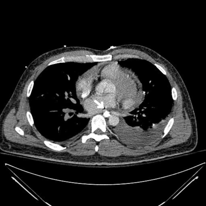
Computed Tomography (CT) Scan: CT scanning, particularly CT of the chest with contrast, is often the initial imaging modality of choice. In the context of atrial esophageal fistula diagnosis, CT can reveal the fistula tract itself, although this may not always be directly visualized. More commonly, CT findings suggestive of AE fistula include the presence of air within the mediastinum or pericardium, esophageal thickening, periesophageal inflammation, and pleural effusion. Specifically, when investigating for AE fistula, a CT of the pulmonary veins can be particularly informative, as this region is the typical site of ablation and fistula formation.
Magnetic Resonance Imaging (MRI): MRI can serve as an alternative or complementary imaging technique to CT. MRI may offer improved soft tissue contrast, potentially aiding in the visualization of the fistula tract and associated inflammatory changes.
Esophagography: An esophagogram, utilizing a water-soluble contrast medium such as gastrografin, can be employed to detect esophageal perforation or injury. In cases of AE fistula, the esophagogram might demonstrate contrast extravasation into the mediastinum or pericardium, indicative of a fistula. As illustrated in Figure 1, contrast collection at the distal esophagus can be a key finding suggestive of esophageal injury, a precursor to fistula formation.
Laboratory Investigations in AE Fistula Diagnosis
While imaging is paramount for anatomical diagnosis, laboratory investigations can provide supportive evidence and assess the systemic impact of an AE fistula.
White Blood Cell Count (WBC): An elevated WBC count is a sensitive, though non-specific, marker that can support the suspicion of an AE fistula, indicating an inflammatory or infectious process.
Blood Cultures: Blood cultures are essential to identify potential bacteremia or sepsis, common complications of AE fistula. The growth of unusual oropharyngeal flora, such as Lactobacillus species, in blood cultures can be a significant clue, pointing towards a breach in the esophageal mucosal barrier and potential fistula formation.
Endoscopic Considerations in Suspected AE Fistula
It is critically important to exercise caution with endoscopic procedures, such as esophagogastroduodenoscopy (EGD) or transesophageal echocardiography (TEE), when an AE fistula is suspected. While EGD may be considered to directly visualize esophageal injury, insufflation of air during endoscopy can pose a significant risk of air embolization through the fistula, potentially leading to severe neurological complications or even death. Therefore, endoscopy in suspected AE fistula cases should only be performed with extreme caution and with a clear plan for immediate intervention if the diagnosis is confirmed. In many instances, less invasive imaging modalities are preferred for initial atrial esophageal fistula diagnosis.
Differential Diagnosis
In the context of post-ablation patients presenting with symptoms suggestive of AE fistula, it is crucial to consider other potential diagnoses. These may include pericarditis, cardiac tamponade, esophageal perforation from other causes, mediastinitis, pneumonia, or other infectious processes. A thorough clinical evaluation, coupled with appropriate investigations, is necessary to differentiate AE fistula from these conditions and ensure accurate atrial esophageal fistula diagnosis.
Management and the Importance of Early Diagnosis
While this article focuses on diagnosis, it is important to briefly touch upon management to underscore the urgency of timely detection. The cornerstone of AE fistula management is surgical repair. Early surgical intervention significantly improves the chances of survival, as untreated AE fistulas carry a near 100% mortality rate. Delayed diagnosis leads to progressive infection, mediastinitis, sepsis, and ultimately, fatal outcomes. Therefore, prompt and accurate atrial esophageal fistula diagnosis is not merely about identifying the condition but is directly linked to enabling timely surgical intervention and improving patient survival.
Conclusion
Atrial esophageal fistula is a devastating complication of atrial fibrillation ablation that demands a high index of suspicion for timely diagnosis. Clinicians must be vigilant in recognizing the varied and often non-specific symptoms in post-ablation patients. A systematic diagnostic approach, utilizing imaging modalities like CT and esophagography, alongside supportive laboratory investigations, is crucial for confirming the diagnosis. Caution should be exercised with endoscopic procedures due to the risk of air embolization. Ultimately, the prompt and accurate atrial esophageal fistula diagnosis is paramount, paving the way for life-saving surgical intervention and improved outcomes in this challenging clinical scenario.