Introduction
The global resurgence of bed bug infestations in recent decades has presented a growing challenge for healthcare professionals. Characterized by an unforeseen increase, this phenomenon is attributed to a confluence of factors including escalating population densities, the surge in international travel, and the worrisome development of insecticide resistance among bed bug populations. For patients sensitized to bed bug saliva, the hallmark of an infestation often manifests as cutaneous symptoms following bites. Therefore, medical practitioners are frequently the first point of contact for individuals experiencing these dermatological reactions. Accurate diagnosis is paramount, not only for patient relief but also to initiate effective eradication measures and mitigate further spread.
However, establishing a definitive diagnosis of bed bug bites is not always straightforward. The clinical presentation can be highly variable, mimicking a wide array of dermatological conditions. These include autoimmune diseases, immune-mediated skin disorders, and reactions to bites or stings from other arthropods. This diagnostic ambiguity can lead to unnecessary investigations, delayed treatment, and persistent patient discomfort. The frustration experienced by patients and the potential for misdiagnosis underscore the critical need for a clear and practical differential diagnostic approach.
This review aims to provide a comprehensive differential diagnostic guide for bed bug bites, supplemented by a visual atlas of clinical presentations. By highlighting the key dermatological features associated with bed bug bites and contrasting them with those of similar conditions, this resource seeks to empower clinicians to confidently differentiate bed bug infestations from other dermatological entities. The ultimate goal is to facilitate prompt and accurate diagnoses, enabling timely initiation of eradication strategies and minimizing patient distress. This focused approach will help avoid superfluous diagnostic procedures and ensure patients receive the appropriate guidance and treatment, addressing both their immediate symptoms and the underlying infestation.
Bed bugs (Cimex lectularius) are small, reddish-brown, wingless insects, typically 5–8 mm in length, making them visible to the naked eye. They are nocturnal feeders, preferring dark, warm environments and commonly hide in mattresses, box springs, bed frames, carpets, behind headboards and wallpaper, and within small cracks in walls. Beyond the bites themselves, indirect signs of infestation can aid in diagnosis, including shed skins, fecal spots, and blood stains on bedding. Bed bugs are most active at night, with peak feeding times around 3 am. They typically target exposed skin areas such as the face, neck, arms, and legs. A characteristic pattern often associated with bed bug bites is the “breakfast, lunch, and dinner” sign – a linear or triangular arrangement of three or more closely spaced bite marks. Notably, adult bed bugs are remarkably resilient and can survive for up to 12 months without feeding, contributing to their widespread prevalence and persistence.
Epidemiology: The Global Resurgence of Bed Bugs
The relationship between humans and bed bugs is a long-standing one, extending back centuries. Historical records from ancient Greece, Rome, and Jewish texts allude to the presence of these persistent pests. The very term “bed bug” (Cimex lectularius) has its roots in ancient Rome, where “cimex” meant insect and “lectularius” referred to a bed. Interestingly, surveys conducted in the early 20th century revealed a significant prevalence of bed bug infestations, with approximately 30% of apartments in major US cities affected. Urban areas in Europe experienced similar levels of infestation during the 1930s, impacting about one-third of residences.
The widespread use of potent pesticides, particularly after World War II, led to a dramatic decline in bed bug populations, bringing infestations to near zero in many developed countries. However, starting around the turn of the millennium, bed bugs began a surprising and significant resurgence. This insidious re-emergence initially went largely unnoticed by public and health authorities, allowing infestations to spread before concerted action was taken. The current bed bug epidemic is now recognized as a serious public health concern, affecting not only developing nations but also developed regions including North America, Europe, and Australia. For example, Australia witnessed an astonishing 4,500% increase in reported infestations between 2000 and 2006.
Several factors contribute to this dramatic resurgence. The exponential growth in international travel and global migration patterns play a significant role in the transportation and spread of bed bugs across geographical boundaries. Changes in pest control management practices, including a shift away from broad-spectrum insecticides and a greater emphasis on targeted pest control, have inadvertently created ecological niches for bed bugs to thrive. Crucially, the development of widespread insecticide resistance in bed bug populations has rendered many traditional chemical control methods less effective. Furthermore, urbanization, densely populated cities, and socioeconomic factors such as low-income housing are recognized as significant predisposing risk factors for infestations. While direct person-to-person transmission is uncommon, bed bugs can readily migrate between dwellings through shared walls, plumbing, and ventilation systems, facilitating rapid spread within apartment buildings and neighboring properties.
The re-emergence of bed bugs as a public health issue prompted early warnings, such as a letter published in the British Medical Journal in 2000, highlighting the potential for a bed bug epidemic outbreak in England. In response to growing concerns, France launched a public health initiative in 2009 to educate the public and healthcare professionals about bed bug identification, bite recognition, and effective control strategies. Around the same time, a bed bug summit was convened in the United States, where current estimates place the infestation rate at approximately 10% of households.
Given their frontline role in patient care, it is paramount to raise awareness about bed bug infestations among family physicians, dermatologists, emergency room personnel, pediatricians, and nurses. These healthcare professionals are crucial in accurately diagnosing bed bug bites, mitigating unwarranted social stigma associated with infestations, and guiding patients through the often complex process of eradication and preventing re-infestation. Their expertise is essential in addressing both the medical and social aspects of this increasingly prevalent public health issue.
Clinical Findings: Recognizing Bed Bug Bites
The bite of a bed bug itself is typically painless, and the initial wheal reaction at the bite site is transient, usually resolving within 3 to 15 minutes. However, in most individuals, proteins present in bed bug saliva trigger immune-mediated reactions, leading to the development of symptomatic skin lesions. The clinical presentation of bed bug bites is highly variable, influenced by individual sensitivity and prior exposure history.
In individuals who have never been exposed to bed bug bites (bite-naive patients), the initial bites often elicit no skin reaction. This lack of immediate response can lead to delayed detection of an infestation. Upon subsequent exposure, sensitized individuals typically develop skin symptoms within 6 to 11 days. With repeated exposures, the latency period for symptom onset decreases progressively. Reactions may appear within 2 to 3 days, and eventually, within hours, leading to skin signs being noticeable upon waking the morning after being bitten. However, it is important to note that not everyone develops a hypersensitivity reaction to bed bug bites. Asymptomatic infestations, representing true insensitivity to bites, are estimated to occur in 4.2% to 25% of individuals.
A survey conducted among residents of infested dwellings in the USA revealed interesting demographic trends in bite reactivity. Females were found to be more likely to react to bed bug bites compared to males. Furthermore, advanced age appeared to be a protective factor, with significantly more individuals over the age of 65 exhibiting no allergic bite reaction compared to younger individuals aged 11 to 65 (42% vs. 26%). Interestingly, mosquito bite sensitivity was also found to correlate with reactivity to bed bug bites, suggesting potential cross-reactivity or shared immune mechanisms.
Several attempts have been made to categorize bed bug bite reactions based on their clinical presentation. Reactions can be broadly classified as cutaneous or systemic. Cutaneous symptoms, based on their frequency, can be further subdivided into usual, common, and complex. A “usual” symptom may be a minimal reaction, characterized by only a small punctum with little to no surrounding inflammation. “Common” symptoms typically present as pruritic (itchy), maculopapular lesions, ranging in size from 2 to 5 mm. These lesions are non-blanching (do not fade upon pressure) and feature a central hemorrhagic punctum surrounded by an erythematous (reddened) border. The rash is typically non-confluent, and weeping or sloughing of the skin is not characteristic.
“Complex” symptoms encompass a broader range of reactions, including wheals (hives), papular urticaria, and vesicles or bullae (blisters) that extend beyond the immediate bite mark. Papular urticaria, considered a hypersensitivity reaction often seen in atopic children, is thought to be mediated by immunoglobulin (Ig) G antibodies. These lesions typically resolve within hours but can persist for days in some cases. While less frequently reported, bullous reactions, potentially representing a type 3 hypersensitivity response to bed bug bites, have been documented in case reports and observed in up to 6% of patients in some studies. These bullous reactions are intensely pruritic, may be hemorrhagic, and can be associated with lymphangitis (inflammation of lymphatic vessels). Importantly, some of these more complex reactions can be delayed in onset, appearing up to a week or even longer after the bite. Furthermore, intense pruritus associated with bed bug bites can lead to excoriations from scratching, increasing the risk of secondary bacterial skin infections (superinfections).
Systemic reactions to bed bug bites are rare. Isolated case reports describe systemic manifestations such as asthma exacerbations, angioedema (swelling of deeper skin layers), hypertension, generalized urticaria, and anaphylaxis (severe allergic reaction). Notably, systemic reactions can occur even in the absence of preceding cutaneous symptoms, as highlighted in one reported case of anaphylaxis. While systemic reactions are uncommon, clinicians should be aware of their potential, particularly in individuals with pre-existing allergic conditions.
Diagnosis: Identifying Bed Bug Bites
The diagnosis of bed bug bites is primarily based on clinical assessment, incorporating a thorough physical examination and a detailed patient history focusing on potential bed bug exposure. Visual evidence of infestation, such as blood spots or fecal stains on bedding, or the direct observation of bed bugs themselves, strongly supports the diagnosis. The characteristic clinical appearance of the bites, often arranged in a linear pattern, coupled with a suggestive medical history, is typically sufficient to establish a diagnosis.
Key historical information that raises suspicion for bed bug bites includes living in or recent stays in environments known to be at higher risk for infestation, such as workers’ hostels, nursing homes, shelters, or hotels. Recent travel, especially international travel, and the acquisition of new or used wooden furniture are also relevant risk factors. Inquiring about similar symptoms in family members or cohabitants can be helpful, although it is not uncommon for bed bugs to preferentially bite only one person in a household, potentially due to variations in individual attractiveness to bed bugs or differences in bite sensitivity. Therefore, a negative family history does not rule out bed bug infestation.
In complex or ambiguous cases, further investigations may be warranted to confirm the diagnosis or exclude other conditions. Immunological tests can be helpful in certain situations. Prick skin testing with bed bug salivary gland extract is a rapid, safe, and effective method to demonstrate both immediate and delayed hypersensitivity reactions. However, this specialized test is not widely available in clinical practice. Serum-specific IgE antibody levels against bed bug salivary proteins, such as nitrophorin, can also be measured, although the clinical utility of this test is still evolving. Serological analysis may not consistently detect systemic infection or the presence of specific IgG autoantibodies in cases with bullous reactions. Histopathological examination of a skin biopsy from a bite lesion is generally not diagnostic for bed bug bites, as there are no pathognomonic histological features. However, skin biopsy can be valuable in excluding other dermatological conditions in the differential diagnosis.
Differential Diagnosis: Distinguishing Bed Bug Bites from Mimicking Conditions
The diverse clinical manifestations of bed bug bites frequently lead to diagnostic uncertainty. The broad range of presentations can mimic numerous dermatological conditions, necessitating a systematic approach to differential diagnosis. To aid clinicians in this process, we have organized a guide encompassing dermatologic diseases that may clinically resemble bed bug bites. This guide provides concise information highlighting both the similarities and the key differentiating features to facilitate accurate identification of the underlying condition. It is important to note that this is not intended to be a comprehensive diagnostic algorithm for each listed disease, as that is beyond the scope of this review. Instead, the aim is to provide clinicians with a practical framework for distinguishing bed bug bites from other skin conditions with potentially overlapping clinical presentations.
Other arthropod bites and stings are important considerations in the differential diagnosis of bed bug bites (Table 1). Furthermore, infestations with ectoparasitic arthropods must also be considered (Table 1). A significant category of conditions to differentiate from bed bug bites are immune-mediated and autoimmune dermatological diseases, which can present with similar cutaneous symptoms (Table 2). Finally, infections and certain psychodermatological conditions may also mimic bed bug bites (Table 1). Tables 1 and 2 summarize the clinical similarities and key distinguishing features of these conditions compared to bed bug bites, complemented by clinical image pairs (Figures 1 and 2) illustrating the clinical variability of bed bug bites and their resemblance to these mimicking skin conditions. Clinical photographs were obtained from the authors’ clinical practice and from our department’s clinical photography database with patient consent.
Table 1. Differential Diagnosis: Stinging/Biting Arthropods, Ectoparasites, and Other Diseases vs. Bed Bug Bites
| Diagnosis | Similarities | Differences |
|---|---|---|
| Stinging and biting arthropods: | ||
| Honeybee, wasp sting | Painful and/or pruritic wheal, flare with a central punctum | Single lesion. Immediate reaction, systemic reactions more common. Patient typically aware of the sting incident. |
| Spider bite | Painful and/or pruritic wheal, flare or papule with a central punctum | Usually single or few lesions, potentially with 1–2 new lesions per day. Lesion size can vary widely (1–40 cm). May present with systemic symptoms depending on spider species (e.g., Latrodectus). |
| Ectoparasites: | ||
| Tick bite | Erythematous papule with a central punctum | Typically a single lesion, often painless initially. History of outdoor exposure in tick-prone areas. Ticks may remain attached to the skin for an extended period if not removed. Lyme disease and other tick-borne illnesses are considerations. |
| Body lice infestation (Pediculosis corporis) | Multiple erythematous papules, excoriations due to scratching | Symptoms primarily localized to areas covered by clothing. Lesion diameter typically 3–5 mm. Body lice and their eggs (nits) may be visible on clothing seams and body hair. Poor hygiene and crowded living conditions are risk factors. |
| Flea bites | Multiple bites on extremities and trunk, may be linear | Often associated with pet ownership or exposure to animals. Lesion diameter typically smaller (maximum 5–10 mm). Bites often concentrated on lower legs and ankles. |
| Scabies infestation | Pruritic erythematous papules in characteristic distribution (interdigital spaces, wrists, sacral area, genitalia) | Lesion diameter 3–5 mm. Intense nocturnal pruritus (itching worse at night). Burrows (thin, wavy lines) may be visible. Symptoms develop 3–6 weeks after initial infestation. Different distribution pattern compared to bed bug bites. |
| Other diseases: | ||
| Bullous erysipelas | Bullae with serous or hemorrhagic fluid, unilateral distribution possible, limbs affected | Typically affects only one limb, always unilateral. Systemic symptoms such as fever, chills, and malaise are common. Extensive erythema with a “flame-like” pattern. Confluent bullae. Signs of bacterial infection are prominent. |
| Delusions of parasitosis (Ekbom syndrome) | Erythematous papules, severe pruritus, excoriations | Spares the central back and face (areas difficult for the patient to reach). Psychiatric history often present. Patient has a fixed, false belief of ongoing infestation despite lack of objective evidence. “Matchbox sign” – patients may bring in skin debris or lint as “evidence” of parasites. |
| Herpes zoster (Shingles) | Painful vesicles on an erythematous base, rarely linear arrangement | Unilateral, grouped vesicles following a dermatomal distribution (nerve pathway), except in disseminated cases. Prodromal pain or paresthesia often precedes rash onset. Tzanck smear or PCR testing can confirm varicella-zoster virus infection. |
| Reactive perforating collagenosis | Erythematous papule with central erosion, usually pruritic, typically on limbs | Acquired form associated with diabetes mellitus or chronic renal failure. Lesions may develop at sites of minor trauma or scratching. Histopathology shows extrusion of collagen and elastic fibers. |
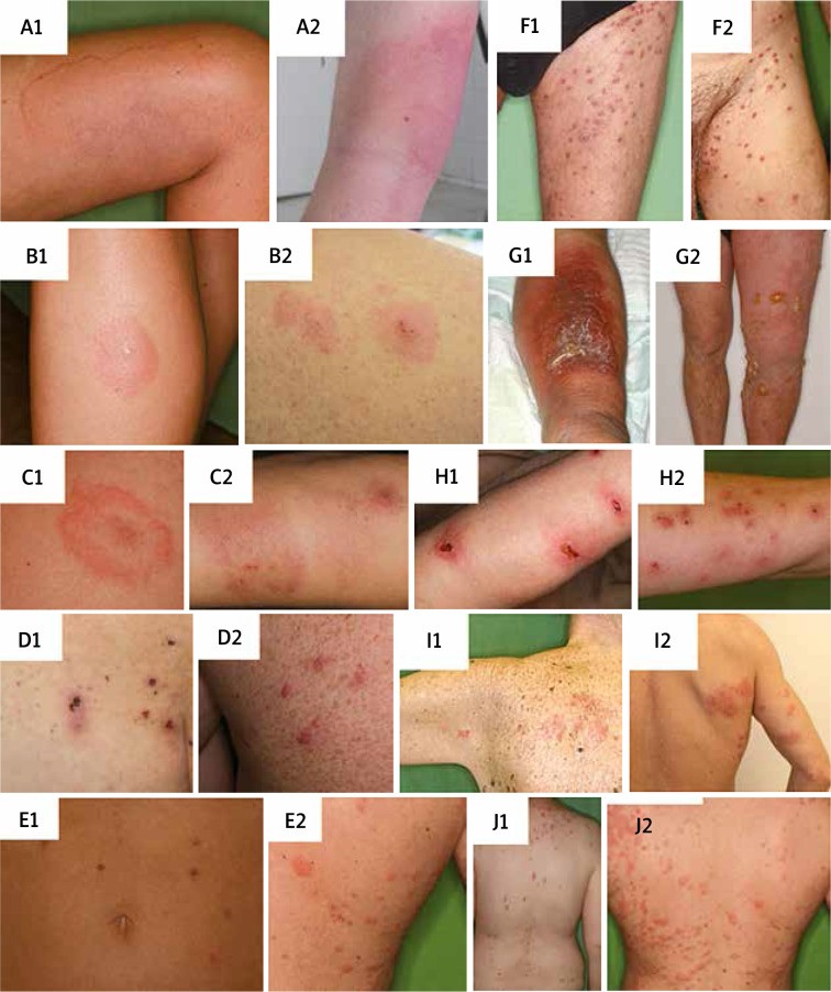
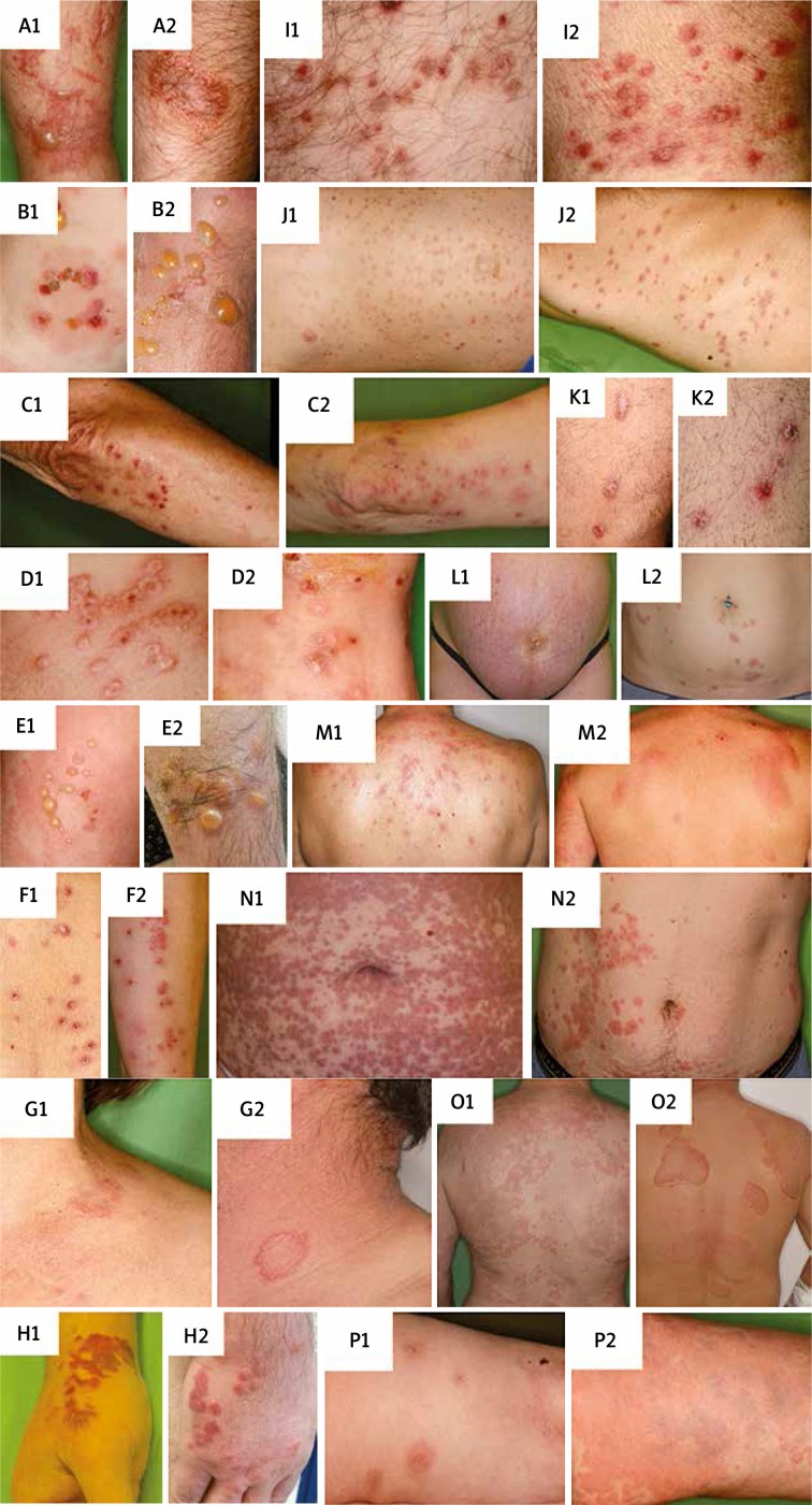
Table 2. Differential Diagnosis: Autoimmune/Immune-Mediated Diseases vs. Bed Bug Bites
| Diagnosis | Similarities | Differences |
|---|---|---|
| Autoimmune/immune-mediated diseases: | ||
| Allergic contact dermatitis | Papules, vesicles, or bullae on an erythematous base with distinct borders. Severe itching | Vesicles tend to coalesce. Distribution pattern often corresponds to the area of contact with the allergen. Patient may be able to identify the causative agent (e.g., poison ivy, nickel). Patch testing can identify specific allergens. |
| Bullous pemphigoid | Tense bullae on an erythematous base, severe itching. Limbs and trunk commonly affected | Primarily affects elderly individuals. Symmetrical distribution. Predilection for flexural areas of limbs and mucous membranes. Nikolsky sign negative (in contrast to pemphigus vulgaris). Direct immunofluorescence of skin biopsy shows linear IgG and/or C3 deposition along the basement membrane zone. |
| Dermatitis herpetiformis | Pruritic papules, vesicles on extensor surfaces (elbows, knees, buttocks, thighs) | Symmetrical and grouped appearance of lesions. Chronic, relapsing course. Strong association with celiac disease (intestinal symptoms in ~20%). Direct immunofluorescence of perilesional skin shows granular IgA deposits at the dermal papillae. |
| Erythema multiforme | Erythematous-blanche “targetoid” lesions on elbows, palms, knees. Vesicles or bullae may develop | Symmetrical distribution. Pain and mucosal involvement (oral, ocular) may occur. Often preceded by herpes simplex virus (HSV) infection or drug exposure. “Target lesions” with concentric rings are characteristic. |
| Linear IgA dermatosis | Tense vesicles and bullae on an erythematous base | Annular (ring-shaped) distribution of lesions. Mucous membrane involvement can occur. Direct immunofluorescence shows linear IgA deposition along the basement membrane zone. |
| Lymphomatoid papulosis | Erythematous/red-brown papulonodules, central ulceration possible, limbs and trunk affected | Single or few lesions at a time. Chronic, recurrent course with lesions appearing in crops. Lesions often heal with scarring. Histopathology shows atypical lymphocytes, but clinical course is usually benign. |
| Papulovesicular polymorphous light eruption (PMLE) | Erythematous blisters, papules, may be pruritic | Monomorphic lesions (similar appearance in a given patient). Eruptions occur after sunlight exposure, predominantly on sun-exposed areas (face, neck, chest, dorsal hands). Phototesting can confirm photosensitivity. |
| Phytophotodermatitis | Blisters, plaques in a linear distribution affecting distal extremities | Occurs after exposure to certain plants (e.g., poison ivy, citrus fruits) followed by sunlight exposure. Lesions often appear in streaks or linear patterns, not solitary papules in a linear arrangement like bed bug bites. History of outdoor activity and plant exposure is key. |
| Pityriasis lichenoides chronica (PLC) | Erythematous papules, nodules, limbs and trunk commonly affected | Prominent scale on lesions, shiny surface after scale removal. Lesions at different stages of development present simultaneously. Chronic, indolent course. |
| Pityriasis lichenoides et varioliformis acuta (PLEVA) | Erythematous papules, vesicles, may be pruritic. Central hemorrhage can mimic punctum | Scaling present, lesions at different stages of development. Predilection for trunk and flexural sites of extremities. Linear pattern less typical. More acute onset and inflammatory than PLC. |
| Prurigo nodularis | Excoriated erythematous papules, nodules, severe itching, extensor surfaces of limbs affected | Symmetrical distribution. Spares the central back (area difficult to scratch). Chronic course with slow development of lesions. Nodules are often intensely pruritic and excoriated, leading to thickened, hyperpigmented lesions. |
| Pruritic urticarial papules and plaques of pregnancy (PUPPP) | Pruritic, erythematous wheals, papules, plaques, seropapules, linear arrangement | Linearity often follows striae (stretch marks) with periumbilical sparing. Proximal limbs are predominantly affected. Onset typically in the third trimester of pregnancy. |
| Sweet syndrome (Acute febrile neutrophilic dermatosis) | Erythematous to purple, tender papules, plaques, vesicles or bullae. Limbs, neck, upper back affected | Systemic symptoms such as fever, neutrophilia (elevated neutrophils in blood). May involve mucosa and other organs. Onset often triggered by infection, drug intake, or underlying chronic illness (e.g., malignancy). |
| Toxicoderma (Drug eruption) | Wheals, maculopapules, vesicles or bullae. Pruritus may be present | History of recent drug intake (1–21 days prior to eruption). Lesions tend to coalesce and become confluent. Distribution often “head-to-toe” (generalized). Temporal relationship to drug initiation is crucial. |
| Urticaria (Hives) | Multiple wheals (hives), intensely pruritic | Wheals vary in size and shape, often with central clearing. Lesions are transient and migratory, changing location within 24-48 hours. Can appear anywhere on the body. Lack of central punctum distinguishes from typical bed bug bites. |
| Urticaria vasculitis | Erythematous wheals, central palpable purpura | Wheals persist for longer than 48 hours (unlike typical urticaria). Residual central hyperpigmentation may be present after lesion resolution. Pattern is not typically linear. Skin biopsy shows leukocytoclastic vasculitis. |
Figure 1. Clinical Images for Differential Diagnosis (Table 1)
Figure 1 Caption: Clinical images illustrating conditions listed in Table 1 for differential diagnosis of bed bug bites. A1 – Honeybee sting: showing a painful wheal and flare reaction. B1 – Spider bite: presenting a painful papule with central punctum. C1 – Tick bite: displaying an erythematous papule with a central punctum, characteristic of a tick bite. D1 – Body lice infestation: multiple erythematous papules and excoriations. E1 – Flea bites: multiple bites on the extremities. F1 – Scabies: pruritic papules in typical distribution. G1 – Bullous Erysipelas: extensive bullae and erythema on the limb. H1 – Delusions of parasitosis (Ekbom syndrome): excoriations from scratching, sparing the central back. I1 – Herpes Zoster: grouped vesicles in a dermatomal distribution. J1 – Reactive perforating collagenosis: erythematous papules with central erosion. A2–J2 – Bed bug bite reactions: demonstrating the variable clinical presentations of bed bug bites for comparison.
Figure 2. Clinical Images for Differential Diagnosis (Table 2)
Figure 2 Caption: Clinical images illustrating conditions listed in Table 2 for differential diagnosis of bed bug bites. A1 – Allergic contact dermatitis: showing vesicles and erythema with distinct borders. B1 – Bullous pemphigoid: tense bullae on an erythematous base. C1 – Dermatitis herpetiformis: grouped vesicles on extensor surfaces. D1 – Erythema multiforme: targetoid lesions on the palm. E1 – Linear IgA Dermatosis: tense vesicles with annular distribution. F1 – Lymphomatoid papulosis: red-brown papulonodules. G1 – Papulovesicular polymorphous light eruption: papules and vesicles on sun-exposed skin. H1 – Phytophotodermatitis: linear plaques and blisters. I1 – Pityriasis lichenoides chronica: papules with scales. J1 – Pityriasis lichenoides et varioliformis acuta: papules and vesicles at different stages. K1 – Prurigo nodularis: excoriated nodules on extensor surfaces. L1 – Pruritic urticarial papules and plaques of pregnancy (PUPPP): linear arrangement along striae. M1 – Sweet-syndrome: erythematous plaques and papules. N1 – Toxicoderma: generalized maculopapular eruption. O1 – Urticaria: multiple wheals of varying sizes. P1 – Urticaria vasculitis: persistent wheals with central purpura. A2–P2 – Bed bug bite reactions: illustrating the diverse clinical presentations of bed bug bites for comparative analysis.
Treatment and Eradication: Addressing Bed Bug Bites and Infestations
In most cases, skin lesions resulting from bed bug bites are self-limiting and spontaneously resolve within 3–10 days. Treatment is primarily symptomatic, focused on alleviating pruritus and preventing secondary infections. While specific standardized treatment guidelines are lacking, several over-the-counter and prescription options are available to manage symptoms.
Topical antipruritic agents such as pramoxine and doxepin creams can provide localized relief from itching. Systemic antihistamines, including both first-generation (e.g., diphenhydramine) and second-generation (e.g., desloratadine, bilastine) formulations, can effectively reduce pruritus, particularly for widespread or intense itching. Topical corticosteroids, such as triamcinolone or mometasone creams, offer a dual benefit by reducing both inflammation and pruritus. They can be used in combination with antihistamines for enhanced symptom control. If secondary bacterial infection develops due to scratching, topical antibiotics like mupirocin or systemic antibiotics may be necessary. Systemic reactions, although rare, should be managed according to the specific symptoms presented. Individuals with extensive or bullous bite reactions may require a short course of oral corticosteroids to reduce inflammation and accelerate healing.
Crucially, successful management of bed bug bites necessitates not only symptomatic treatment but also the complete eradication of bed bugs from the patient’s environment. Eradication is often the most challenging aspect of managing bed bug infestations. Numerous commercially available products and specialized pest control companies offer eradication services. Integrated pest management (IPM) approaches are considered the most effective strategy, particularly in multi-dwelling settings, as they combine various methods to eliminate bed bugs while minimizing pesticide use and environmental impact.
Until professional eradication is achieved, personal protective measures can help reduce bites. Topical repellents containing 5% permethrin cream or 40% diethyltoluamide (DEET), applied to exposed skin, can provide temporary protection against bed bug bites. In 2016, a stepwise American eradication protocol was established to provide guidance for homeowners and pest control professionals. European, American, and Australian guidelines are also available, addressing bed bug management in various settings, including military, industrial, and healthcare facilities.
For less extensive infestations, physical control methods alone may be sufficient. Room treatments include thorough vacuuming, steam cleaning, dry ice application, and heat treatment. For movable items, disinfestation can be achieved through laundering, tumble drying on high heat, portable heating units, freezing, or oxygen removal. In cases of widespread or established infestations, chemical control methods are often necessary. However, the increasing prevalence of insecticide resistance necessitates the use of professional pest control services that employ synergistic combinations of pesticides, such as pyrethroids and carbamates, in various formulations to achieve effective extermination.
Prevention remains the most effective strategy for avoiding bed bug infestations. When traveling or purchasing furniture, particularly used items, careful inspection for signs of bed bugs is crucial. In the home, moving beds slightly away from walls and preventing blankets from touching the floor can reduce bed bug access. In the event of known infestation in a lodging or travel destination, thorough inspection of luggage before departure is essential to prevent bringing bed bugs home.
Discussion: Public Health and Clinical Significance of Bed Bug Bites
The rising incidence of bed bug infestations has significant public health implications. The costs associated with eradication, potential lost workdays in severe cases, and the psychological impact on affected individuals contribute to a substantial societal burden. The mental health consequences of bed bug infestations are increasingly recognized, although the precise nature and extent of this relationship are still being investigated. Case reports have documented severe psychological distress, even leading to suicide in individuals with uncontrolled infestations and underlying psychiatric vulnerabilities. Studies have demonstrated a significant association between bed bug exposure and increased anxiety and sleep disturbances in affected individuals compared to unexposed controls. Surveys of residents in infested dwellings have reported sleep disruption, insomnia, nervousness, and emotional distress in 20–29% of respondents. A literature review examining the psychological impact of bed bugs found that over half of the reviewed articles mentioned psychological associations, with chronic infestations linked to anxiety, phobia, post-traumatic stress disorder (PTSD), depression, and even psychosis.
Beyond mental health, chronic bed bug infestations have been anecdotally linked to physical health consequences such as diarrhea and iron deficiency anemia, although more research is needed to confirm these associations. Social stigma is a significant aspect of the patient experience. Many individuals perceive infestation as a sign of poor hygiene, leading to feelings of shame, social isolation, and occupational dysfunction. The substantial costs associated with professional extermination, furniture replacement, and re-clothing further exacerbate the economic burden on affected individuals and families. Eradication costs in the USA, for example, can range from USD 800 to USD 1,200 for a single dwelling. Furthermore, the economic costs extend to the healthcare system, including expenses related to unnecessary medical diagnostic tests performed before bed bug bites are correctly diagnosed. The aim of this review is to provide a clinically focused overview of bed bug bites, with particular emphasis on differential diagnosis, to increase awareness among medical practitioners and facilitate earlier, more accurate diagnoses and appropriate management. By improving diagnostic accuracy and promoting timely eradication, we can mitigate the multifaceted burden of bed bug infestations on individuals and public health.
Acknowledgments
Norbert Wikonkál and András Bánvölgyi share senior authorship.
The work should be attributed to the Department of Dermatology, Venerology and Dermatooncology, Semmelweis University, Hungary, Mária street 41, 1085 Budapest. Head of the Institution: Prof. Miklós Sárdy, MD PhD.
We are grateful to Rita Mátrahegyi, clinical photographer of our department for capturing clinical images for this article.
Conflict of interest
The authors declare no conflict of interest.