Introduction
Intracranial calcifications (IC), the presence of calcium deposits within the brain, are commonly observed in non-contrast computed tomography (NCCT) scans across all age groups. These radiological findings, while sometimes benign, can also indicate a wide spectrum of pathological conditions. As automotive repair specialists at xentrydiagnosis.store, understanding the intricacies of diagnostic tools and medical imaging, particularly in the context of complex systems, shares parallels with diagnosing intricate vehicle issues. Just as we meticulously analyze diagnostic reports to pinpoint automotive problems, medical professionals rely on imaging to decipher the complexities of the human body. This article, tailored for an English-speaking audience and optimized for search engines, delves into the differential diagnosis of brain calcifications, aiming to provide a comprehensive overview that surpasses the original review in depth and SEO value. This enhanced guide will aid in appreciating the complexities of medical diagnostics, drawing parallels to the detailed diagnostic processes we employ in automotive repair.
Decoding Intracranial Calcifications: A Diagnostic Spectrum
Intracranial calcifications are not diseases in themselves but rather radiological signs that can stem from a variety of underlying causes. Their detection on NCCT scans necessitates a systematic approach to differential diagnosis, considering factors such as patient age, clinical presentation, calcification pattern, and location. Distinguishing between benign, age-related calcifications and those indicative of pathology is crucial. This section will explore the broad categories of intracranial calcifications, setting the stage for a detailed discussion of their differential diagnosis.
Physiologic vs. Pathologic Calcifications: The Initial Differentiation
The first step in differential diagnosis is to differentiate between physiologic and pathologic intracranial calcifications. Physiologic calcifications are age-related, incidental findings that typically require no further investigation. Pathologic calcifications, conversely, are associated with underlying diseases and necessitate further diagnostic workup.
Physiologic Intracranial Calcifications: Normal Age-Related Findings
Physiologic calcifications are common, particularly with increasing age. They are typically located in specific areas of the brain and exhibit characteristic patterns.
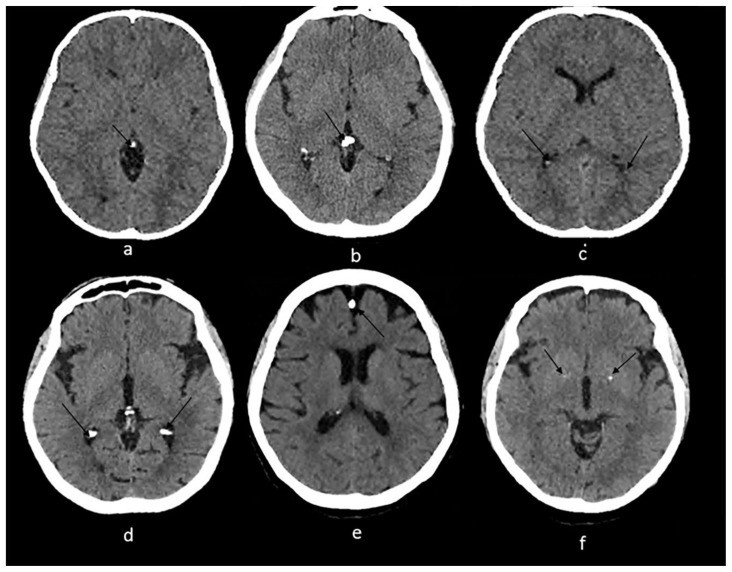
- Pineal Gland Calcifications: The pineal gland is the most frequent site of physiologic calcification. These are often coarse and compact, appearing as dots on CT scans. While common, especially in adults, larger or numerous pineal calcifications in younger children may warrant further investigation to rule out neoplasms.
-
Choroid Plexus Calcifications: Commonly seen in the atria of the lateral ventricles, choroid plexus calcifications are usually punctate and considered physiologic. However, atypical locations, such as the bodies of the lateral ventricles or the third ventricle roof, may suggest pathology.
-
Habenular Calcifications: Located anterior to the pineal gland, habenular calcifications often present a curvilinear pattern. Their association with conditions like schizophrenia has been noted, although they are generally considered physiologic in many adults.
-
Dural Calcifications: Calcifications of the falx cerebri and tentorium cerebelli are also age-related findings. While more prevalent in older individuals, they can occasionally be seen in children.
-
Basal Ganglia Calcifications: While basal ganglia calcifications can be pathologic (as in Fahr’s disease), isolated, faint calcifications in the basal ganglia can be considered physiologic, especially in older adults.
Pathologic Intracranial Calcifications: Identifying Underlying Diseases
Pathologic intracranial calcifications are associated with a wide range of conditions, broadly categorized as:
- Genetic and Developmental Disorders
- Congenital and Acquired Infections
- Vascular Diseases
- Neoplastic Lesions
- Metabolic and Endocrine Disorders
- Inflammatory Conditions
- Neurotoxic Exposures
The subsequent sections will delve into each of these categories, detailing the specific patterns, locations, and associated clinical features to aid in differential diagnosis.
Genetic and Developmental Disorders: Brain Calcification as a Key Marker
Certain genetic and developmental disorders are strongly associated with intracranial calcifications. Recognizing the characteristic patterns of calcification in these conditions is crucial for diagnosis.
- Sturge-Weber Syndrome (SWS): This neurocutaneous syndrome is characterized by a facial port-wine stain, leptomeningeal angiomatosis, and distinctive “tram-track” or gyriform calcifications, reflecting cortical involvement. These calcifications may not be evident in early infancy but develop over time.
-
Tuberous Sclerosis (TS): TS is another neurocutaneous syndrome featuring subependymal nodules and cortical tubers. Calcified subependymal nodules along the caudothalamic groove are pathognomonic, aiding in the diagnosis of TS.
-
Neurofibromatosis (NF): While less consistently associated with calcifications compared to SWS and TS, NF can present with choroid plexus calcifications and cerebellar nodular calcifications. Calcifications in NF2 are often tumor-related, particularly in meningiomas.
-
Cockayne Syndrome (CS): This rare genetic disorder often manifests with bilateral basal ganglia calcifications, described as “rock-like” or spot-like. Gyral, cerebellar, and thalamic calcifications can also occur.
-
Krabbe Disease: This leukodystrophy can exhibit blush-like calcifications in the internal capsule and corona radiata, areas affected by white matter abnormalities.
-
Aicardi-Goutières Syndrome (AGS): AGS is characterized by symmetrical, spot-like calcifications in the basal ganglia and deep white matter, mimicking congenital infections but with a genetic etiology.
-
Fahr Disease: Fahr disease, or idiopathic basal ganglia calcification, is marked by symmetrical calcifications in the basal ganglia, thalamus, dentate nucleus, and cerebral cortex. It’s a diagnosis of exclusion, requiring ruling out metabolic causes like hypoparathyroidism.
Congenital and Acquired Infections: Calcifications as Sequelae
Intracranial calcifications are a significant feature of both congenital and acquired brain infections. The pattern and location of calcifications often provide clues to the specific infectious agent.
Congenital Infections: TORCH and Beyond
Congenital infections, particularly those in the TORCH group (Toxoplasmosis, Others, Rubella, Cytomegalovirus, Herpes), are notorious for causing brain calcifications in newborns.
- Congenital Cytomegalovirus (CMV) Infection: CMV is the most common congenital infection associated with brain calcifications. These are typically thick, chunky periventricular calcifications and faint punctate basal ganglia calcifications. Periventricular calcifications are strongly linked to developmental delay and intellectual disability.
- Congenital Toxoplasmosis: Toxoplasmosis calcifications are nodular and periventricular, with curvilinear calcifications in the thalamus and basal ganglia. These calcifications may decrease or resolve with treatment.
-
Neonatal Herpes: Herpes infections can cause extensive brain damage and scattered calcifications, often associated with multicystic encephalomalacia.
-
Congenital Rubella: Rubella calcifications are typically located in the basal ganglia and periventricular areas.
-
Congenital Zika Virus: Zika virus infection in utero is linked to punctate calcifications between the cortex and subcortical white matter.
-
Congenital HIV Encephalitis: HIV can cause symmetrical calcifications in the basal ganglia and subcortical white matter in congenital infections.
Acquired Infections: Post-Infectious Calcification
Acquired infections can also lead to intracranial calcifications, often as a late sequela of the infection.
- Neurocysticercosis: This parasitic infection, caused by Taenia solium, results in eccentric calcified nodules within peripherally calcified cysts in the brain parenchyma and subarachnoid space.
- Tuberculoma: Calcification within a tuberculoma can create a pathognomonic target sign on CT scans.
- Cryptococcus neoformans Infection: Calcifications from Cryptococcus are rare and appear late in the disease as punctate lesions in the brain parenchyma and leptomeninges.
Vascular Calcifications: Atherosclerosis and Vascular Malformations
Calcifications in the context of vascular diseases can be broadly divided into atherosclerosis and vascular malformations.
- Vascular Atherosclerosis: Intracranial atherosclerosis is a major cause of stroke, and vascular calcifications are a common feature, especially in the internal carotid, vertebral, and middle cerebral arteries. These calcifications are often scattered and stippled.
- Vascular Malformations: Cavernous angiomas, arteriovenous malformations (AVMs), dural arteriovenous fistulas (AVF), and aneurysms can all exhibit calcifications. Cavernous angiomas often show stippled calcifications. AVM calcifications can be dystrophic or located along the vessels. AVFs may present with symmetrical subcortical calcifications. Aneurysm calcifications are common in thrombosed aneurysms.
Neoplastic Calcifications: Brain Tumors and Calcium Deposits
Brain tumors, both intra-axial and extra-axial, can calcify, and the pattern of calcification is diagnostically relevant.
Intra-axial Neoplasms: Calcification within the Brain Parenchyma
-
Astrocytomas: While various astrocytomas can calcify, pilocytic astrocytomas are notable for calcification, particularly in their solid components. Subependymal giant cell astrocytomas (SEGAs), associated with TS, may also show heterogeneous calcifications.
-
Oligodendrogliomas: These tumors are highly prone to calcification, often exhibiting nodular and clumped patterns. Calcifications can extend beyond the tumor margins into the surrounding brain.
-
Gangliogliomas: These tumors frequently present with a mural calcified nodule.
-
Medulloblastomas: Calcifications in medulloblastomas are typically tiny, scattered dots or clumps within the posterior fossa tumor.
- Metastatic Lesions: Metastases rarely calcify, with exceptions like lung, breast, osteosarcoma, and mucinous adenocarcinoma metastases.
Extra-axial Neoplasms: Calcification Outside the Brain Parenchyma
-
Meningiomas: Meningiomas frequently calcify, exhibiting diverse patterns such as sand-like, sunburst, rim, and globular calcifications.
-
Craniopharyngiomas: These suprasellar tumors, especially the adamantinomatous type, commonly calcify. Calcifications can be circumferential or chunky.
- Pineal Tumors: Pineal tumors, including pineocytomas and pineoblastomas, often calcify. Calcifications can be peripheral, creating an “exploded” pattern.
-
Germ Cell Tumors: Teratomas, a type of germ cell tumor, often exhibit heterogeneous calcifications alongside fat and soft tissue components.
-
Lipomas: Lipomas can display egg-shell or central calcification patterns.
Intraventricular Neoplasms: Tumors within the Ventricular System
Intraventricular tumors that may calcify include ependymomas, choroid plexus tumors, central neurocytomas, and intraventricular meningiomas. Ependymomas can have dot-like or mass-like calcifications. Central neurocytomas may show calcifications ranging from dots to large masses. Intraventricular meningiomas mimic extra-axial meningioma calcification patterns.
Metabolic and Endocrine Disorders: Disrupting Calcium Homeostasis
Disorders affecting calcium homeostasis can lead to brain calcifications.
- Hypoparathyroidism: This condition is strongly associated with basal ganglia calcifications, particularly in the globus pallidus. Calcifications can also occur in the dentate nucleus, thalamus, and subcortical white matter. Symmetrical basal ganglia calcifications necessitate differentiating hypoparathyroidism from Fahr’s disease.
- Hypothyroidism: Hypothyroidism, to a lesser extent, can also be associated with intracranial calcifications, often described as small, linear calcifications in the cerebellum and periventricular white matter.
- Hyperparathyroidism: While less common than hypoparathyroidism in causing brain calcifications, hyperparathyroidism can also lead to calcium deposition in the brain.
Inflammatory Conditions: Autoimmunity and Granulomatous Disease
Inflammatory conditions, both autoimmune and granulomatous, can be associated with intracranial calcifications.
-
Systemic Lupus Erythematosus (SLE): SLE can involve the central nervous system and, in some cases, lead to intracranial calcifications. These are typically symmetrical and bilateral, most commonly affecting the cerebellum, centrum semiovale, and basal ganglia.
-
Sarcoidosis: Neurosarcoidosis can rarely cause intracranial calcifications, with reported locations including the suprasellar, hypothalamic, and cerebellar regions.
Neurotoxicity: Environmental and Industrial Exposures
Exposure to certain neurotoxins can result in brain calcifications.
- Lead and Carbon Monoxide Poisoning: Chronic lead exposure and carbon monoxide poisoning can cause punctiform, curvilinear, speck-like, and diffuse calcifications, particularly in the subcortical areas, basal ganglia, and cerebellum.
Conclusion: A Pattern-Based Approach to Differential Diagnosis
Intracranial calcifications, while frequently encountered, represent a diagnostic puzzle requiring careful consideration of their radiological characteristics alongside clinical context. This review has highlighted the diverse etiologies of brain calcifications, emphasizing the importance of pattern recognition – location, morphology, and distribution – in formulating a differential diagnosis. Just as automotive diagnostics relies on interpreting fault codes and sensor data, the diagnosis of brain calcifications hinges on the meticulous analysis of imaging findings in conjunction with patient history and clinical presentation. By systematically considering the categories discussed – physiologic, genetic, infectious, vascular, neoplastic, metabolic, inflammatory, and toxic – clinicians can navigate the complexities of Brain Calcification Differential Diagnosis, leading to accurate diagnoses and appropriate patient management.
TEACHING POINT
A detailed description of the radiological phenotype of intracranial calcifications – including size, location, and shape – is paramount for accurate diagnosis. This, combined with the patient’s age and clinical presentation, forms the cornerstone of effective differential diagnosis and clinical decision-making.
ACKNOWLEDGEMENTS
We extend our gratitude to Dr. Mohammad Rawashdeh, Mr. Youssef Annous, and Mr. Amer Traboulsi for their valuable contributions to the original review upon which this enhanced guide is based.
ABBREVIATIONS
AGS Aicardi-Goutières syndrome
AIDS Acquired immunodeficiency syndrome
AVF Arterio-venous fistula(s)
AVM Arterio-venous malformation(s)
BGC Basal ganglia calcification(s)
CMV Cytomegalovirus
CNS Central nervous system
CS Cockayne syndrome
CT Computed tomography
GGCT Germinomatous germ cell tumor(s)
HIV Human immunodeficiency virus
IC Intracranial calcification(s)
MRI Magnetic Resonance Imaging
NCCT Non-contrast computed tomography
NF1 Neurofibromatosis type 1
NF2 Neurofibromatosis type 2
NGGCT Non-germinomatous germ cell tumor(s)
PTH Parathyroid hormone
SEGA Subependymal giant cell astrocytoma
SWB Sturge-Weber syndrome
TORCH Toxoplasma, others, rubella, cytomegalovirus and herpes
TS Tuberous sclerosis
REFERENCES
References from the original article should be included here, maintaining the original numbering. (Please manually copy the references from the original article to ensure accuracy).
