Mammograms are a crucial tool in breast health, acting as an X-ray of the breast. While routine screening mammograms are vital for women without apparent symptoms to detect early signs of breast cancer, diagnostic mammograms serve a different, yet equally important purpose. They come into play when a screening mammogram reveals suspicious findings or when a woman experiences signs of breast cancer that warrant a more thorough investigation.
These concerning signs and symptoms can include:
- The discovery of a lump in the breast.
- Unexplained breast pain.
- Nipple discharge.
- Noticeable thickening of the breast skin.
- Changes in skin texture, such as enlarged pores on the breast.
- Alterations in breast size or shape.
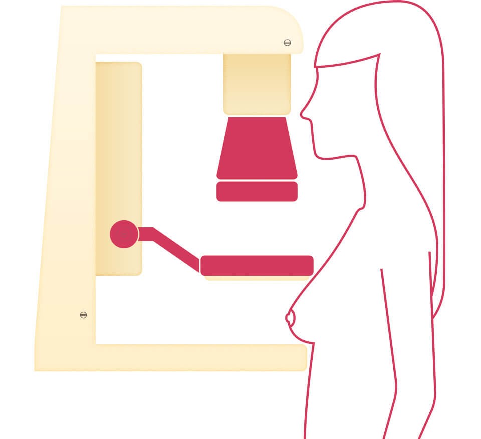
Alt text: A woman stands beside a diagnostic mammogram machine in a medical setting.
A diagnostic mammogram is instrumental in determining whether these symptoms are indicative of breast cancer. Unlike screening mammograms, it offers a more detailed examination of breast tissue, employing specialized techniques and taking multiple views. Diagnostic mammograms are also essential in specific situations, such as for patients who have breast implants, ensuring comprehensive breast health assessment.
It’s also important to be aware of breast density. Starting in late 2024, radiologists will be required to report breast density levels. Dense breast tissue is recognized as a risk factor for breast cancer and can also complicate mammogram interpretation. If you are found to have dense breasts, discussing supplementary imaging options like ultrasound or breast MRI with your healthcare provider is recommended for a more complete evaluation.
What to Expect During a Diagnostic Mammogram
If your physician recommends a diagnostic mammogram, it’s important to know that it will typically be a longer procedure than a routine screening mammogram. This is because a diagnostic mammogram involves taking more X-ray images, providing a comprehensive view of the breast from various angles. The radiologist may also focus on specific areas of concern identified in previous screenings or during physical examinations, magnifying these areas to gain a clearer image of the tissue and facilitate a more accurate breast cancer diagnosis.
Beyond detecting masses too small to be felt during a self-exam or clinical breast exam, mammograms are also effective in identifying ductal carcinoma in situ (DCIS). DCIS refers to abnormal cells within the milk ducts of the breast that, while not currently invasive, have the potential to develop into invasive cancer in some individuals.
DCIS often doesn’t present as a palpable mass. Instead, it frequently appears on mammograms as tiny calcium deposits known as microcalcifications. Clusters of microcalcifications or those arranged in a linear pattern can be a sign of DCIS and require further investigation. It’s crucial to understand that not all cases of DCIS will progress to invasive cancer, and ongoing research is dedicated to determining which DCIS cases are more likely to become invasive. This research aims to help doctors personalize treatment plans based on the specific characteristics of a woman’s DCIS.
The Reliability of Mammograms in Breast Cancer Detection
The effectiveness of a mammogram in detecting breast cancer can vary depending on several factors, including the size of a potential tumor, the density of the breast tissue, and the expertise of the radiologist performing and interpreting the mammogram. Mammography tends to be less effective in younger women (under 50) compared to older women, primarily because younger women often have denser breast tissue. Dense tissue appears white on a mammogram, similar to how tumors appear, which can make tumor detection more challenging.
Alt text: A modern mammography machine used for breast cancer screening and diagnosis.
Significant advancements in mammogram technology have occurred over the last two decades. 3D mammography, also known as tomosynthesis, represents a major improvement. This advanced technology is proven to detect breast cancer up to 28% more accurately than traditional 2D mammograms.
When scheduling your mammogram, it is advisable to inquire whether the facility offers 3D mammography. Furthermore, asking if a breast imaging radiologist will be interpreting your results is also beneficial, as their specialized expertise can enhance the accuracy of the mammogram reading.
If you have had previous mammograms at a different facility, ensure these prior images are available to the radiologist at your current facility. Comparing current and prior mammograms is a critical step in identifying any changes in breast tissue over time and contributing to a more accurate breast cancer diagnosis.
Related Resources:
- Breast Ultrasound
- Healthy Breast Habits