Cutaneous blisters, known medically as bullae when larger than 0.5 cm, are a concerning skin manifestation that can arise from a diverse range of underlying conditions. Accurately determining the cause of these bullous skin lesions is critical for effective treatment and patient management. This article provides a detailed overview of the differential diagnosis of bullous skin lesions, drawing upon a compelling case study to illustrate the complexities involved in diagnosis and treatment.
Understanding Bullous Skin Lesions
What are Bullous Lesions?
Bullous lesions are characterized by fluid-filled sacs that appear on the skin’s surface. They are essentially large blisters, differing from smaller vesicles primarily by size. These lesions can vary in size, shape, distribution, and the type of fluid they contain, all of which can offer crucial clues to the underlying cause. Bullae form due to a separation within the skin layers, which can be caused by various pathological processes including autoimmune reactions, infections, genetic disorders, and drug reactions.
Why Differential Diagnosis is Crucial?
The appearance of bullous skin lesions is not specific to any single disease. A wide array of conditions, from relatively benign to life-threatening, can present with blistering. Therefore, a thorough differential diagnosis is paramount. This process involves systematically considering and ruling out various potential causes based on clinical presentation, patient history, and diagnostic tests. An accurate diagnosis is essential because it dictates the appropriate treatment strategy, which can range from simple topical remedies to aggressive systemic therapies. Misdiagnosis or delayed diagnosis can lead to prolonged suffering, complications, and in some cases, increased morbidity and mortality.
Broad Spectrum of Differential Diagnoses for Bullous Skin Lesions
The differential diagnosis for bullous skin lesions is extensive and encompasses several categories of diseases.
Autoimmune Bullous Diseases
Autoimmune bullous diseases are a group of conditions where the body’s immune system mistakenly attacks healthy skin components, leading to blister formation. Key examples include:
- Bullous Pemphigoid: Characterized by tense bullae on an erythematous base, often in the elderly. It typically involves the trunk and extremities and is associated with autoantibodies against hemidesmosomal proteins.
- Pemphigus Vulgaris: A potentially life-threatening condition featuring flaccid bullae and erosions affecting the skin and mucous membranes. Autoantibodies target desmosomal proteins, causing epidermal cell separation (acantholysis).
- Linear IgA Bullous Dermatosis (LABD): Characterized by blisters arranged in a “string of pearls” pattern, with linear IgA deposits at the basement membrane. LABD can be idiopathic or drug-induced, as highlighted in our case study.
- Dermatitis Herpetiformis: An intensely itchy, chronic blistering skin condition strongly associated with celiac disease. Small vesicles and papules appear symmetrically, especially on extensor surfaces.
- Epidermolysis Bullosa Acquisita: A rare, chronic blistering disease associated with autoantibodies against type VII collagen, a major component of anchoring fibrils in the skin.
- Paraneoplastic Pemphigus: A severe form of pemphigus associated with underlying malignancy. It presents with painful mucocutaneous blisters and erosions.
Infectious Causes
Infections can also manifest with bullous lesions:
- Viral Infections: Herpes simplex virus (HSV) and varicella-zoster virus (VZV) can cause vesicles that may coalesce into bullae. Hand, foot, and mouth disease, caused by coxsackievirus, can also present with blisters.
- Bacterial Infections: Bullous impetigo, caused by Staphylococcus aureus, is characterized by fragile bullae that rupture easily, leaving behind a honey-colored crust.
Drug-Induced Bullous Lesions
Adverse drug reactions are a significant cause of bullous skin lesions. These reactions can range from mild to severe and include:
- Stevens-Johnson Syndrome (SJS) / Toxic Epidermal Necrolysis (TEN): Severe, life-threatening reactions usually triggered by medications. SJS/TEN involves widespread mucocutaneous blistering and epidermal detachment.
- Drug Reaction with Eosinophilia and Systemic Symptoms (DRESS) Syndrome: Another severe drug reaction characterized by rash, fever, internal organ involvement, and eosinophilia. Bullous lesions can be a feature.
- Drug-Induced Linear IgA Bullous Dermatosis (LABD): As exemplified by our case study, certain drugs like vancomycin can induce LABD.
- Fixed Drug Eruption: Recurrent lesions at the same skin site with each exposure to the offending drug. Bullae can form in the center of these lesions.
- Non-Severe Cutaneous Adverse Reaction (SCAR) T-cell mediated systemic drug eruptions: A broader category encompassing various drug-induced skin reactions, some of which can be bullous.
Genetic Disorders
Genetic conditions can also predispose individuals to bullous skin lesions:
- Epidermolysis Bullosa (EB): A group of inherited disorders characterized by skin fragility and blister formation in response to minor trauma. There are various subtypes of EB, ranging in severity.
Other Causes
Several other conditions can be included in the differential diagnosis:
- Erythema Multiforme: An acute, self-limited condition often triggered by infections (especially HSV). It presents with target-like lesions, and bullae may form in the center.
- Friction Blisters: Common blisters caused by repetitive mechanical friction.
- Bullous Diabeticorum: Spontaneous, painless bullae that develop in individuals with diabetes mellitus.
- T-cell mediated Contact Dermatitis: Severe allergic contact dermatitis can sometimes present with bullae.
Case Study: Vancomycin-Induced Linear IgA Bullous Dermatosis (LABD)
To illustrate the diagnostic process and the importance of considering drug-induced causes, let’s delve into a case of a 62-year-old man who developed bullous skin lesions.
Patient Presentation
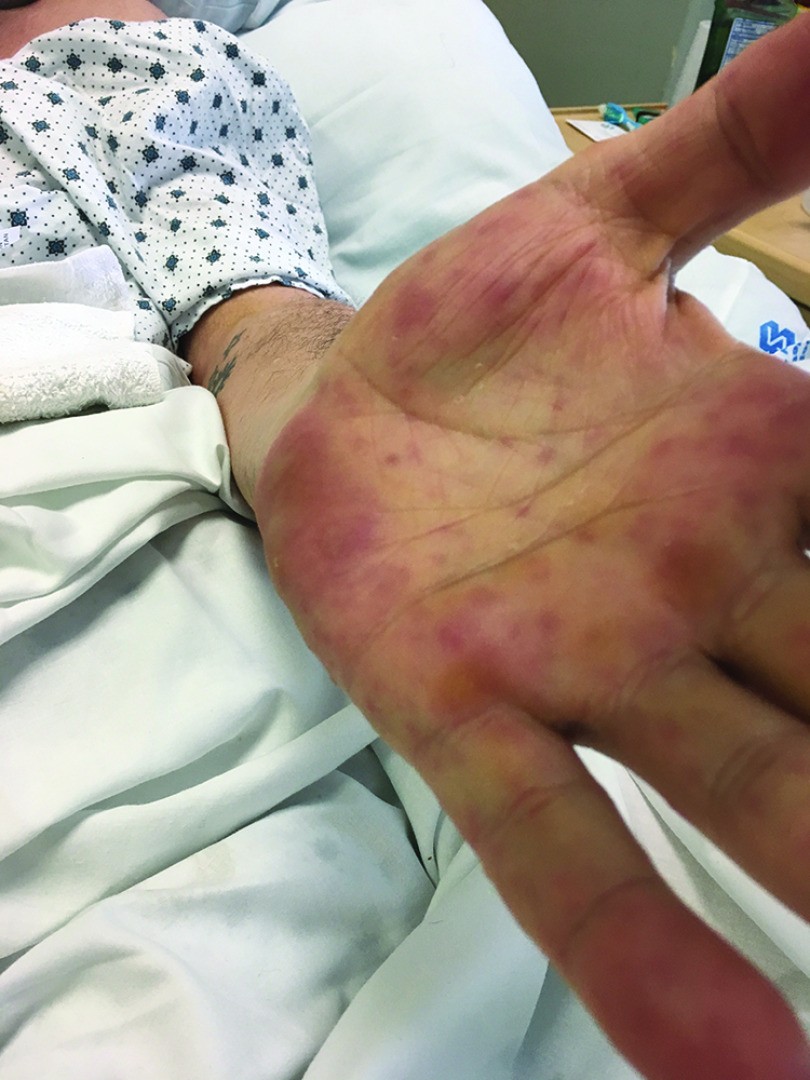
A 62-year-old man with pre-existing psoriasis and multiple sclerosis, admitted for a gangrenous leg infection, developed a sudden onset bullous rash. He had received piperacillin-tazobactam and vancomycin. Ten days after the initial antibiotics, he received vancomycin again as premedication for a cystoscopy. Within hours, he experienced perioral tingling, lip swelling, and an erythematous rash that progressed to painful lesions on his palms and mouth blisters.
Diagnostic Journey
Physical examination revealed tense bullae on his chin, palms, and inner thighs. He also had mouth pain and difficulty swallowing. Initial laboratory tests were unremarkable for infection. Given the temporal association with vancomycin administration, a drug reaction was suspected. To narrow down the differential diagnosis, a skin biopsy was performed.
Diagnosis and Treatment
The skin biopsy showed a subepidermal bulla with linear IgA deposition along the dermoepidermal junction on direct immunofluorescence (DIF). This confirmed the diagnosis of vancomycin-induced LABD. Vancomycin was discontinued, and the patient was treated with a short course of prednisone and topical triamcinolone. His lesions improved significantly within days, and no new lesions developed.
This case underscores the importance of considering drug reactions in the differential diagnosis of bullous skin lesions, even with commonly used antibiotics like vancomycin. LABD, although rare, is a recognized adverse effect of vancomycin and other medications.
Diagnostic Approach to Bullous Skin Lesions
Diagnosing the cause of bullous skin lesions requires a systematic approach:
Clinical Evaluation
A detailed history and physical examination are crucial first steps. This includes:
- History: Medication history (including recent changes and over-the-counter drugs), past medical history (especially autoimmune conditions and infections), onset and progression of lesions, symptoms (pain, itching), and any potential triggers.
- Physical Examination: Assessment of the morphology, distribution, and Nikolsky sign (in pemphigus vulgaris and SJS/TEN, gentle pressure can cause blister spread or new blister formation) of the bullous lesions, as well as examination of mucous membranes and overall systemic involvement.
Diagnostic Tests
Specific diagnostic tests help to confirm or exclude various conditions:
- Skin Biopsy: Essential for histopathology and direct immunofluorescence (DIF). Histopathology can reveal the level of blister formation (epidermal or subepidermal) and inflammatory patterns. DIF is crucial for diagnosing autoimmune bullous diseases, identifying specific antibody deposits (IgG, IgA, C3) and their patterns (linear, intercellular).
- Serology: Blood tests to detect circulating autoantibodies, such as anti-desmoglein antibodies in pemphigus vulgaris and anti-BP180/BP230 antibodies in bullous pemphigoid. Serology can support the diagnosis when biopsy is not feasible or DIF is negative.
- Tzanck Smear or Viral Culture: For suspected herpes infections, Tzanck smear can show multinucleated giant cells, and viral culture or PCR can confirm the specific virus.
- Blood Tests: Complete blood count, metabolic panel, and eosinophil count can help evaluate for systemic involvement and drug reactions like DRESS syndrome.
- Dermatopathology consultation: Seeking expert dermatopathology review is often beneficial for complex cases.
Management and Treatment Strategies
Treatment of bullous skin lesions is directed at the underlying cause and symptom relief:
Addressing the Underlying Cause
- Drug-Induced: Discontinuation of the offending drug is paramount.
- Infections: Antiviral or antibiotic therapy as appropriate for the specific pathogen.
- Autoimmune Diseases: Immunosuppressive agents such as corticosteroids, dapsone, methotrexate, azathioprine, rituximab, or intravenous immunoglobulin (IVIG) may be used, depending on the specific disease and severity.
- Genetic Disorders: Management of epidermolysis bullosa is primarily supportive, focusing on wound care and preventing secondary infections.
Symptomatic Relief
- Wound Care: Gentle cleansing, debridement of bullae, and application of non-adherent dressings to promote healing and prevent infection.
- Pain Management: Analgesics for pain relief.
- Pruritus Control: Topical corticosteroids or systemic antihistamines to manage itching.
- Fluid and Electrolyte Management: Important in severe cases like SJS/TEN to address fluid loss and electrolyte imbalances.
Conclusion
The differential diagnosis of bullous skin lesions is broad, requiring a meticulous approach to identify the underlying etiology. From autoimmune disorders and infections to drug reactions and genetic conditions, the causes are diverse. A thorough clinical evaluation combined with appropriate diagnostic testing, particularly skin biopsy and immunofluorescence, is essential for accurate diagnosis. Prompt and targeted treatment, addressing both the cause and symptoms, is crucial for optimal patient outcomes. If you or someone you know develops bullous skin lesions, seeking prompt medical evaluation from a qualified healthcare professional is highly recommended to ensure timely and appropriate management.
References
[List of references from the original article would be placed here, maintaining the original numbering and links if possible, or re-sourced with standard formatting.]
(Note: The references from the original article should be listed here in the final output. Since they are provided in the original article, they can be directly copied and formatted if needed.)
Table 1. Differential diagnosis for bullous skin disease
SJS = Stevens-Johnson syndrome; TEN = toxic epidermal necrolysis.