Carbohydrate antigen 19-9 (CA 19-9) is a glycoprotein complex found on the surface of cells, initially identified in 1979. Produced by ductal cells in organs such as the pancreas, biliary system, stomach, colon, uterus, and salivary glands, CA 19-9 expression is linked to Lewis blood group antigens, specifically observed in individuals with Le A−B+ or Le A+B− blood types. Notably, a significant portion of the population (up to 6% of Caucasians and 22% of non-Caucasians) are genotypically Le A−B− and consequently do not produce CA 19-9.
While CA 19-9 is recognized as a tumor marker, particularly in pancreatic cancer, it’s crucial to understand its elevation can occur in both benign and malignant conditions across gastrointestinal and extra-gastrointestinal sites. Elevated CA 19-9 levels are most notably associated with pancreatic ductal adenocarcinoma and intraductal papillary mucinous neoplasm (IPMN). However, increased levels can also be seen in cancers of the biliary tract, liver, stomach, colon, urological system, lungs, gynecological organs, thyroid, and salivary glands. Benign conditions associated with CA 19-9 elevation include pancreatitis, pancreatic cysts, diabetes mellitus, liver fibrosis, benign cholestatic diseases, and various non-cancerous conditions affecting the urological, pulmonary, and gynecological systems.
This article aims to explore the complexities of Ca 19-9 Differential Diagnosis by presenting a unique case of markedly elevated CA 19-9 in an asymptomatic individual without a clear underlying cause. Furthermore, it will review the clinical applications and diagnostic implications of CA 19-9 testing.
Case Report: Asymptomatic Patient with Exuberant CA 19-9 Elevation
A 52-year-old male patient was referred for gastroenterological consultation due to an unexpectedly high CA 19-9 level detected during routine bloodwork. The patient reported no gastrointestinal symptoms, denying any history of abdominal pain, jaundice, itching, fever, or any biliary issues. He had experienced a minor weight gain of 0.5 kg recently. His primary care physician had included serum CA 19-9 in a routine laboratory panel. The initial CA 19-9 result was remarkably elevated at 96,544.3 U/mL. Subsequent investigations, including abdominal computed tomography (CT) scan (Figure 1), magnetic resonance imaging with cholangiopancreatography, and positron emission tomography (PET)-CT (Figure 2), were performed to investigate potential underlying malignancy. However, these imaging studies revealed no abnormalities. An endoscopic ultrasound showed minimal dilation of the ventral pancreatic duct near the papilla, but no definitive lesions were identified.
Figure 1 -. Abdominal computed tomography showing a normal image of the pancreas in a patient with elevated CA 19-9.
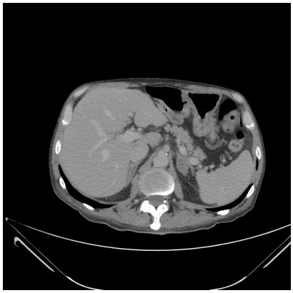 Abdominal CT scan showing normal pancreas in patient with elevated CA 19-9, diagnostic imaging for CA 19-9 differential diagnosis.
Figure 2 -. PET-CT scan showing normal glycolytic metabolism in patient with elevated CA 19-9, further investigation in differential diagnosis.
Given the absence of any identifiable cause for the extreme CA 19-9 elevation, a strategy of observation with serial tumor marker measurements was adopted. Over the next few months, the patient’s CA 19-9 levels demonstrated a spontaneous decline. One month after the initial test, the level was 2822 U/mL, followed by 343 U/mL the next month, and 48.3 U/mL in the subsequent month. The most recent measurement showed a level of 20.8 U/mL, approaching the normal range.
Discussion: CA 19-9 in Clinical Practice and Differential Diagnosis
CA 19-9 is a widely recognized tumor marker, particularly in the context of pancreatic ductal adenocarcinoma. Pancreatic cancer remains a significant global health concern, ranking as the fourth leading cause of cancer-related deaths worldwide, with a poor 5-year survival rate of less than 7%. Elevated CA 19-9 levels are observed in over 80% of patients with advanced pancreatic cancer. However, despite its association with pancreatic cancer, international guidelines emphasize that CA 19-9 should be used in conjunction with radiological imaging, such as pancreas protocol CT, which is considered the gold standard for diagnosis. Systematic reviews have established threshold levels for elevated CA 19-9 in pancreatic cancer diagnosis at >37-40 U/mL, with a reported sensitivity of 79.0%, specificity of 82.0%, positive predictive value of 72.0%, and negative predictive value of 81.0%.
It is critical to recognize that CA 19-9 elevation is not specific to malignancy. Benign pancreatic conditions, such as pancreatitis, and pre-malignant lesions, including IPMNs, can also lead to increased CA 19-9 levels in 10-50% of cases. This overlap underscores why CA 19-9 should not be employed as a standalone screening tool for pancreatic cancer. Its limited screening utility extends even to high-risk populations, like individuals with familial pancreatic cancer or Peutz-Jeghers syndrome, where normal CA 19-9 results can occur despite the presence of preinvasive lesions detected on imaging. Therefore, CA 19-9 is not indicated for pancreatic cancer screening in asymptomatic individuals.
However, once pancreatic cancer is diagnosed, CA 19-9 becomes a valuable tool for staging and guiding treatment decisions. Preoperative CA 19-9 levels have been shown to correlate with pancreatic cancer prognosis. Current biological staging approaches for pancreatic cancer treatment planning consider CA 19-9 levels, with recommendations for neoadjuvant therapy in patients with anatomically resectable pancreatic cancer but preoperative CA 19-9 exceeding 500 IU/mL. This cutoff is supported by studies demonstrating reduced resectability and survival rates in patients with markedly elevated preoperative CA 19-9.
Post-surgery, monitoring CA 19-9 levels after resection and before adjuvant chemotherapy initiation is crucial for prognosis and assessing treatment response. In post-treatment follow-up, rising CA 19-9 levels can precede clinical or radiological recurrence by up to six months, highlighting its role in recurrence monitoring.
In the context of IPMNs, a pre-malignant pancreatic cystic neoplasm, elevated serum CA 19-9 is considered a “worrisome feature.” While CA 19-9 alone cannot reliably distinguish malignant from benign pancreatic cysts, it is useful when combined with other factors like imaging findings and cyst size greater than 3 cm. Serum CA 19-9 levels above 37 U/mL are considered a relative indication for IPMN resection according to European guidelines. Interestingly, CA 19-9 levels measured in cyst fluid obtained via endoscopic ultrasound-guided fine-needle aspiration are less accurate than other cyst fluid tumor markers, such as CEA and CA 125, in differentiating pancreatic cystic lesions.
CA 19-9 also plays a significant role in the diagnosis, staging, and follow-up of cholangiocarcinoma (bile duct cancer). In cases of biliary strictures with concerning imaging features, CA 19-9 levels exceeding 100 U/mL are suggestive of perihilar cholangiocarcinoma. Furthermore, CA 19-9 levels correlate with prognosis in gastric cancer, including tumor stage, vascular invasion, and lymph node and distant metastasis. However, CA 19-9 is not recommended as a standalone staging marker for gastric cancer and should be used in conjunction with CEA and CA 72-4.
Beyond malignancies, benign diseases can also cause elevated serum CA 19-9 levels. Cholestatic conditions, such as choledocholithiasis (gallstones in the bile duct) and cholangitis (bile duct inflammation), can raise CA 19-9 to very high levels in the absence of cancer. Case reports have documented Mirizzi syndrome, a complication of gallstones, associated with CA 19-9 levels as high as 21,068 U/mL, posing a significant challenge in differentiating it from cholangiocarcinoma.
Differential Diagnosis of Elevated CA 19-9
The differential diagnosis for elevated CA 19-9 is broad and includes both malignant and benign conditions. A systematic approach is essential to determine the underlying cause, especially in asymptomatic patients.
Malignant Conditions:
- Pancreatic cancer (ductal adenocarcinoma, IPMN)
- Biliary tract cancers (cholangiocarcinoma, gallbladder cancer)
- Gastric cancer
- Colorectal cancer
- Hepatocellular carcinoma
- Lung cancer
- Ovarian cancer
- Thyroid cancer
- Salivary gland cancers
Benign Conditions:
- Pancreatitis (acute and chronic)
- Pancreatic cysts (non-neoplastic)
- Cholestasis (choledocholithiasis, primary sclerosing cholangitis, primary biliary cholangitis)
- Cholangitis
- Liver cirrhosis and fibrosis
- Benign biliary strictures
- Diabetes mellitus
- Pulmonary diseases (e.g., pneumonia, COPD)
- Gynecological conditions (e.g., endometriosis, pelvic inflammatory disease)
- Thyroid diseases (benign thyroid nodules, thyroiditis)
- Inflammatory bowel disease
- Cystic fibrosis
Conclusions: Clinical Implications for CA 19-9 Testing
Clinicians must be aware of the limitations of CA 19-9 as a screening test for pancreatic malignancies. Elevated CA 19-9 levels can be observed in various benign conditions, and importantly, individuals with the Le A−B− blood group will not produce CA 19-9, even in the presence of cancer. Indiscriminate use of CA 19-9 as a screening tool in asymptomatic patients can lead to unnecessary, costly, and potentially invasive diagnostic procedures. Therefore, CA 19-9 testing should be strategically utilized in the appropriate clinical context, considering the patient’s symptoms, risk factors, and in conjunction with radiological and other diagnostic modalities for accurate differential diagnosis and patient management.
REFERENCES
[1] How to cite this article: Meira-Júnior JD, Costa TN, Montagnini AL, Nahas SC, Jukemura J. Elevated CA 19-9 in an asymptomatic patient: what does it mean? ABCD Arq Bras Cir Dig. 2022;35:e1687. https://doi.org/10.1590/0102-672020220002e1687
[2] Financial Source: None