Introduction
In the daily practice of automotive repair, encountering unusual soft tissue masses can be perplexing. While the vast majority of these lesions in vehicles prove to be benign, differentiating them from potentially “malignant” issues—those that can severely impact vehicle performance and safety—is crucial for any expert technician. Just as in human medicine, where radiologists discern benign from malignant soft tissue lesions in patients, automotive technicians must develop a keen eye to identify and diagnose abnormalities within a vehicle’s soft tissue components. Understanding the nature of these masses, particularly if they exhibit calcification or ossification, is paramount for accurate diagnosis and effective repair strategies.
The presence of calcifications or ossification within a soft tissue mass in a vehicle can significantly narrow down the potential diagnoses. This is because certain automotive pathologies are more prone to producing ossifications (bone-like formations), while others tend to result in calcifications (mineral deposits). This article serves as a comprehensive guide for auto repair experts, focusing on the Calcified Soft Tissue Mass Differential Diagnosis. We will explore various benign conditions that can manifest as calcified or ossified masses, mimicking more serious problems and potentially leading to misdiagnosis and unnecessary interventions. Our aim is to equip you with the knowledge to confidently differentiate these benign entities from more concerning issues, ensuring optimal vehicle health and longevity.
Calcifications Versus Ossification in Automotive Soft Tissue
Distinguishing between calcifications and ossification is a foundational step in accurately diagnosing soft tissue masses in vehicles. While sophisticated imaging modalities like radiography, CT scans, MRI, and ultrasound used in human medicine are not routinely applied in auto repair, understanding the underlying principles of how these modalities differentiate calcification and ossification can inform our diagnostic approach.
In essence, calcifications in automotive soft tissues typically appear as mineralized densities. Imagine them as hardened deposits of minerals within the material. Ossification, on the other hand, suggests the formation of bone-like structures, often exhibiting a more organized structure similar to bone itself.
Although we primarily rely on visual inspection and tactile examination in auto repair, understanding how these phenomena are differentiated in medical imaging provides a valuable conceptual framework:
-
Radiography (X-rays): In medical contexts, radiography is often the initial imaging technique. Mature bone is distinguished by its outer cortex and inner trabecular pattern, whereas calcifications appear as less structured mineral densities. While we don’t use X-rays on car parts routinely, this principle highlights the structural difference between organized bone and simpler mineral deposits.
-
Computed Tomography (CT): CT scans offer greater sensitivity in detecting calcifications and ossification due to their ability to measure tissue density in Hounsfield Units (HU). Bone exhibits much higher HU values compared to general calcifications. This illustrates that density is a key differentiator – ossified material will generally be denser and more bone-like in its composition.
-
Magnetic Resonance Imaging (MRI): MRI in medicine shows calcifications typically as areas of uniformly low signal intensity across different sequences. Mature bone, especially with fatty marrow, has a distinct appearance. While MRI isn’t used on cars, it underscores that the internal structure and composition differ between simple calcification and true ossification.
-
Ultrasound: In medical imaging, ultrasound can sometimes struggle to differentiate between calcifications and ossification, especially if acoustic shadowing obscures details. This is a reminder that even with advanced techniques, differentiation can be nuanced.
In the context of auto repair, we must rely on our expertise to assess the visual and tactile characteristics of the mass. Is it a gritty, stone-like deposit (suggesting calcification)? Or does it feel and appear more like bone, potentially with a more organized structure (suggesting ossification)? This distinction, even if made without advanced imaging, is a vital first step in formulating a calcified soft tissue mass differential diagnosis.
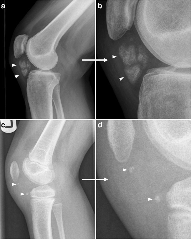
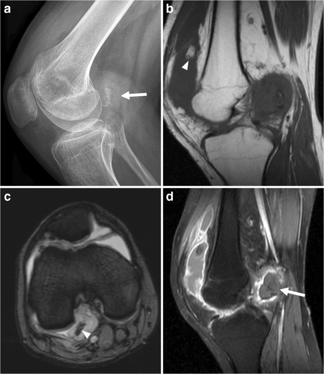
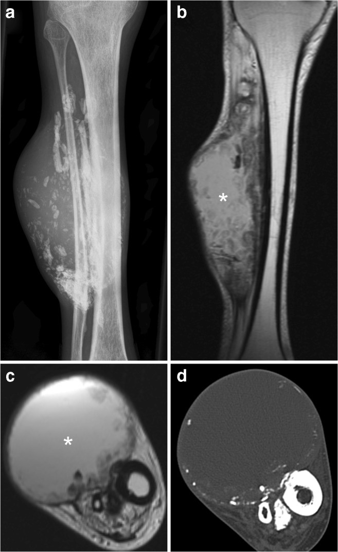
Fig. 1. Ossification versus Calcification: Conceptual Automotive Analogy
Alt Text: Automotive components analogy illustrating ossification versus calcification. Image a and b show a structured, bone-like automotive part representing ossification. Image c and d show mineral deposits on a car part, representing calcification.
Note: While this image is from the original medical article, the concept of structured vs unstructured mineral deposits is analogous to automotive scenarios.
Common Benign Calcified or Ossified Soft Tissue Masses in Vehicles
Now, let’s delve into specific benign conditions in vehicles that can present as calcified or ossified soft tissue masses, potentially mimicking more serious problems. Understanding these conditions is crucial for developing a robust calcified soft tissue mass differential diagnosis.
1. Myositis Ossificans Analogy: “Mechanic’s Muscle Ossification”
While the term “myositis ossificans” is medical, we can draw an analogy for automotive contexts. Imagine a scenario of “Mechanic’s Muscle Ossification”. This would be akin to heterotopic ossification in humans, where bone tissue forms outside of the skeletal system. In vehicles, this could occur in:
- Rubber Mounts and Bushings: Repeated stress, vibration, or blunt force trauma (like impacts from road debris) could theoretically lead to ossification within rubber components designed for flexibility and shock absorption.
- Reinforced Hoses: In hoses with fabric or steel reinforcement, trauma or degradation could, hypothetically, trigger a process resembling ossification within the hose structure itself.
Clinical Presentation Analogy:
- Initial Stage: Similar to the early stages of myositis ossificans in humans, we might observe swelling, stiffness, or restricted movement in the affected component. For instance, a rubber engine mount might appear swollen and less pliable.
- Intermediate Stage: Calcifications could begin to appear. In our automotive analogy, this might manifest as hard, gritty deposits within the rubber or hose material.
- Mature Stage: Ossification becomes more defined, potentially forming a bone-like rim or structure. The component becomes significantly less flexible and may exhibit cracking or failure due to rigidity.
Imaging Analogy (Conceptual):
- Radiography/CT analogy: Mature “Mechanic’s Muscle Ossification” might show a peripheral rim of dense material resembling cortical bone, with a less dense center.
- MRI analogy: Early stages could show edema (swelling) in the surrounding material. Mature stages might reveal internal trabecular-like structures (again, conceptually).
Differential Diagnosis Analogy:
- “Parosteal Osteosarcoma” Analogy: This is the malignant mimic in the medical context. In auto repair, we might consider this analogous to severe structural degradation or material failure that requires immediate and extensive repair or replacement.
- Soft Tissue Sarcoma Analogy: This could be analogous to advanced material breakdown or corrosion that has spread and compromised surrounding components.
- Soft Tissue Abscess Analogy: This relates to fluid or contaminant buildup within a component, which is less relevant to ossification but part of the broader differential for soft tissue masses.
Key Automotive Diagnostic Considerations:
- History: Look for history of impacts, heavy use, or extreme conditions that could have stressed the component.
- Location: Common stress points, areas prone to impact damage.
- Progression: Has the stiffness or hardness developed over time?
- Physical Examination: Tactile examination to assess hardness, structure.
Management Analogy:
- Just as close medical surveillance or biopsy is needed in uncertain cases of myositis ossificans, in auto repair, careful monitoring of the component’s condition and potential replacement are crucial when “Mechanic’s Muscle Ossification” is suspected.
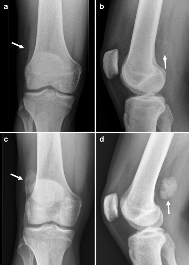
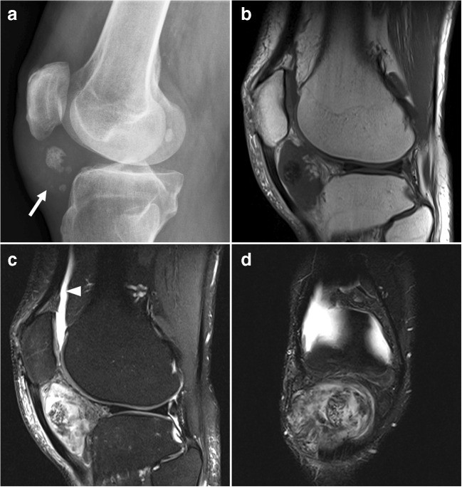
Fig. 2. “Mechanic’s Muscle Ossification” Stages Analogy
Alt Text: Automotive component analogy for stages of “Mechanic’s Muscle Ossification”. Image a and b represent early/intermediate stage with amorphous deposits. Image c and d show a later stage with more defined ossification in an automotive part.
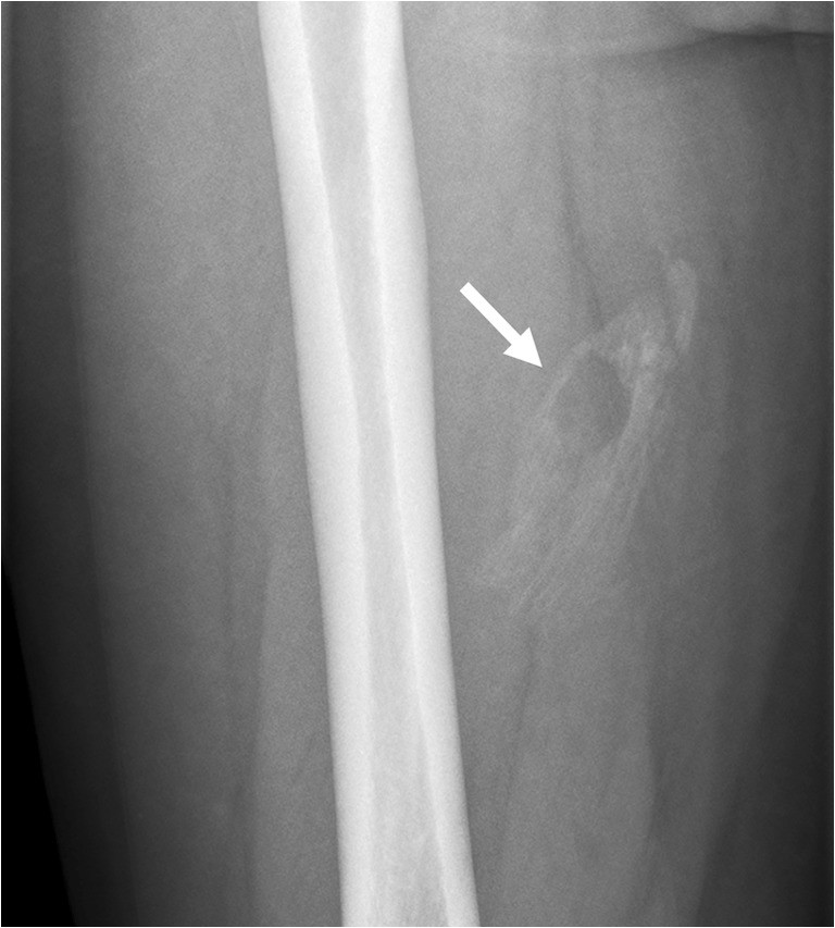
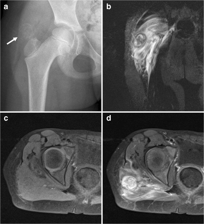
Fig. 3. Mature “Mechanic’s Muscle Ossification” Analogy
Alt Text: Mature “Mechanic’s Muscle Ossification” analogy in an automotive context. Image shows a well-defined lesion with peripheral ossification in a vehicle’s soft tissue component.
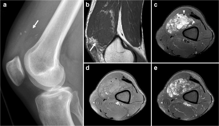
Fig. 4. “Mechanic’s Muscle Ossification” with Surrounding Edema Analogy
Alt Text: “Mechanic’s Muscle Ossification” analogy showing surrounding edema in automotive soft tissue. Image a shows a deposit. Images b, c, and d conceptually represent swelling and altered material properties around the lesion.
2. Tophaceous Gout Analogy: “Corrosion Crystal Deposits”
In vehicles, we encounter corrosion, and certain types of corrosion products could, in an analogous way to tophaceous gout, form crystal-like deposits. Let’s call this “Corrosion Crystal Deposits”.
Automotive Locations Analogy:
- Battery Terminals and Cables: Acid corrosion can lead to crystalline buildup around battery terminals and cable connections.
- Exhaust System Joints: Corrosion at exhaust joints can produce deposits that mimic calcifications.
- Coolant System Components: Dried coolant residue, especially if mixed with hard water minerals, can form hard, crystalline deposits.
Clinical Presentation Analogy:
- Swelling: Visible buildup of crystalline material.
- Hardness: The deposits are typically hard and can be gritty.
- Color: Often white, bluish-white, or greenish, depending on the corrosion type.
Imaging Analogy (Conceptual):
- Radiography/CT analogy: Hyperdense soft tissue swelling with potential dense calcifications.
- MRI analogy: Variable signal intensity, potentially with enhancement conceptually representing the corrosive process.
Differential Diagnosis Analogy:
- Synovial Sarcoma Analogy: Again, this is the malignant mimic in the medical article. In auto repair, this might be analogous to severe, rapidly spreading corrosion that resembles a more aggressive issue.
Key Automotive Diagnostic Considerations:
- Location: Sites prone to corrosion (electrical connections, exhaust, coolant systems).
- Visual Appearance: Crystalline structure, color.
- Chemical Tests: Identifying the nature of the deposit (e.g., battery acid residue, coolant residue).
Management Analogy:
- Similar to biopsies in gout diagnosis, chemical analysis or careful removal and examination of the deposit can help confirm the diagnosis of “Corrosion Crystal Deposits.”
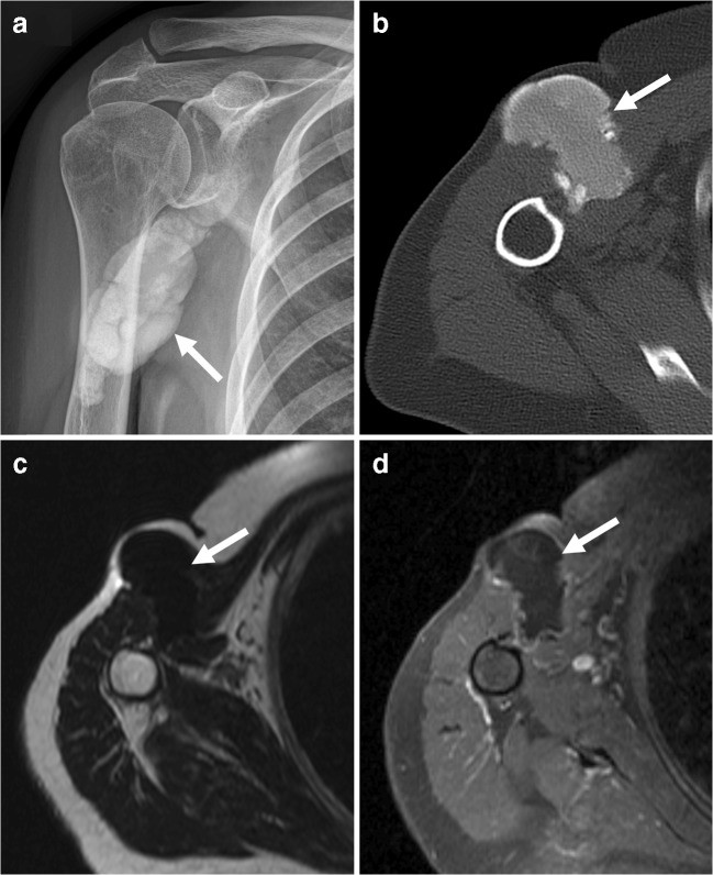
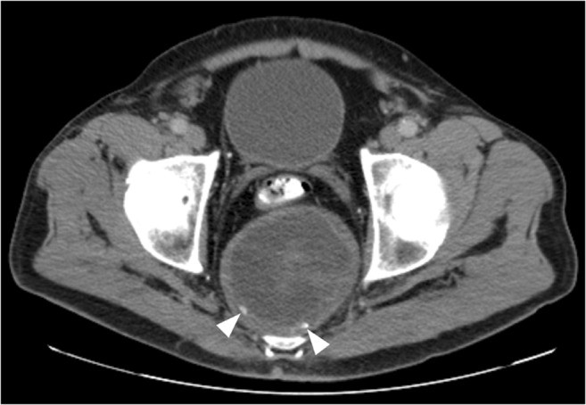
Fig. 5. “Corrosion Crystal Deposits” Analogy
Alt Text: “Corrosion Crystal Deposits” analogy in automotive context. Image a and b show deposits on a car part, resembling calcifications. Image c conceptually shows enhancement related to corrosion.
3. Benign Vascular Lesions Analogy: “Fluid Accumulation with Mineral Concretions”
Venous malformations in medicine can be analogous to fluid accumulations within vehicle components, sometimes with mineral concretions forming within. Let’s call this “Fluid Accumulation with Mineral Concretions”.
Automotive Locations Analogy:
- Power Steering Hoses: Bulges or swellings in power steering hoses can sometimes contain solidified fluid and mineral deposits.
- Brake Lines: Corrosion within brake lines could lead to localized swellings with internal mineral buildup.
- Coolant Hoses: Similar to power steering hoses, coolant hoses can develop bulges containing solidified coolant and minerals.
Clinical Presentation Analogy:
- Soft, Compressible Masses: Initially, the swelling might be soft and compressible, like a venous malformation.
- Nonpulsatile: Unlike arterial issues, these swellings are typically nonpulsatile.
- Phleboliths Analogy: Mineral concretions form within the fluid accumulation, analogous to phleboliths in venous malformations.
Imaging Analogy (Conceptual):
- Radiography/CT analogy: Detection of mineral concretions within the mass.
- Ultrasound analogy: “Sponge-like” appearance on ultrasound (if applicable to automotive materials), compressibility.
- MRI analogy: Septated lobulated mass, fluid-fluid levels (conceptually), slow gradual enhancement representing fluid dynamics.
Differential Diagnosis Analogy:
- Other vascular malformations analogy: Other types of fluid leaks or swellings in vehicle systems.
Key Automotive Diagnostic Considerations:
- Location: Hoses and lines prone to fluid pressure and corrosion.
- Palpation: Softness, compressibility, presence of hard concretions.
- Fluid Type: Identifying the fluid within the swelling (power steering fluid, coolant, brake fluid).
Management Analogy:
- Similar to avoiding biopsy for venous malformations in medicine due to bleeding risk, in auto repair, puncture or aggressive probing of these swellings might be avoided initially to prevent fluid release or further damage.
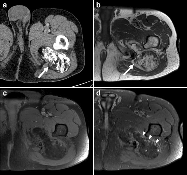
Fig. 6. “Fluid Accumulation with Mineral Concretions” Analogy
Alt Text: “Fluid Accumulation with Mineral Concretions” analogy in automotive context. Image a shows mineral concretions in a car part. Images b, c, d, and e conceptually represent fluid accumulation and internal structures.
4. Calcium Hydroxyapatite Deposition Disease Analogy: “Lubricant Calcification with Joint Involvement”
Calcific tendinopathy in medicine, especially with bone involvement, can be analogous to lubricant calcification affecting joints or moving parts in vehicles. Let’s call this “Lubricant Calcification with Joint Involvement”.
Automotive Locations Analogy:
- Ball Joints and Suspension Joints: Dried, hardened grease or lubricant in ball joints, tie rod ends, or other suspension joints.
- Wheel Bearings: Degraded grease in wheel bearings.
- CV Joints: Hardened grease in constant velocity joints.
Clinical Presentation Analogy:
- Pain/Restriction of Motion: Stiffness, restricted movement, noises (creaking, grinding) from the affected joint.
- Aggressive Imaging Appearance Analogy: In medical imaging, this condition can mimic malignancy. In auto repair, severely affected joints might present with alarming symptoms suggesting serious mechanical failure.
- Bone Marrow Edema Analogy: In the medical context, bone marrow edema is a key MRI finding. In our automotive analogy, this could be related to excessive wear, heat buildup, and damage to the joint surfaces.
Imaging Analogy (Conceptual):
- Radiography/CT analogy: Calcifications around the joint area.
- MRI analogy: “Bone marrow edema” analogy – signs of wear, damage, and altered material properties in the joint components.
Differential Diagnosis Analogy:
- Malignancy analogy (soft tissue sarcoma, periosteal sarcoma, metastatic disease): Serious mechanical failure, structural damage requiring major component replacement.
- Infection analogy: Contamination or foreign material entry into the joint causing inflammation and dysfunction.
Key Automotive Diagnostic Considerations:
- Location: Joints and bearings that rely on lubrication.
- Symptoms: Stiffness, noise, restricted movement, play in the joint.
- Grease Condition: Examination of the grease – hardened, gritty, discolored?
Management Analogy:
- Just as correlating MRI with radiography is important in medical diagnosis, in auto repair, combining physical examination with inspection of the lubricant and joint components is crucial for diagnosis.
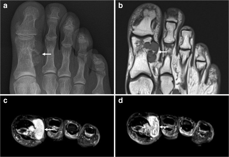
Fig. 7. “Lubricant Calcification with Joint Involvement” Analogy
Alt Text: “Lubricant Calcification with Joint Involvement” analogy in automotive context. Image a shows deposits around a joint. Images b and c conceptually represent intra-joint extension of calcification and surrounding damage.
5. Periosteal Chondroma Analogy: “Surface Rust with Concretion”
Periosteal chondroma, a benign bone tumor arising from the periosteum, can be conceptually related to surface rust on metal components that develops a hard, concretion-like outer layer. Let’s call this “Surface Rust with Concretion”.
Automotive Locations Analogy:
- Frame Rails and Chassis Components: Surface rust on frame rails or other structural components that develops a hard, layered scale.
- Exhaust System Components: Heavy surface rust on exhaust pipes or mufflers with a thick, hardened outer layer.
- Suspension Components (Metal Parts): Rust formation on metal suspension parts that becomes thick and concretion-like.
Clinical Presentation Analogy:
- Focal Swelling: The rust concretion creates a raised area on the metal surface.
- Hardness: The concretion is typically hard and firmly attached.
- Pain Analogy: While rust itself isn’t painful, severe rust can compromise structural integrity, potentially leading to noises, vibrations, or functional issues that could be considered analogous to “pain.”
Imaging Analogy (Conceptual):
- Radiography/CT analogy: Calcified chondroid matrix analogy – the rust concretion itself. “Scalloping and overhanging edges of cortex” analogy – the way the rust layer conforms to the underlying metal surface.
- MRI analogy: “Lobulated configuration, no edema” analogy – the defined shape of the rust concretion without widespread surrounding damage (unless rust is very advanced). “Septal enhancement pattern” analogy – conceptual representation of different layers or zones within the rust concretion.
Differential Diagnosis Analogy:
- Periosteal chondrosarcoma analogy: More aggressive, penetrating rust that deeply compromises structural integrity.
- Periosteal/parosteal osteosarcoma analogy: Rust formation that is more overtly destructive and bone-like (conceptually).
Key Automotive Diagnostic Considerations:
- Location: Areas prone to rust (exposed metal, areas with moisture accumulation).
- Appearance: Layered, concretion-like rust.
- Extent: Is it just surface rust, or is it deeply penetrating?
- Structural Integrity: Has the rust compromised the component’s strength?
Management Analogy:
- Similar to surgical excision for periosteal chondroma, removal of surface rust and protective treatment of the underlying metal are common management strategies.
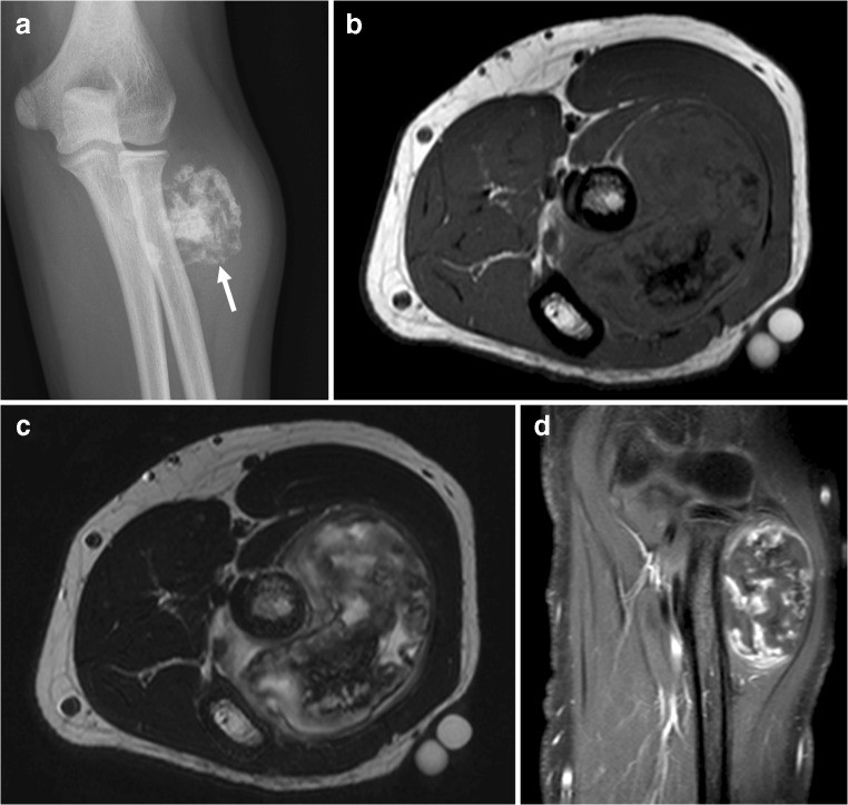
Fig. 8. “Surface Rust with Concretion” Analogy
Alt Text: “Surface Rust with Concretion” analogy in automotive context. Image a shows rust concretion on a metal part. Images b, c, and d conceptually represent the layered structure and attachment to the underlying metal.
Fig. 9. “Surface Rust with Concretion” Detail Analogy
Alt Text: Detailed “Surface Rust with Concretion” analogy. Image a shows rust concretion on a metal part. Images b, c, and d conceptually represent the rust layer and its relationship to the metal surface.
6. Primary Synovial Chondromatosis Analogy: “Joint Debris Accumulation”
Primary synovial chondromatosis, involving cartilage nodule development in joints, can be analogized to debris accumulation within vehicle joints. Let’s call this “Joint Debris Accumulation”.
Automotive Locations Analogy:
- Constant Velocity (CV) Joints: Accumulation of debris, broken grease, or wear particles within a CV joint.
- Ball Joints: Buildup of contaminants and wear debris in ball joints.
- Steering Rack Joints: Debris accumulation in steering rack joints.
Clinical Presentation Analogy:
- Pain, Swelling, Restriction of Motion Analogy: Noises (clicking, popping), stiffness, limited movement in the affected joint.
- Intraarticular Calcifications Analogy: Debris particles within the joint that could become hardened or mineralized over time, resembling calcifications.
- Chondroid Ring-and-Arc Mineralization Pattern Analogy: Conceptual representation of debris particles clumped together in ring-like or arc-like patterns within the joint.
Imaging Analogy (Conceptual):
- Radiography/CT analogy: Detection of debris or mineralized particles within the joint space.
- MRI analogy: Variable signal characteristics depending on the nature of the debris. “Enhancement” analogy – conceptual representation of inflammation or reaction within the joint due to debris.
Differential Diagnosis Analogy:
- Malignant transformation to synovial chondrosarcoma analogy: Severe joint damage, component failure requiring major repair.
Key Automotive Diagnostic Considerations:
- Location: Joints prone to debris accumulation (exposed joints, joints with damaged seals).
- Symptoms: Joint noises, stiffness, restricted movement.
- Lubricant Condition: Inspection of lubricant for debris and contamination.
Management Analogy:
- Similar to surgical removal of cartilage nodules in synovial chondromatosis, cleaning and replacing contaminated or debris-filled joints is a common automotive repair.
Fig. 10. “Joint Debris Accumulation” Analogy
Alt Text: “Joint Debris Accumulation” analogy in automotive context. Image a shows debris within a joint-like space. Images b, c, and d conceptually represent debris fragments and joint reaction.
7. Hoffa’s Disease Analogy: “Fat Pad Impingement with Ossification”
Hoffa’s disease, affecting the fat pad in the knee, can be analogized to fat pad impingement and potential ossification in automotive components. Let’s call this “Component Fat Pad Impingement”.
Automotive Locations Analogy:
- Rubber Bump Stops: Over-compression and damage to rubber bump stops in suspension systems.
- Body Mounts: Compression and degradation of body mounts.
- Engine/Transmission Mounts (Rubber Portions): Damage to rubber sections of engine or transmission mounts due to excessive stress or age.
Clinical Presentation Analogy:
- Pain Analogy: Noises, vibrations, harshness in ride quality, or drivetrain vibrations.
- Acute/Repetitive Trauma Analogy: Rough driving, heavy loads, worn suspension components.
- Edema and Hemorrhage Analogy: Initial swelling and damage to the “fat pad” component (rubber mount).
- Ossification Analogy: Over time, the damaged rubber mount could become hardened, cracked, or even develop ossification-like changes.
Imaging Analogy (Conceptual):
- Radiography/CT analogy: Ossified fragments within the “fat pad” component.
- MRI analogy: “Edema” – initial swelling, increased signal intensity. “Fibrin and hemosiderin” analogy – later stages with hardened or degraded material (low signal intensity).
Differential Diagnosis Analogy:
- Synovial sarcoma analogy: Severe structural failure of the mount, requiring complete replacement.
- Venous malformation analogy: Fluid buildup within the mount (less relevant to ossification focus).
Key Automotive Diagnostic Considerations:
- Location: Rubber mounts and bump stops in suspension and drivetrain.
- Symptoms: Noises, vibrations, harshness, sagging.
- Visual Inspection: Cracks, tears, compression, hardening of the rubber component.
Management Analogy:
- Just as differentiating calcification from ossification is important in Hoffa’s disease diagnosis, in auto repair, assessing the nature of the hardening (is it simple degradation or something more like ossification?) is relevant for determining repair strategy.
Fig. 11. “Component Fat Pad Impingement” Analogy
Alt Text: “Component Fat Pad Impingement” analogy in automotive context. Image a shows ossified fragments in a component “fat pad”. Images b, c, and d conceptually represent the hardened component and surrounding damage.
8. Tumoral Calcinosis Analogy: “Mineral Accumulation in Void Spaces”
Tumoral calcinosis, with its massive periarticular calcinosis, can be analogized to mineral accumulation within void spaces in vehicle components. Let’s call this “Void Space Mineral Accumulation”.
Automotive Locations Analogy:
- Frame Rails (Internal Voids): Mineral buildup within hollow frame rails or chassis members due to water ingress and evaporation.
- Body Panels (Double-Layered Areas): Accumulation of rust and mineral deposits between double-layered body panels.
- Rocker Panels (Internal Cavities): Mineral buildup inside rocker panels.
Clinical Presentation Analogy:
- Painless Mass(es) Analogy: Increased weight, muffled noises, potential for rust breakthrough.
- Periarticular Analogy: “Peri-component” – mineral buildup around structural components.
- Chalky White Matter Analogy: Flaking rust or mineral deposits that might extrude from corroded areas.
Imaging Analogy (Conceptual):
- Radiography/CT analogy: Lobulated calcific soft tissue mass analogy – mineral mass within the void space. “Cystic” analogy – the void space itself. “Fluid-calcium levels” analogy – conceptual representation of layers of mineral deposits and trapped moisture.
- MRI analogy: Heterogeneous lesion, low T1/T2 signal intensity analogy – dense mineral material. “Enhancement” analogy – hypervascularity of the surrounding corroding metal (conceptually).
Differential Diagnosis Analogy:
- Calcinosis associated with chronic renal failure analogy: Widespread corrosion due to prolonged exposure to harsh conditions.
- Soft tissue sarcoma analogy: Severe, rapidly spreading rust that deeply compromises structural integrity.
Key Automotive Diagnostic Considerations:
- Location: Enclosed void spaces in structural components.
- Appearance: Bulging, rust breakthrough, increased weight.
- Rust Extent: Is it localized or widespread?
- Drainage Holes: Are drainage holes blocked, contributing to moisture trapping?
Management Analogy:
- Similar to checking calcium-phosphate levels and renal function in tumoral calcinosis, in auto repair, assessing the cause of mineral buildup (water leaks, blocked drains, etc.) is crucial for effective repair.
Fig. 12. “Void Space Mineral Accumulation” Analogy
Alt Text: “Void Space Mineral Accumulation” analogy in automotive context. Image a shows mineral mass in a void space. Images b, c, and d conceptually represent the location within a component and material properties.
9. Lipoma with Metaplasia Analogy: “Grease Accumulation with Hardening”
Lipoma with metaplastic bone, a rare lipoma variant with bone formation, can be analogized to grease accumulation in vehicles that hardens and develops bone-like concretions. Let’s call this “Hardened Grease Concretion”.
Automotive Locations Analogy:
- Chassis Lubrication Points: Old, hardened grease buildup around chassis lubrication fittings.
- Suspension Component Grease Points: Dried, hardened grease around suspension grease points.
- Door Hinges and Latches: Old, hardened grease or lubricant on door hinges or latches.
Clinical Presentation Analogy:
- Soft Tissue Mass Analogy: Initial grease accumulation.
- Painful Analogy: Stiffness, restricted movement of the lubricated part.
- Metaplastic Bone Analogy: Hardening and concretion formation within the grease, resembling bone-like material.
Imaging Analogy (Conceptual):
- Radiography/CT analogy: Fat, bone, and calcifications analogy – visualization of hardened grease and any mineral concretions within it.
- MRI analogy: “Fatty” component – the grease itself. “Bone” component – the hardened concretions. “Enhancement” analogy – conceptual representation of the interface between the hardened grease and surrounding components.
Differential Diagnosis Analogy:
- Soft tissue sarcoma (liposarcoma) analogy: Severe binding or seizing of the lubricated part, requiring disassembly and extensive cleaning or replacement.
Key Automotive Diagnostic Considerations:
- Location: Grease points and lubrication areas.
- Appearance: Hardened, concretion-like grease.
- Function: Is the lubricated part moving freely, or is it stiff or binding?
Management Analogy:
- Similar to surgical excision for lipoma with metaplasia, removal of hardened grease and relubrication are the typical automotive “surgical” approach.
Fig. 13. “Hardened Grease Concretion” Analogy
Alt Text: “Hardened Grease Concretion” analogy in automotive context. Image a shows hardened grease with concretions. Images b, c, and d conceptually represent the grease, hardened areas, and surrounding components.
10. Calcifying Aponeurotic Fibroma Analogy: “Hardened Sealant/Adhesive”
Calcifying aponeurotic fibroma, a rare fibrous tumor that can calcify, can be analogized to hardened sealant or adhesive in vehicles that develops calcifications. Let’s call this “Hardened Sealant Calcification”.
Automotive Locations Analogy:
- Windshield Sealant: Hardened, cracked windshield sealant that develops mineral deposits.
- Body Seam Sealers: Old, hardened body seam sealer that becomes brittle and calcified-looking.
- Undercoating (Hardened Areas): Hardened, cracked undercoating that develops mineral deposits or a calcified appearance.
Clinical Presentation Analogy:
- Slow-Growing Mass Analogy: Gradual hardening and cracking of sealant/adhesive.
- Poorly Circumscribed Analogy: The hardened sealant often blends into surrounding areas.
- Calcifications Analogy: Mineral deposits and a calcified appearance within the hardened sealant.
Imaging Analogy (Conceptual):
- Radiography/CT analogy: Soft tissue mass with stippled calcifications analogy – hardened sealant with mineral deposits.
- MRI analogy: Heterogeneous signal intensity, intense enhancement analogy – conceptual representation of the complex material properties of hardened sealant and surrounding areas.
Differential Diagnosis Analogy:
- Soft tissue sarcoma analogy (synovial sarcoma, undifferentiated pleomorphic sarcoma): Severe sealant failure, water leaks, rust formation requiring extensive bodywork.
Key Automotive Diagnostic Considerations:
- Location: Sealed seams, windshield perimeters, underbody areas.
- Appearance: Hardened, cracked sealant with mineral deposits.
- Function: Is the sealant still effectively sealing, or is it leaking?
Management Analogy:
- Similar to biopsy for calcifying aponeurotic fibroma, careful examination and potentially removal of the hardened sealant are needed for diagnosis and repair.
Fig. 14. “Hardened Sealant Calcification” Analogy
Alt Text: “Hardened Sealant Calcification” analogy in automotive context. Image a shows hardened sealant with mineral reflections. Images b and c conceptually represent the sealant material and surrounding areas.
11. Calcific Myonecrosis Analogy: “Compartment Fluid with Peripheral Calcification”
Calcific myonecrosis, a posttraumatic muscle condition with liquefaction and peripheral calcification, can be analogized to fluid buildup in a vehicle compartment with peripheral mineral deposits. Let’s call this “Compartment Fluid Mineralization”.
Automotive Locations Analogy:
- Door Cavities (Water Accumulation): Water trapped in door cavities leading to rust and mineral buildup around the edges.
- Rocker Panels (Internal Fluid): Fluid trapped in rocker panels causing internal rust and peripheral mineral deposits.
- Frame Rails (Fluid Ingress): Fluid entry into frame rails with mineral deposition along the inner walls.
Clinical Presentation Analogy:
- Posttraumatic Analogy: Water ingress due to damage, blocked drains, or environmental exposure.
- Slowly Enlarging Mass Analogy: Gradual accumulation of fluid and mineral deposits.
- Painful Analogy: Increased weight, potential for rust breakthrough, noises from fluid sloshing.
Imaging Analogy (Conceptual):
- Radiography/CT analogy: Mass with peripherally oriented plaque-like calcifications analogy – fluid-filled compartment with peripheral mineral deposits. “Liquid-appearing center” analogy – the fluid within the compartment.
- MRI analogy: Homogeneous intermediate T1, heterogeneously high T2 signal intensity analogy – conceptual representation of fluid properties. No enhancement analogy – unless there’s active corrosion.
Differential Diagnosis Analogy:
- Soft tissue sarcoma analogy: Severe structural rust, requiring extensive bodywork and component replacement.
- Infection analogy: Contamination within the fluid, leading to corrosion and material degradation.
Key Automotive Diagnostic Considerations:
- Location: Enclosed compartments prone to fluid trapping.
- Symptoms: Increased weight, sloshing noises, rust breakthrough.
- Drainage: Are drainage holes blocked?
- Fluid Type: Is it just water, or is it contaminated?
Management Analogy:
- Similar to the long time interval after trauma in calcific myonecrosis, “Compartment Fluid Mineralization” often develops over years. Diagnosis relies on recognizing the pattern of fluid accumulation and peripheral mineralization.
Fig. 15. “Compartment Fluid Mineralization” Analogy
Alt Text: “Compartment Fluid Mineralization” analogy in automotive context. Image a shows peripheral mineral deposits in a compartment. Images b, c, and d conceptually represent the fluid center and peripheral mineralization.
12. Ancient Schwannoma Analogy: “Degenerated Wire Insulation”
Ancient schwannomas, degenerated nerve sheath tumors, can be analogized to long-standing, degenerated wire insulation in vehicles that develops calcifications. Let’s call this “Degenerated Insulation Mineralization”.
Automotive Locations Analogy:
- Wiring Harness Insulation (Engine Bay): Old, heat-exposed wire insulation in the engine bay that becomes brittle, cracked, and develops mineral deposits.
- Wiring Insulation (Underbody): Wiring insulation exposed to road debris and moisture that degrades and mineralizes.
- Wiring Insulation (Door Jambs): Wiring insulation in door jambs that experiences repeated flexing and wear, leading to degradation and mineralization.
Clinical Presentation Analogy:
- Long-Standing, Slow-Growing Analogy: Gradual degradation of wire insulation over time.
- Deep Location Analogy: Wiring often runs within harnesses or protected areas.
- Cystic Necrosis and Calcifications Analogy: Degeneration of insulation material, cracking, and formation of mineral deposits within the insulation.
Imaging Analogy (Conceptual):
- Radiography/CT analogy: Calcifications within the wire insulation. “Cystic necrotic areas” analogy – cracked or hollowed-out insulation.
- MRI analogy: “Enhancing fibrous capsule” analogy – conceptual representation of the remaining outer layer of insulation. “Split fat sign” analogy – less relevant in this context.
Differential Diagnosis Analogy:
- Sarcoma or malignant peripheral nerve sheath tumor analogy: Severe wiring damage, electrical shorts, requiring extensive wiring harness replacement.
Key Automotive Diagnostic Considerations:
- Location: Areas of wiring exposed to heat, moisture, or physical stress.
- Appearance: Brittle, cracked insulation with mineral deposits.
- Electrical Function: Are there any electrical issues related to the wiring (shorts, intermittent faults)?
Management Analogy:
- Similar to biopsy for ancient schwannoma, careful examination and potentially replacement of the degraded wiring section are needed.
Fig. 16. “Degenerated Insulation Mineralization” Analogy
Alt Text: “Degenerated Insulation Mineralization” analogy in automotive context. Image shows a component with degraded insulation and mineral deposits.
13. Castleman Disease Analogy: “Localized Corrosion Nodule”
Castleman disease, a lymphoproliferative disorder, can be analogized to localized corrosion nodules in vehicles that develop calcifications. Let’s call this “Localized Corrosion Nodule”.
Automotive Locations Analogy:
- Body Panels (Localized Rust Nodule): A small, raised nodule of rust forming on a body panel, potentially with mineral deposits.
- Frame Rails (Isolated Rust Spot): A localized rust spot on a frame rail that forms a raised, nodular lesion.
- Exhaust System (Small Rust Nodule): A small rust nodule on an exhaust component.
Clinical Presentation Analogy:
- Localized Form Analogy: Isolated rust nodule in a specific location.
- Asymptomatic Analogy: Initially, the rust nodule might be unnoticed.
- Internal Calcifications Analogy: Mineral deposits within the rust nodule.
Imaging Analogy (Conceptual):
- Radiography/CT analogy: Internal calcifications within the nodule. “Homogeneous and intense enhancement” analogy – conceptual representation of the rust nodule and surrounding corrosion.
Differential Diagnosis Analogy:
- Lymphoma analogy: Widespread, severe corrosion affecting multiple areas.
- Soft tissue sarcoma analogy: Aggressive, penetrating rust that deeply compromises structural integrity.
Key Automotive Diagnostic Considerations:
- Location: Isolated rust spot on metal components.
- Appearance: Small, raised nodule of rust.
- Extent: Is it truly localized, or is there more widespread rust nearby?
Management Analogy:
- Similar to biopsy for Castleman disease, careful examination and removal of the rust nodule are needed to assess its nature and extent.
Fig. 17. “Localized Corrosion Nodule” Analogy
Alt Text: “Localized Corrosion Nodule” analogy in automotive context. Image shows a localized corrosion nodule on a car part.
Summary Table: Automotive Calcified Soft Tissue Mass Differential Diagnosis
To aid in your calcified soft tissue mass differential diagnosis in automotive contexts, consider this summary table, adapting the medical table to our automotive analogies:
Table 1. Automotive Calcified or Ossified “Soft Tissue Lesions”: Clinical and Diagnostic Features
| Automotive “Lesion” Analogy | Typical Automotive Features | Key Diagnostic Features | Main Differential Considerations |
|---|---|---|---|
| “Mechanic’s Muscle Ossification” | Stress, trauma to rubber mounts/bushings/hoses | Peripheral ossification rim, progressive hardening | Severe structural degradation |
| “Corrosion Crystal Deposits” | Corrosion on battery terminals, exhaust, coolant systems | Crystalline deposits, location at corrosion-prone sites | Rapidly spreading corrosion |
| “Fluid Accumulation with Mineral Concretions” | Swelling in hoses/lines, fluid leaks | Mineral concretions within fluid accumulation, soft compressible mass initially | Other fluid leaks, swellings |
| “Lubricant Calcification with Joint Involvement” | Stiff joints, noises, lubrication points, bearings | Hardened lubricant, joint stiffness, noise, location at lubrication points | Severe mechanical failure, contamination |
| “Surface Rust with Concretion” | Surface rust on frame/chassis/exhaust | Layered rust concretion, surface rust appearance | Deeply penetrating rust |
| “Joint Debris Accumulation” | Noisy joints, stiffness, CV joints, ball joints | Debris within joint space, joint noises, stiffness | Severe joint damage |
| “Component Fat Pad Impingement” | Rubber mounts/bump stops, harsh ride, vibrations | Hardened/cracked rubber mount, vibrations, noises, location at mounts/bump stops | Structural mount failure |
| “Void Space Mineral Accumulation” | Frame/body voids, weight increase, rust breakthrough | Mineral mass within void space, fluid levels (conceptually), location in void spaces | Widespread corrosion |
| “Hardened Grease Concretion” | Chassis/suspension grease points, stiff lubrication points | Hardened grease, concretions, stiff movement, location at grease points | Severe binding/seizing |
| “Hardened Sealant Calcification” | Windshield/body sealant, cracked sealant, leaks | Hardened sealant, mineral deposits, location at sealant areas | Severe sealant failure, leaks, rust |
| “Compartment Fluid Mineralization” | Door/rocker panels, fluid trapping, weight increase | Fluid-filled compartment, peripheral mineral deposits, location in enclosed compartments | Structural rust, contamination |
| “Degenerated Insulation Mineralization” | Wiring harnesses, engine bay, old wiring | Brittle/cracked insulation, mineral deposits, location on wiring | Severe wiring damage, electrical shorts |
| “Localized Corrosion Nodule” | Body panels, frame, exhaust, isolated rust spots | Small rust nodule, localized rust, internal mineralization (conceptually) | Widespread corrosion, aggressive rust |
* “Tumoral Calcinosis Analogy” and “Calcinosis Associated with Chronic Renal Failure Analogy” both relate to widespread mineral accumulation due to different underlying causes (inherent component material issue vs. environmental exposure). In automotive contexts, distinguishing the cause of the mineral buildup is crucial for appropriate repair strategies.
Conclusion: Mastering the Calcified Soft Tissue Mass Differential Diagnosis in Auto Repair
Navigating the complexities of calcified soft tissue mass differential diagnosis in auto repair requires a blend of technical expertise, observational skills, and a conceptual framework to understand the various benign conditions that can mimic more serious issues. By understanding the analogies to medical conditions and focusing on location, appearance, history, and functional impact, you can develop a refined approach to diagnosing these automotive “lesions.”
Remember, while we’ve drawn analogies to medical diagnoses, the goal in auto repair is always to ensure vehicle safety, reliability, and longevity. A thorough assessment, combined with a systematic approach to differential diagnosis, will empower you to make informed decisions, recommend appropriate repairs, and provide exceptional service to your customers. This guide provides a starting point for enhancing your diagnostic skills in this often-overlooked area of automotive expertise.