Introduction
Cementoblastoma is recognized as a rare benign odontogenic tumor originating from cementoblasts, the cells responsible for producing cementum, which is the specialized calcified tissue covering the roots of teeth. Characterized by its attachment to the tooth root and its propensity to form a radiopaque mass, cementoblastoma, while benign, necessitates careful diagnosis and differentiation from other lesions that present with similar clinical, radiographic, and histological features. Accurate diagnosis is crucial for appropriate treatment planning and to avoid misdiagnosis with more aggressive or malignant entities. This article delves into the differential diagnosis of cementoblastoma, providing a comprehensive guide for dental professionals to confidently navigate this diagnostic challenge.
Clinical Presentation of Cementoblastoma
Cementoblastomas typically manifest in young adults, with a significant proportion of cases occurring in individuals under 30 years of age, often during the second and third decades of life. While no definitive gender predilection is established, studies suggest possible slight variations in prevalence between males and females. A notable characteristic is its predilection for the mandibular permanent first molar, frequently affecting a vital tooth. Though less common, cementoblastomas can occur in association with primary or impacted teeth.
Patients may present with varying symptoms, ranging from being asymptomatic, with the lesion discovered as an incidental radiographic finding, to experiencing pain and swelling. Pain arises from the expansion of the lesion, affecting the buccal and lingual cortical plates of the alveolar ridges.
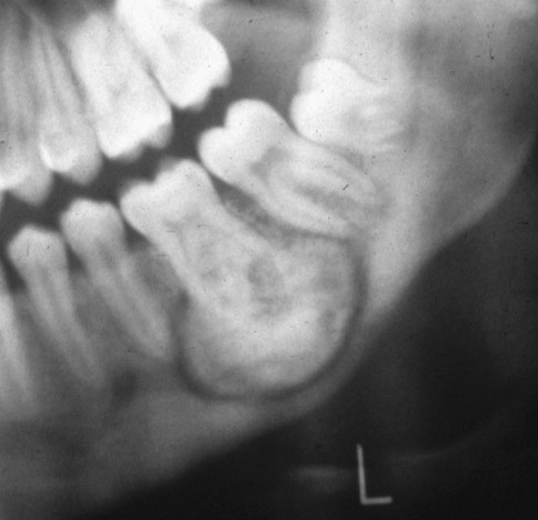
Radiographic Features
Radiographically, cementoblastoma typically appears as a well-circumscribed radiopaque mass directly attached to the root of the affected tooth. A key feature is a thin radiolucent rim surrounding the radiopaque mass, representing unmineralized tissue at the periphery of the lesion. This attachment to the tooth root is a critical radiographic indicator, often considered nearly pathognomonic for cementoblastoma.
Additional radiographic signs can include root resorption, blurring of the root outline, potential invasion into the root canal, expansion of the bone, displacement or involvement of adjacent teeth, erosion of the cortical bone, and obliteration of the periodontal ligament space.
Histopathological Features
Histologically, cementoblastoma is defined by masses of cementum that are generally hypocellular, embedded within a fibrovascular stroma. A hallmark microscopic feature is prominent cementoblastic rimming, where a layer of plump cementoblasts borders the cementum masses. Another distinctive characteristic is the presence of basophilic reversal lines within the cementum, creating a Pagetoid-like appearance. The fibrovascular stroma may contain multinucleated osteoclast-type giant cells and plump cementoblasts. At the periphery, radiating columns of unmineralized tissue contribute to the radiolucent rim seen radiographically. While cementoblasts and cementoclasts, especially in the peripheral areas, may exhibit pleomorphism, mitotic figures are typically absent.
Cementoblastoma Differential Diagnosis
The differential diagnosis of cementoblastoma is critical and includes several lesions that share overlapping features. Key entities to consider are:
1. Osteoblastoma
Osteoblastoma is a benign bone-forming tumor that is histologically remarkably similar to cementoblastoma. In fact, the primary distinguishing feature is the attachment to a tooth root, which is characteristic of cementoblastoma and absent in osteoblastoma. When an osteoblastic lesion is found in the jaws and is not attached to a tooth root, osteoblastoma becomes the more likely diagnosis. Clinical and radiographic correlation is essential to differentiate between these two entities, especially when the lesion is in proximity to dental roots.
2. Osteosarcoma
Osteosarcoma, particularly the osteoblastic variant, can present with radiopaque features and may histologically resemble cementoblastoma due to the presence of osteoblasts and bone formation. However, osteosarcoma is a malignant tumor and typically exhibits more aggressive clinical and radiographic features. Histologically, the presence of mitotic figures and a more disorganized pattern of bone formation in osteosarcoma helps to differentiate it from cementoblastoma, where mitotic activity is not a feature. Clinical context, patient age, and radiographic evidence of aggressive growth are crucial in distinguishing osteosarcoma from cementoblastoma.
3. Hypercementosis
Hypercementosis is an excessive deposition of cementum on the root of a tooth. Radiographically, it appears as a thickening or blunting of the root apex. Unlike cementoblastoma, hypercementosis follows the root contour and does not present as a distinct mass attached to the root. Histologically, hypercementosis shows an increase in normal cementum, lacking the cellular and structural complexity of cementoblastoma. Clinically, hypercementosis is often an incidental finding on radiographs and is not associated with pain or expansion like cementoblastoma.
4. Periapical Cemento-osseous Dysplasia (PCOD)
Periapical Cemento-osseous Dysplasia (PCOD) is a fibro-osseous lesion that occurs in the periapical region of teeth, commonly in the mandibular anterior area. PCOD progresses through stages, starting radiolucent, mixed radiolucent/radiopaque, and finally radiopaque in the mature stage. While mature PCOD can be radiopaque, it is typically not attached to the tooth root and often involves multiple teeth. Histologically, PCOD exhibits a mixture of fibrous tissue, bone, and cementum-like material, but lacks the prominent cementoblastic rimming and organized cementum masses of cementoblastoma. Location, involvement of multiple teeth, and the lack of root attachment radiographically are key differentiators.
5. Benign Cementoblastoma vs. Other Benign Odontogenic Tumors
While cementoblastoma itself is benign, it’s important to differentiate it from other benign odontogenic tumors that may present with radiopaque lesions in the jaws. These can include odontomas, ameloblastic fibro-odontoma, and calcifying odontogenic cyst (Gorlin cyst) with calcifications. Careful radiographic and histological evaluation is necessary. The attachment to the tooth root and the specific histological features of cementum with cementoblastic rimming and basophilic reversal lines are crucial in confirming cementoblastoma over other benign odontogenic tumors.
Diagnosis and Clinical Correlation
The definitive diagnosis of cementoblastoma relies on a synthesis of clinical, radiographic, and histopathological findings. Radiographic evidence of a radiopaque mass attached to a tooth root, particularly in the mandibular molar region of a young adult, is highly suggestive. Histopathological examination is essential to confirm the diagnosis, identifying the characteristic features of cementum masses, cementoblastic rimming, and basophilic reversal lines. Correlation with clinical presentation, including patient age, symptoms, and location, is vital to accurately differentiate cementoblastoma from its mimics.
Treatment
The treatment of choice for cementoblastoma is complete surgical excision of the lesion, which typically includes extraction of the involved tooth due to the tumor’s attachment to the root. Enucleation and curettage are performed to ensure complete removal of the tumor. While cementoblastoma is benign, incomplete removal can lead to recurrence. Some clinicians advocate for curettage of the bony cavity after tooth extraction to minimize recurrence risk.
Conclusion
The differential diagnosis of cementoblastoma is a nuanced process requiring careful consideration of clinical, radiographic, and histological features. Distinguishing cementoblastoma from osteoblastoma, osteosarcoma, hypercementosis, PCOD, and other benign odontogenic tumors is critical for accurate diagnosis and appropriate patient management. A comprehensive approach, integrating all diagnostic modalities and understanding the subtle nuances of each entity in the differential, ensures optimal patient care and avoids potential misdiagnoses. For dental professionals, a thorough understanding of Cementoblastoma Differential Diagnosis is paramount in the realm of oral and maxillofacial pathology.