Introduction
Cementomas are benign tumors of the periodontal tissues, presenting a diagnostic challenge due to their varied forms and resemblance to other fibro-osseous lesions of the jaw. Accurate diagnosis is crucial for appropriate management and to avoid unnecessary interventions. This article provides a comprehensive guide to the Cementoma Differential Diagnosis, focusing on distinguishing cementomas, particularly gigantiform cementoma, from other conditions with similar clinical, radiographic, and histological features. Understanding the nuances of each diagnostic modality is essential for clinicians in oral and maxillofacial surgery, pathology, and radiology to ensure optimal patient care.
Understanding Cementomas: Types and Characteristics
Cementomas are classified into four main types, each with distinct characteristics that aid in differential diagnosis:
-
Cementoblastoma (True Cementoma): This benign neoplasm is characterized by the proliferation of cementoblasts, forming a mass of cementum fused to the root(s) of a tooth, typically a mandibular molar or premolar. Radiographically, it appears as a dense radiopaque mass with a radiolucent halo, often obliterating the root outline. Histologically, it shows sheets of cementoblasts and irregular cementum masses.
-
Cementifying Fibroma: A benign fibro-osseous neoplasm containing varying amounts of fibrous tissue, bone, and cementum-like material. It is well-circumscribed and can occur in any tooth-bearing area, more commonly in the mandible. Radiographically, it presents as a unilocular or multilocular lesion, initially radiolucent, becoming progressively radiopaque as mineralization occurs. Histopathology reveals fibrous stroma with varying amounts of bone and cementum.
-
Periapical Cemental Dysplasia (PCD) / Periapical Osseous Dysplasia: This is a reactive, non-neoplastic condition, predominantly affecting the periapical bone of mandibular anterior teeth in middle-aged Black women. It progresses through three stages: osteolytic (radiolucent), cementoblastic (mixed), and mature (radiopaque). It is typically asymptomatic and teeth are vital. Radiographically, it starts as a radiolucent lesion mimicking a periapical cyst or granuloma, evolving into a mixed and finally a radiopaque lesion. Histologically, it shows fibrous tissue with bone and cementum-like particles.
-
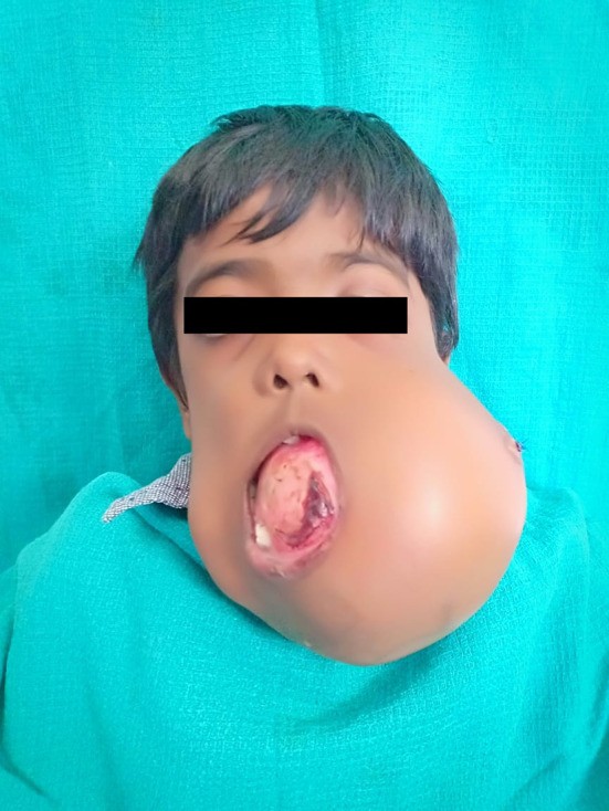
Gigantiform Cementoma: A rare, autosomal dominant condition characterized by massive, expansile, multifocal cemento-osseous lesions affecting the jaws. It typically presents in the first two decades of life and can cause significant facial deformity. Radiographically, it appears as large, irregular, radiopaque masses, often crossing the midline and involving multiple quadrants. Histologically, it is composed of fibrous tissue with abundant cementum deposition and less bone formation.
Differential Diagnosis of Gigantiform Cementoma
Gigantiform cementoma, due to its aggressive growth and radiographic appearance, requires careful differentiation from other fibro-osseous lesions and bone tumors. The cementoma differential diagnosis for gigantiform cementoma includes:
Radiographic Differential Diagnosis
-
Ossifying Fibroma: While ossifying fibroma can also be expansile and radiopaque, it is usually solitary and well-encapsulated. Gigantiform cementoma, in contrast, is typically multifocal and less well-defined radiographically, often crossing the midline. Ossifying fibroma usually presents as a more consistently mixed radiolucent-radiopaque lesion, whereas gigantiform cementoma tends to be predominantly radiopaque, especially in later stages.
-
Osteoma: Osteomas are benign bone tumors, typically slow-growing and solitary. They are densely radiopaque and well-circumscribed, often pedunculated or sessile, arising from the cortical bone. Unlike gigantiform cementoma, osteomas are not expansile in the same manner and lack the lobular, irregular radiopaque appearance.
-
Fibrous Dysplasia: Fibrous dysplasia can be monostotic or polyostotic, causing bone expansion and deformity. Radiographically, it exhibits a characteristic “ground-glass” appearance, which is different from the dense radiopacity of gigantiform cementoma. While polyostotic fibrous dysplasia can involve multiple jaw quadrants, it usually does not present with the same massive, cementum-rich radiopacity as gigantiform cementoma.
-
Cementoblastoma: Cementoblastoma, while radiopaque, is typically associated with a single tooth root and is not as expansile or multifocal as gigantiform cementoma. Cementoblastoma is also characterized by a radiolucent rim, which may be less pronounced or absent in gigantiform cementoma. The location and extent of the lesion are key differentiators.
-
Osteosarcoma: Although less common in the jaws compared to long bones, osteosarcoma is a malignant bone tumor that can present with radiopacity and bone expansion. However, osteosarcoma typically exhibits more aggressive radiographic features, such as ill-defined borders, periosteal reaction (sunburst or Codman’s triangle), and rapid growth. Gigantiform cementoma, despite its size, is benign and lacks these aggressive radiographic signs. Clinical history and progression are crucial in differentiating these conditions.
Histopathological Differential Diagnosis
-
Fibrous Dysplasia: Histologically, fibrous dysplasia consists of irregular trabeculae of woven bone within a fibrous stroma, lacking osteoblastic rimming (“Chinese character bone”). Gigantiform cementoma, on the other hand, shows more prominent cementum-like material in irregular masses within a fibrous stroma. While both can have a fibrous component, the dominant mineralized tissue differs significantly.
-
Cherubism: Cherubism is a genetic disorder causing bilateral, multilocular radiolucencies in the jaws, primarily in children. Histologically, it is characterized by fibrous tissue with multinucleated giant cells. Gigantiform cementoma lacks the prominent giant cell component and presents with radiopaque masses rather than radiolucencies. Clinical presentation and age of onset are also important differentiators.
-
Psammomatoid Juvenile Ossifying Fibroma (PsJOF): PsJOF can histologically resemble gigantiform cementoma due to the presence of cementicles and ossifying material within a fibrous stroma. However, PsJOF typically presents as a more circumscribed lesion and clinically behaves differently from the expansile, aggressive nature of gigantiform cementoma. Grossly, gigantiform cementoma is hard and not easily cleavable, unlike PsJOF.
Clinical Presentation and Familial History
- Age of Onset: Gigantiform cementoma typically presents in the first two decades of life, whereas PCD is more common in middle-aged individuals. Cementoblastoma and ossifying fibroma can occur across a wider age range.
- Location and Extent: Gigantiform cementoma is often multifocal and can cross the midline, involving multiple quadrants of the jaws. Other cementomas are typically more localized.
- Familial History: The familial nature of gigantiform cementoma, with autosomal dominant inheritance, is a significant diagnostic clue. A detailed family history of similar jaw swellings is crucial.
- Growth Pattern: Gigantiform cementoma is characterized by rapid, expansile growth, causing significant facial deformity if untreated. PCD is typically static and asymptomatic. Cementoblastoma and ossifying fibroma grow more slowly and are less extensive than gigantiform cementoma.
Diagnostic Approach to Cementomas
Establishing an accurate cementoma differential diagnosis requires a systematic approach integrating clinical, radiographic, and histological findings, along with patient history:
-
Clinical Examination: Thorough extraoral and intraoral examination to assess swelling, location, size, consistency, and any associated symptoms. Documenting the patient’s age, gender, and family history is critical.
-
Radiographic Evaluation:
- Panoramic Radiographs: Initial screening to assess the extent and location of the lesion, especially for multifocal involvement.
- Periapical Radiographs: Detailed view of individual teeth and periapical areas, useful for differentiating PCD and cementoblastoma.
- Cone-Beam Computed Tomography (CBCT) or Computed Tomography (CT): Essential for evaluating the three-dimensional extent of the lesion, cortical bone involvement, and relationship to adjacent structures, particularly for large lesions like gigantiform cementoma. CT is superior for assessing bone detail and expansile nature. Radiographic features to analyze include radiopacity, borders, internal structure, and effect on surrounding structures.
-
Histopathological Examination: Biopsy is mandatory for definitive diagnosis. Incisional biopsy is usually sufficient, but excisional biopsy may be performed for smaller lesions. Histological analysis helps to differentiate cementomas from other fibro-osseous lesions based on the type and pattern of mineralization, cellular components, and presence of specific features (e.g., giant cells in cherubism, woven bone in fibrous dysplasia, cementum masses in cementoblastoma and gigantiform cementoma).
-
Laboratory Investigations: Alkaline phosphatase levels may be elevated in some cases of gigantiform cementoma and can be monitored pre- and post-operatively, although this is not a specific diagnostic test.
-
Genetic Counseling and Family Screening: In cases suggestive of familial gigantiform cementoma, genetic counseling should be offered, and family screening may be indicated to identify other affected individuals.
Management Considerations Based on Diagnosis
The management of cementomas varies depending on the type, size, location, and clinical behavior. Accurate cementoma differential diagnosis is essential for determining the appropriate treatment strategy:
-
Periapical Cemental Dysplasia (PCD): Typically requires no treatment, as it is a reactive condition and asymptomatic. Diagnosis is crucial to avoid unnecessary endodontic treatment or surgery.
-
Cementifying Fibroma: Surgical excision is the treatment of choice. Enucleation and curettage are usually sufficient due to its well-circumscribed nature.
-
Cementoblastoma: Surgical excision, including extraction of the involved tooth, is the standard treatment. Complete removal usually results in a good prognosis with low recurrence.
-
Gigantiform Cementoma: Management is challenging due to its extensive size, rapid growth, and potential for recurrence. Treatment options range from observation for slow-growing, asymptomatic lesions to aggressive surgical resection for symptomatic or rapidly expanding lesions. Extensive resection, as demonstrated in the case report, may be necessary to prevent recurrence and manage airway obstruction in severe cases. Regular follow-up is essential to monitor for recurrence.
Conclusion
The cementoma differential diagnosis is a complex process requiring careful consideration of clinical, radiographic, and histological findings. Distinguishing cementomas, particularly gigantiform cementoma, from other fibro-osseous lesions and bone tumors is crucial for appropriate patient management. A systematic diagnostic approach, integrating these modalities and considering familial history, ensures accurate diagnosis and guides effective treatment strategies, ultimately improving patient outcomes and quality of life. For gigantiform cementoma, early diagnosis and prompt intervention are vital to manage this rare and potentially debilitating condition.