INTRODUCTION
As automotive repair experts at xentrydiagnosis.store, we often encounter unique challenges that require a systematic and thorough diagnostic approach. While our primary focus is vehicle diagnostics, the principles of differential diagnosis are universally applicable. This article, inspired by a medical case study, explores the concept of “Cheek Mass Differential Diagnosis,” providing a framework and relevant examples that resonate with our problem-solving mindset in automotive repair. Just as medical professionals meticulously differentiate between various conditions presenting with similar symptoms, we, as automotive diagnosticians, must employ a similar process to pinpoint the root cause of complex vehicle issues. This article will guide you through a structured approach, mirroring the medical methodology, to enhance your diagnostic skills.
CASE PRESENTATION: A Diagnostic Puzzle
Imagine a scenario: A 55-year-old vehicle owner reports to your repair shop complaining of a peculiar issue – a growing “swelling” on the right side of their vehicle, specifically near the front fender area. This “swelling” has been progressively increasing over the last two months. Initially, it was just a minor visual anomaly, but recently, it has become more pronounced, causing some discomfort and even slightly hindering the vehicle’s maneuverability, described as a “reduced mouth opening” in the original medical context. The owner mentions experiencing some “pain and discomfort” in the area for the past week, coinciding with the increased “swelling.”
Upon initial inspection, you observe an asymmetrical appearance of the vehicle’s front end, with a noticeable bulge in the right fender area, approximately 1.5 cm anterior to the wheel well trim – analogous to the tragus in the medical case. The outer paint surface appears normal, with no signs of damage or discoloration. The vehicle owner is not reporting any other immediate issues like overheating or warning lights on the dashboard. The “mouth opening,” in automotive terms, can be interpreted as the steering angle or turning radius, which is slightly reduced.
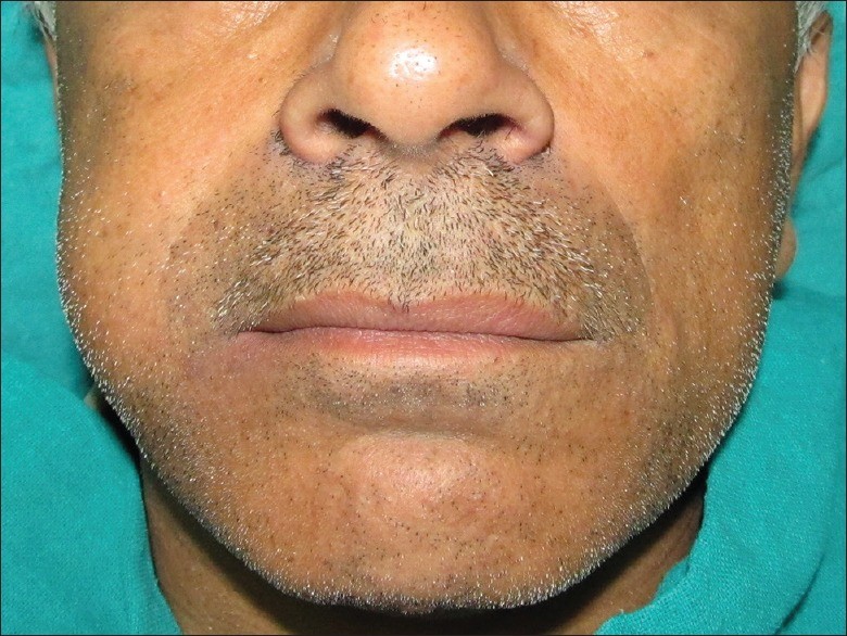
Figure 1.
Visual representation of a cheek swelling, illustrating a localized, dome-shaped mass. This image helps to conceptualize the initial symptom as a focal point for diagnosis.
A preliminary examination reveals a small, smooth, dome-shaped “swelling.” The “overlying skin,” or in this case, the vehicle’s body panel, is of normal color and texture. There is no indication of overheating or immediate mechanical failure. The “mouth opening,” or steering range, is limited to 25 degrees in one direction compared to the other. Good overall vehicle condition is noted, with no obvious issues in other areas. No signs of recent accidents or external damage are apparent. The “oral soft tissues,” representing the interior components, appear normal upon initial visual check. Palpation – or a physical check – reveals a diffuse, somewhat firm lesion within the fender area, and the outer body panel is unaffected. The mass feels soft to firm and smooth. No unusual vibrations or fluid sounds are detected. All fluid levels and basic system checks appear normal. No unusual noises from the engine or suspension are evident during a brief static test.
Further investigation, akin to aspiration in the medical case, might involve a non-invasive diagnostic scan. Initial scans reveal an anomaly in the described area. More advanced diagnostic imaging, comparable to an MRI, shows a well-defined mass located near the vehicle’s frame rail and suspension components, with some surrounding areas exhibiting signs of stress or minor deformation.
Figure 2.
Coronal MRI image illustrating a well-defined cheek mass. In automotive terms, this represents advanced diagnostic imaging revealing a localized issue requiring further differential diagnosis.
DIFFERENTIAL DIAGNOSIS: Ruling Out Possibilities in Automotive Repair
Just as in the medical field, when faced with a cheek mass, a range of potential diagnoses must be considered and systematically evaluated – a process known as differential diagnosis. In our automotive context, this translates to considering various potential causes for the observed “swelling” and related symptoms.
The most common causes of “swelling” in a vehicle’s fender area could include:
-
Abscess (Localized Corrosion/Rust): Similar to an abscess caused by infection, localized corrosion or rust buildup beneath the body panel can create a swelling. This is often associated with moisture accumulation and can lead to structural weakness. This would be analogous to an acute onset issue with pain and swelling. However, the slow progression reported by the owner makes this less likely as the primary cause initially. We would need to rule out external damage or paint issues first.
-
Lipomas (Body Filler/Improper Repair): Lipomas, benign tumors of fatty tissue, can be likened to improperly applied body filler or a poorly executed previous repair. This could create a smooth, dome-shaped bulge. However, the diagnostic imaging suggests a different density than typical body filler, making this less probable.
-
Salivary Gland Tumors (Component Malfunction/Leakage): Salivary gland tumors, affecting saliva production, could be compared to malfunctions or leaks in vehicle fluid systems. However, in this case, there’s no evidence of fluid leakage or any related system warnings, reducing the likelihood of this diagnosis. The two-month history and normal fluid levels argue against this.
-
Lymphadenopathy (Electrical Wiring/Harness Issues): Enlarged lymph nodes, indicative of immune response, could be paralleled to issues within the vehicle’s electrical wiring or harness. A localized swelling could be caused by wiring damage or a malfunctioning electrical component. However, the diagnostic scan doesn’t suggest an electrical fault as the primary cause.
-
Cysts (Fluid Accumulation/Blocked Drainage): Cysts, fluid-filled sacs, can be similar to fluid accumulation due to blocked drainage channels within the vehicle’s body or components. This could present as a slow-growing swelling. However, aspiration was negative in the medical case, and a similar non-invasive test in our automotive scenario didn’t reveal fluid accumulation.
-
Muscular Tumors (Structural Component Deformation/Failure): Tumors of muscular origin can be compared to deformations or failures in structural components, such as frame rails or suspension parts, especially those near the wheel well. This is a more serious consideration, given the location and the diagnostic imaging results. The involvement of “masticatory muscles” in the medical case can be related to load-bearing or movement-related components in the vehicle.
-
Neurogenic Tumors (Sensor/Wiring Anomaly): Neurogenic tumors, arising from nerve sheath cells, could be analogous to anomalies in sensors or wiring related to vehicle control and monitoring systems. While less common, these could potentially cause localized issues and might not be immediately apparent through standard diagnostics.
Considering the slow-growing nature of the “swelling,” a benign cause is initially more likely. However, malignant possibilities must also be kept in mind during the diagnostic process.
DIAGNOSTIC APPROACH: From Initial Assessment to Advanced Techniques
In the medical case, initial diagnostic steps included physical examination, aspiration, OPG (orthopantomography), ultrasonography, and MRI. In our automotive scenario, we follow a similar structured approach:
-
Initial Inspection and History Taking: This mirrors the patient history and physical exam. We gather information from the vehicle owner about the onset, progression, and associated symptoms. Visual inspection and basic checks form the initial assessment.
-
Non-invasive Diagnostics (OPG/Ultrasonography Analogy): Similar to OPG and ultrasonography, we use initial diagnostic scans – perhaps a general system scan or visual inspection with specialized tools – to get a broader picture. This helps rule out obvious issues and narrow down the possibilities.
-
Advanced Diagnostic Imaging (MRI Analogy): Just as MRI provides detailed soft tissue imaging, advanced diagnostic tools, such as detailed component scans or even disassembly for physical inspection, are used to get a clearer picture of the “mass.” This is crucial for understanding the nature and extent of the problem.
-
Aspiration/Sampling (Component Testing/Fluid Analysis): While direct aspiration might not be applicable, component testing or fluid analysis (if relevant) can provide further insights. This is akin to taking a sample for lab analysis in the medical field.
-
Differential Diagnosis Refinement: Based on the accumulated data, we refine our differential diagnosis list, prioritizing the most likely causes and systematically ruling out others.
MANAGEMENT AND TREATMENT: From Diagnosis to Resolution
In the medical case, excisional biopsy was performed under general anesthesia, followed by pathological diagnosis and follow-up. In automotive repair, our “excisional biopsy” translates to a more invasive diagnostic and repair procedure.
-
Excisional Biopsy Analogy (Component Removal/Inspection): To definitively diagnose and treat the “cheek mass,” we might need to remove the affected component or body panel section for closer inspection. This is our “excisional biopsy.”
-
Pathological Diagnosis Analogy (Detailed Component Analysis): The removed component is then thoroughly examined to determine the exact nature of the “mass.” Is it corrosion, body filler, structural deformation, or something else? This is our “pathological diagnosis.”
-
Treatment (Repair/Replacement): Based on the diagnosis, the appropriate treatment is implemented. This could involve rust repair, bodywork correction, component replacement, or structural repair.
-
Follow-up (Post-Repair Inspection and Testing): Just as in the medical case, follow-up is crucial. Post-repair inspection and testing ensure the issue is resolved and the vehicle is functioning correctly. This is analogous to the one-year follow-up in the medical case to confirm no recurrence.
Figure 3.
Post-operative image showing resolution of cheek swelling. In automotive terms, this represents the successful repair and restoration of the vehicle’s body and function after diagnosis and treatment.
Figure 4.
Post-operative MRI confirming normal tissue structure after mass removal. Analogously, this shows the successful restoration of structural integrity and component normalcy after automotive repair.
PATHOLOGICAL FINDINGS: Identifying the Root Cause
In the medical case, the pathological diagnosis revealed a schwannoma, a benign nerve sheath tumor. In our automotive scenario, the “pathological findings” would be the specific cause identified during the detailed component analysis. It might be:
- Extensive Corrosion: Indicating rust as the root cause.
- Improper Body Filler Application: Confirming a previous poor repair.
- Structural Deformation: Revealing damage to a frame rail or support member.
- Accumulated Debris/Blockage: Identifying a drainage issue.
Figure 5.
Microscopic image of schwannoma cells. This represents the detailed analysis of a removed automotive component, revealing the specific cause of the issue, whether corrosion, material fatigue, or manufacturing defect.
DISCUSSION: Applying Differential Diagnosis in Automotive Expertise
The medical case highlights the importance of a systematic differential diagnosis approach, even for seemingly unusual presentations like a cheek mass. Similarly, in automotive repair, we must apply this rigorous methodology to diagnose complex and atypical vehicle issues.
Schwannomas, while rare in the cheek, are a recognized possibility in medical differential diagnosis. Analogously, seemingly unusual or rare vehicle problems can occur and should be considered, not immediately dismissed. The slow-growing nature of the schwannoma in the medical case mirrors the progressive development of some automotive issues, emphasizing the need to consider long-term changes and owner history.
The discussion in the medical paper delves into the characteristics of schwannomas, their presentation, and diagnostic methods. For automotive experts, this underscores the importance of continuous learning and staying updated on various potential vehicle problems, diagnostic techniques, and repair procedures. Just as medical professionals consider various tumor types, we must be aware of a wide range of potential mechanical, electrical, and structural faults in vehicles.
The successful surgical excision of the schwannoma in the medical case emphasizes the importance of accurate diagnosis leading to effective treatment. In automotive repair, a precise diagnosis is equally crucial for implementing the correct repair strategy and ensuring vehicle longevity and safety.
CONCLUSION: Mastering the Diagnostic Process
This exploration of “cheek mass differential diagnosis,” inspired by a medical case study, provides a valuable framework for automotive repair professionals. By adopting a systematic and thorough approach to differential diagnosis, we can enhance our problem-solving skills, accurately identify the root causes of complex vehicle issues, and provide effective and lasting repairs. Just as considering rare conditions like schwannoma is vital in medicine, being open to less common but possible causes is crucial in automotive diagnostics. Mastering the art of differential diagnosis is a hallmark of expertise in both medicine and automotive repair, leading to better outcomes and customer satisfaction.
REFERENCES
The references from the original medical article are retained as a testament to the source material and for potential medical professional readers who may find them relevant.
[1] Enzinger FM, Weiss SW. Soft Tissue Tumors. 3rd ed. St. Louis: Mosby; 1995. Lipoma; pp. 389–430.
[2] Som PM, Braun IF, Shapiro MD, Reede DL, Curtin HD, Zimmerman RA. Lesions of the parapharyngeal space: improved diagnosis and patient management. Radiol Clin North Am. 1984;22:467–76. [PubMed]
[3] Gnepp DR. Diagnostic Surgical Pathology of the Head and Neck. Philadelphia: WB Saunders; 2001. Tumors of the salivary glands; pp. 541–674.
[4] Auclair PL. Surgical Pathology of the Salivary Glands. Philadelphia: WB Saunders; 1991. Benign epithelial salivary gland tumors; pp. 167–244.
[5] Sato I, Nakajima T, Yagi T, Tanaka N, Hoshi K. Swelling of the buccal lymph node mimicking a tumor in the buccal region: report of 5 cases. J Oral Maxillofac Surg. 2001;59:211–4. [PubMed]
[6] Robbins KT, Clayman G, Levine PA, Medina J, Sessions R, Shaha A, et al. Neck dissection classification update: revisions proposed by the American Head and Neck Society and the American Academy of Otolaryngology-Head and Neck Surgery. Arch Otolaryngol Head Neck Surg. 2002;128:751–8. [PubMed]
[7] Banks P, Castro IO. Hodgkin’s disease of the tonsil. Cancer. 1970;26:230–44. [PubMed]
[8] Vokes EE, Weichselbaum RR, Lippman SM, Hong WK. Head and neck cancer. N Engl J Med. 1993;328:184–94. [PubMed]
[9] Arora VK, Nijhawan R, Arora R. Cysticercus cellulosae in masseter muscle: a cytodiagnostic pitfall on fine needle aspiration. Cytopathology. 2004;15:367–9. [PubMed]
[10] Badmus TA, Adelusola OO, Ogun GO. Leiomyosarcoma of the oral cavity and pharynx: a clinicopathologic study of 13 cases and review of the literature. Head Neck Pathol. 2007;1:161–8. [PMC free article] [PubMed]
[11] Verocay J. Multiple Geschwülste der Nervenscheiden (Neurinome) In: Festschrift fúr H. Ribbert. Bonn: Friedrich Cohen; 1910. pp. 378–415.
[12] White SW, Cerullo LJ, Jones RA. Neurilemmoma of the trigeminal nerve. Surg Neurol. 1977;8:385–9. [PubMed]
[13] Das Gupta TK, Brasfield RD, Strong EW, Hajdu SI. Benign solitary schwannomas (neurilemomas) Cancer. 1969;24:355–66. [PubMed]
[14] Lusk MD, McCarthy JG, Graham WP. Neurilemmoma of the face: case report and review of the literature. J Craniofac Surg. 1992;3:106–10. [PubMed]
[15] Conley J, Janecka IP. Neurilemmoma of the facial nerve. Plast Reconstr Surg. 1973;52:55–60. [PubMed]
[16] D’Agostino AN, Soule EH, Miller RH. Primary malignant neoplasms of nerves (malignant neurilemomas) in patients with neurofibromatosis (von Recklinghausen’s disease) Cancer. 1963;16:1003–14. [PubMed]
[17] Ellis GL, Abrams AM, Auclair PL. Tumors of the salivary glands. In: Atlas of Tumor Pathology. Armed Forces Institute of Pathology; Washington: 1996. pp. 261–94.
[18] Farr HW, Gray GF, Jr, Vrana TP, Panio M. Intracranial neurilemoma and neurofibroma. Report of five cases and review of literature. Cancer. 1967;20:727–31. [PubMed]
[19] MacCarty CS, Piepgras DG, Baker HL, Jr, O’Duffy BM. Trigeminal neurinomas (schwannomas) J Neurosurg. 1970;32:386–95. [PubMed]
[20] Woodruff JM, Godwin TA, Erlandson RA, Susin M, Martini N. Cellular schwannoma. A variety of benign and malignant nerve sheath tumor. Am J Surg Pathol. 1981;5:733–44. [PubMed]
[21] Enzinger FM, Weiss SW. Soft Tissue Tumors. 3rd ed. St. Louis: Mosby; 1995. Neurilemmoma; pp. 799–830.
[22] Daimaru Y, Hashimoto H, Enzinger FM, Swift FT. Benign schwannoma of peripheral soft tissues. Am J Surg Pathol. 1983;7:415–22. [PubMed]
[23] Ghosh BC, Ghosh L, Huvos AG, Fortner JG. Neurogenic sarcomas. Clinicopathologic study of 94 cases. Ann Surg. 1973;178:100–9. [PMC free article] [PubMed]
[24] Ducatman BS, Scheithauer BW, Piepgras DG, Reiman HM, Ilstrup DM. Malignant peripheral nerve sheath tumors. A clinicopathologic study of 120 cases. Cancer. 1986;57:2006–21. [PubMed]
[25] Fletcher CD, Bridge JA, Hogendoorn PC, Mertens F. World Health Organization Classification of Tumours. Pathology and Genetics of Tumours of Soft Tissue and Bone. Lyon: IARC Press; 2002. pp. 147–50.
[26] Asaumi J, Hisatomi M, Asaumi R, Konouchi H, Shigehara T, Tanaka Y. Intraosseous schwannoma of the mandible: report of 2 cases and review of the literature. Oral Surg Oral Med Oral Pathol Oral Radiol Endod. 2004;98:91–5. [PubMed]
[27] Shirasuna K, Sato M, Sugiyama M, Miyazaki T. Central schwannoma of the mandible: review of the literature and report of 2 cases. J Oral Maxillofac Surg. 2003;61:743–6. [PubMed]
[28] Imagama S, Matsuyama Y, Sakai T, Katayama Y, Nakamura H, Kawakami N, et al. Dumbbell-shaped tumors of the cervical spine: surgical strategy and outcome in 22 patients. Spine. 2007;32:E717–24. [PubMed]
[29] Eeles RA, Snaith L, Fisher C, Horwich A, Easton DF, Macdonald AM, et al. Malignant transformation of a benign schwannoma in neurofibromatosis type 2 confirmed by genetic analysis. Clin Neuropathol. 1996;15:299–303. [PubMed]
[30] Wanebo JE, Malik J, VandenBerg SR, Wanebo HJ, Sternlicht MD. Malignant peripheral nerve sheath tumors. Cancer. 1993;71:1247–53.