Introduction
The discovery of a solitary pulmonary coin lesion on chest radiography often initiates a complex diagnostic journey. These lesions, characterized by their round or oval shape and well-defined borders on imaging, represent a wide spectrum of potential etiologies, ranging from benign to malignant conditions. The differential diagnosis of a coin lesion in the lung is extensive, encompassing infectious diseases, inflammatory processes, benign tumors, and primary or metastatic malignancies. Among the rarer entities in this differential, human pulmonary dirofilariasis (HPD) stands out as an often-unexpected finding.
Human pulmonary dirofilariasis is caused by infection with parasites of the Dirofilaria genus, most commonly Dirofilaria immitis (DI), the same nematode responsible for heartworm disease in dogs. While dogs are the definitive hosts, humans can become accidental hosts through mosquito bites. In humans, Dirofilaria typically does not complete its life cycle. Instead, the parasite often migrates to the pulmonary arteries, where its death triggers an inflammatory response leading to the formation of a pulmonary nodule. This nodule, clinically presenting as a coin lesion, can be easily mistaken for more common pulmonary pathologies, particularly lung cancer.
This article aims to delve into the differential diagnosis of coin lesions in the lung, with a specific focus on highlighting human pulmonary dirofilariasis as a crucial, albeit rare, consideration. We will draw upon a case study where a pulmonary nodule, initially suspected of malignancy, was ultimately diagnosed as HPD, emphasizing the diagnostic challenges and the importance of including this parasitic infection in the differential workup of solitary pulmonary nodules.
Case Presentation
To illustrate the diagnostic complexities and the unexpected nature of HPD, we present a case of a 62-year-old female patient who presented to our clinic with a pulmonary nodule detected during a routine chest X-ray.
Medical History
The patient, a 62-year-old female, was referred to our hospital following an abnormal finding on a routine chest X-ray. Her medical history was unremarkable, except for a history of trigeminal neuralgia. She was a non-smoker and reported no respiratory symptoms such as cough, shortness of breath, or chest pain. She had no known exposure to pets recently diagnosed with heartworm, nor had she traveled to areas highly endemic for dirofilariasis.
Preoperative Assessment
Physical examination revealed no significant findings. Her vital signs were stable, and auscultation of the chest was clear. Laboratory investigations, including a complete blood count and serum biochemistry, were within normal limits. Specifically, the white blood cell count was 4.97×10³/µL, hemoglobin 13.2 g/dL, and platelet count 276×10³/µL. Liver function tests, renal function, and electrolytes were also normal. Pulmonary function testing demonstrated normal spirometry with a forced expiratory volume in one second (FEV1) of 2.42 L (102% of predicted) and vital capacity (VC) of 3.00 L (102% of predicted). Arterial blood gas analysis was also normal, with PaCO2 at 35 mmHg, PaO2 at 74 mmHg, and pH at 7.39.
A subsequent chest CT scan revealed a well-defined pulmonary nodule, measuring 1.54 cm × 1.46 cm, located in the apical segment of the right lower lobe (Figure 1). The nodule exhibited smooth margins and no calcification or cavitation. To further evaluate the nodule’s nature and assess for malignancy, a positron emission tomography-computed tomography (PET/CT) scan was performed. Surprisingly, the PET/CT showed no significant fludeoxyglucose (FDG) uptake in the pulmonary nodule, suggesting a low likelihood of malignancy. Despite the benign features on PET/CT, the possibility of early-stage lung cancer could not be entirely excluded, and further diagnostic evaluation was deemed necessary. A video-assisted thoracic surgical (VATS) wedge resection was planned for both diagnostic confirmation and potential therapeutic intervention.
Figure 1 The preoperative CT scan demonstrates a subpleural human pulmonary dirofilariasis (HPD) pulmonary nodule located in the apical segment of the right lower lobe, characterized by well-defined margins. CT, computed tomography.
Procedure and Histopathology
Preoperative bronchoscopy was performed and showed no endobronchial lesions or abnormalities. The patient underwent VATS wedge resection of the pulmonary nodule. Intraoperatively, the pleural cavity was unremarkable, with no inflammatory adhesions or pleural effusion. Frozen section analysis of the resected nodule revealed tissue necrosis with granulomatous inflammation, but no immediate evidence of malignancy. A chest drain was placed, and the patient was extubated without complications and transferred to the intermediate care unit postoperatively.
The postoperative course was smooth. The chest drain was removed on the first postoperative day, and the patient was discharged on the third postoperative day without any complications. Histopathological examination of the permanently fixed specimen confirmed a completely resected, encapsulated nodule measuring 1.5 cm × 1.2 cm × 1.2 cm. Microscopic examination revealed a granulomatous inflammatory wall surrounding a central necrotic area (Figure 2). Isolated eosinophils were observed in the periphery of the nodule. Importantly, sections stained for parasites revealed thread-like worm fragments within a small arterial vessel in the necrotic center. Based on morphological characteristics, the parasite was identified as Dirofilaria immitis. The surrounding lung parenchyma was normal, and the visceral pleura was intact. Following the definitive diagnosis of HPD, no further medical treatment was deemed necessary. The patient remained asymptomatic and without recurrence during subsequent follow-up.
Figure 2 Histological examination of the resected pulmonary nodule. (A) Demonstrates an infarcted area with peripheral granulomatous inflammation. (B, C) Show the parasite Dirofilaria immitis within an arterial vessel. (D) Higher magnification of the parasite (stain: A, B, D, hematoxylin & eosin; C, Elastica van Gieson. Magnification: A, B, C, 2×; D, 10×). These images confirm the presence of Dirofilaria immitis and the characteristic inflammatory response.
Discussion: Differential Diagnosis of Coin Lesions and the Role of HPD
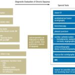
Solitary pulmonary nodules, or coin lesions, pose a significant diagnostic challenge in clinical practice. Their discovery necessitates a systematic approach to differential diagnosis to distinguish between benign and malignant etiologies. The differential diagnosis is broad and can be categorized into several major groups:
Malignant Neoplasms
- Primary Lung Cancer: Adenocarcinoma, squamous cell carcinoma, large cell carcinoma, and small cell lung cancer are the most concerning possibilities, especially in smokers and older individuals. These often present as enlarging nodules, sometimes with irregular borders, spiculation, or associated lymphadenopathy.
- Metastatic Disease: Metastases from other primary cancers (e.g., breast, colon, kidney, melanoma) can manifest as solitary or multiple pulmonary nodules. History of extra-pulmonary malignancy is crucial in this context.
- Carcinoid Tumors: These are neuroendocrine tumors that can present as pulmonary nodules, often centrally located.
Benign Neoplasms
- Hamartomas: These are the most common benign lung tumors, often containing fat, cartilage, and calcification in a “popcorn” pattern on CT.
- Granulomas: Resulting from prior infections like tuberculosis or fungal infections (histoplasmosis, coccidioidomycosis), granulomas are common benign nodules, often showing calcification or cavitation.
- Fibromas, Lipomas, Chondromas: These are less common benign tumors of the lung parenchyma.
Infectious and Inflammatory Conditions
- Infections: Bacterial abscesses, fungal infections (aspergillosis, cryptococcosis), and parasitic infections (including HPD) can present as pulmonary nodules.
- Inflammatory Granulomatous Diseases: Sarcoidosis and Wegener’s granulomatosis (now Granulomatosis with polyangiitis) can manifest with lung nodules.
- Rheumatoid Nodules: In patients with rheumatoid arthritis, lung nodules can develop.
Vascular and Miscellaneous Conditions
- Arteriovenous Malformations (AVMs): These vascular abnormalities can appear as nodules on chest imaging.
- Pulmonary Infarcts: Embolic events can lead to pulmonary infarcts, which may present as nodules.
- Organizing Pneumonia: This inflammatory condition can sometimes present as nodular opacities.
Human Pulmonary Dirofilariasis (HPD) in the Differential
Within this broad differential, HPD, while rare, is a significant consideration, particularly because it can mimic malignancy both clinically and radiologically. As highlighted in our case, HPD typically presents as a solitary pulmonary nodule, often subpleural and well-defined, resembling early-stage lung cancer.
Epidemiology and Risk Factors:
HPD is geographically distributed worldwide, with higher prevalence in areas where canine heartworm disease is endemic and mosquito vectors are abundant. Regions like the southeastern United States, the Gulf Coast, parts of Europe, Japan, and Australia have reported cases. While dog ownership was initially considered a risk factor, it is now understood that broader environmental factors, such as the density of infected mosquito populations and human exposure to these vectors, are more critical. Interestingly, risk may increase following natural disasters that disrupt mosquito control and increase human-vector interaction.
Clinical Presentation and Diagnosis:
The majority of HPD cases are asymptomatic, with nodules discovered incidentally on routine chest imaging, as in our presented case. When symptoms occur, they are non-specific and can include chest pain, cough, hemoptysis, and fever, potentially arising from the inflammatory response to the dying parasite within the pulmonary artery. Eosinophilia, a common finding in parasitic infections, is surprisingly infrequent in HPD, occurring in only a minority of cases. Serological tests for Dirofilaria antigens lack sensitivity and specificity due to cross-reactivity with other parasites, limiting their diagnostic utility.
Radiologically, HPD typically manifests as a solitary, well-circumscribed pulmonary nodule, often 1-3 cm in size, predominantly in the lower lobes, especially the right lower lobe. CT scans usually confirm the well-defined nature of the nodule. While PET/CT is often employed to assess malignancy risk, HPD nodules typically show low or no FDG uptake, as seen in our case, but mild uptake has been reported in some instances, further complicating differentiation from malignancy. Other radiological findings, such as pleural thickening or infiltrates, are less specific.
Diagnostic Confirmation and Management:
Due to the non-specific clinical and radiological features, HPD often necessitates tissue diagnosis for definitive confirmation. While bronchoscopy, sputum cytology, and needle aspiration are generally not helpful, CT-guided percutaneous needle biopsy has been used successfully in some cases. However, surgical wedge resection, typically via VATS, remains the gold standard for both diagnosis and treatment. Histopathological examination of the resected nodule, revealing the characteristic necrotic center, granulomatous inflammation, and identification of Dirofilaria fragments, confirms the diagnosis. Molecular techniques like PCR and ELISA can be used to enhance diagnostic accuracy in challenging cases.
Surgical resection is considered curative for HPD. As humans are dead-end hosts for Dirofilaria, and microfilaremia does not occur, no further anti-parasitic medical treatment is necessary after nodule resection. In cases where HPD is strongly suspected based on clinical and radiological features, and malignancy is deemed highly unlikely, a conservative approach with close radiological follow-up might be considered in the future to avoid surgery, although this remains investigational. The VATS approach, as performed in our case, offers a minimally invasive and effective method for both diagnosing and treating HPD, particularly when it is part of the differential diagnosis of a suspicious pulmonary nodule.
Conclusion
Human pulmonary dirofilariasis, although a rare cause of pulmonary coin lesions, should be included in the differential diagnosis of solitary pulmonary nodules, especially in individuals residing in or traveling to endemic areas. Its ability to mimic lung cancer clinically and radiologically underscores the importance of considering less common etiologies in the diagnostic workup. VATS wedge resection provides a safe and effective approach for both definitive diagnosis and curative treatment of HPD in cases presenting as pulmonary coin lesions, ensuring accurate diagnosis and appropriate management while addressing the broader differential diagnosis of pulmonary nodules.
Acknowledgements
None.
Footnote
Conflicts of Interest: The authors have no conflicts of interest to declare.
Informed Consent: Written informed consent was obtained from the patient for publication of this manuscript and any accompanying images.
References
Cite this article as: Grapatsas K, Kayser G, Passlick B, Wiesemann S. Pulmonary coin lesion mimicking lung cancer reveals an unexpected finding: Dirofilaria immitis. J Thorac Dis 2018;10(6):3879-3882. doi: 10.21037/jtd.2018.05.137