Understanding Cor Pulmonale and Its Diagnosis
Cor pulmonale, often referred to as “pulmonary heart,” describes a condition characterized by the alteration in the structure and function of the right ventricle due to primary respiratory system disorders leading to pulmonary hypertension. While its definition continues to evolve within the medical field, understanding its diagnosis is crucial, especially when considering the broader context of health and related fields. This article delves into the diagnosis criteria for cor pulmonale, aiming to provide a comprehensive overview for professionals and anyone seeking to understand this complex condition better.
Etiology and Pathophysiology of Cor Pulmonale
Cor pulmonale arises as a consequence of pulmonary hypertension, which itself is linked to a spectrum of diseases affecting the lungs, pulmonary vasculature, upper airways, and chest wall. Chronic obstructive pulmonary disease (COPD), interstitial lung disease, idiopathic pulmonary arterial hypertension, obstructive sleep apnea, and kyphoscoliosis are among the primary culprits. Conditions that inflict lung damage, such as autoimmune diseases like scleroderma, cystic fibrosis, and obesity hypoventilation syndrome, also contribute to the development of pulmonary hypertension and subsequently, cor pulmonale. In acute settings, massive pulmonary embolism stands out as the most frequent cause of acute cor pulmonale, often mimicking myocardial infarction in its presentation.
The underlying pathophysiology is rooted in the increased resistance within the pulmonary vasculature. Normally, the right ventricle operates against a low-resistance system. However, chronic hypoxemia triggers vasoconstriction, leading to smooth muscle proliferation in the small pulmonary arteries and structural remodeling. This hypoxemia also disrupts the balance of vascular mediators like nitric oxide, endothelin-1, and platelet-derived growth factors, further exacerbating pulmonary hypertension. The escalating pulmonary vascular resistance elevates pulmonary arterial pressure, forcing the right ventricle to work harder, ultimately leading to right ventricular enlargement, encompassing thickening, dilation, or both.
Clinical Presentation and Symptoms Relevant to Diagnosis
Recognizing cor pulmonale involves understanding its clinical manifestations. Patients commonly present with dyspnea on exertion, fatigue, and lethargy. Exertional syncope, exertional chest pain, abdominal edema, and lower extremity edema are also frequently reported symptoms. However, it’s important to note that clinical signs often emerge in the later stages of the disease, well after pulmonary hypertension has developed.
Physical examination findings can provide crucial diagnostic clues:
- Jugular Venous Distension (JVD): A prominent jugular V wave may indicate tricuspid regurgitation, a common feature of cor pulmonale.
- Peripheral Edema: Ankle edema, while not specific to cor pulmonale, is a significant sign of right heart failure.
- Cardiovascular Examination: Findings may include a palpable parasternal lift, a loud S2 heart sound (accentuation of the pulmonary component), a narrow splitting of S2, a holosystolic murmur of tricuspid regurgitation, and a right-sided S4 heart sound.
- Abdominal Examination: Hepatomegaly and ascites can be indicative of right heart failure associated with cor pulmonale.
Cor Pulmonale Diagnosis Criteria: A Detailed Overview
Diagnosing cor pulmonale requires a comprehensive approach, integrating clinical evaluation, physical examination, and various diagnostic tests. There isn’t a single definitive criterion, but rather a constellation of findings that, when considered together, establish the diagnosis.
Clinical Evaluation and History
A thorough medical history is paramount. Clinicians will assess for pre-existing respiratory conditions such as COPD, interstitial lung disease, or sleep apnea. Symptoms like chronic cough, shortness of breath, wheezing, and known diagnoses of pulmonary diseases significantly raise suspicion for cor pulmonale in patients presenting with signs of right heart failure. Occupational history, smoking habits, and exposure to environmental pollutants are also crucial aspects of the clinical history as they are risk factors for pulmonary diseases that can lead to cor pulmonale.
Physical Examination Findings
As discussed earlier, physical findings like JVD, peripheral edema, and specific cardiac auscultation findings are important indicators. The presence of these signs, especially in a patient with a known or suspected respiratory condition, strengthens the clinical suspicion of cor pulmonale.
Laboratory and Imaging Studies
Several laboratory and imaging modalities play a vital role in confirming the diagnosis of cor pulmonale and excluding other conditions.
Electrocardiogram (ECG)
While not highly sensitive or specific, an ECG can reveal signs of right ventricular hypertrophy or enlargement. Common ECG findings in cor pulmonale include right axis deviation, right ventricular hypertrophy criteria (e.g., tall R waves in V1-V3, right atrial enlargement), and sometimes, signs of right ventricular strain. However, a normal ECG does not rule out cor pulmonale.
Chest Radiograph
A chest X-ray may show enlargement of the pulmonary arteries and right ventricle, although these findings can be subtle in early stages. Cardiomegaly, predominantly affecting the right ventricle, can be visualized. The chest radiograph is also useful in identifying underlying pulmonary diseases such as emphysema or interstitial fibrosis that may be the cause of cor pulmonale.
Doppler Echocardiography
Echocardiography is a cornerstone in the non-invasive diagnosis of pulmonary hypertension and cor pulmonale. Doppler echocardiography allows for the estimation of pulmonary artery systolic pressure (PASP) by measuring the velocity of tricuspid regurgitation. Right ventricular size and function can also be assessed. Echocardiographic findings suggestive of cor pulmonale include:
- Elevated PASP (estimated systolic pulmonary artery pressure >35-40 mmHg is often used as a threshold)
- Right ventricular dilatation and hypertrophy
- Right ventricular systolic dysfunction
- Tricuspid regurgitation
- Paradoxical septal motion
It’s important to note that echocardiography is operator-dependent and the accuracy of PASP estimation can be affected by the quality of the tricuspid regurgitant jet.
Chest CT Angiography
CT angiography is primarily used to rule out pulmonary thromboembolism, especially in acute cor pulmonale. It can also provide detailed anatomical information about the pulmonary arteries and lung parenchyma, aiding in the identification of underlying lung diseases. Measurements of the main pulmonary artery diameter greater than 29 mm can suggest pulmonary hypertension, although this is not a definitive diagnostic criterion for cor pulmonale itself.
Ventilation/Perfusion (V/Q) Scanning
V/Q scanning is particularly valuable when chronic thromboembolic pulmonary hypertension (CTEPH) is suspected as the cause of cor pulmonale. It helps assess for mismatched perfusion defects, indicative of thromboembolic disease.
Magnetic Resonance Imaging (MRI)
Cardiac MRI offers highly accurate assessments of right ventricular size, function, and mass. While not routinely used for initial diagnosis due to cost and availability, MRI can be valuable in complex cases or when echocardiographic images are suboptimal. It is considered a more accurate modality for assessing right ventricular volumes and ejection fraction compared to echocardiography.
Pulmonary Function Tests (PFTs) and 6-Minute Walk Test
PFTs are essential to assess the severity and type of underlying lung disease contributing to cor pulmonale. They can help differentiate between obstructive and restrictive lung diseases. The 6-minute walk test evaluates exercise capacity and functional limitations, providing insights into the overall impact of the disease on the patient’s daily life.
Right Heart Catheterization: The Gold Standard
Right heart catheterization is considered the gold standard for confirming pulmonary hypertension and diagnosing cor pulmonale. It directly measures pressures in the right atrium, right ventricle, pulmonary artery, and pulmonary capillary wedge pressure (PCWP). Diagnostic criteria based on right heart catheterization include:
- Mean Pulmonary Artery Pressure (mPAP) ≥ 25 mmHg at rest (This is the hemodynamic definition of pulmonary hypertension).
- Pulmonary Capillary Wedge Pressure (PCWP) ≤ 15 mmHg (This differentiates pulmonary hypertension due to lung disease from pulmonary hypertension secondary to left heart failure).
- Evidence of right ventricular dysfunction, which may be inferred from elevated right atrial pressure and reduced cardiac output.
Right heart catheterization is invasive and carries risks, so it is typically reserved for cases where non-invasive tests are inconclusive or when detailed hemodynamic assessment is necessary for treatment planning.
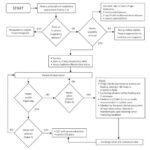
Diagnostic Algorithms and Guidelines
Clinical guidelines for pulmonary hypertension and cor pulmonale emphasize a stepwise diagnostic approach. Initial screening often involves echocardiography in patients with suspected symptoms or risk factors. If echocardiography suggests pulmonary hypertension, further investigations, including PFTs, V/Q scan (if CTEPH is suspected), and ultimately right heart catheterization, are pursued to confirm the diagnosis and determine the underlying cause. These guidelines often incorporate algorithms to streamline the diagnostic process based on clinical probability and initial test results.
Differentiating Cor Pulmonale from Other Conditions
Differential diagnosis is crucial to distinguish cor pulmonale from other conditions that can mimic its symptoms. These include:
- Left-sided heart failure: Differentiated by PCWP > 15 mmHg in left heart failure.
- Primary pulmonary hypertension (Pulmonary Arterial Hypertension): Diagnosis of exclusion after ruling out secondary causes, including lung diseases.
- Congenital heart diseases with right-to-left shunts.
- Valvular heart diseases, particularly tricuspid and pulmonic valve disease.
- Pericardial diseases such as constrictive pericarditis.
- Other causes of right heart failure, such as right ventricular infarction or arrhythmogenic right ventricular cardiomyopathy.
Treatment and Management Strategies
The primary goal of cor pulmonale treatment is to manage the underlying respiratory condition and alleviate pulmonary hypertension. Oxygen therapy is crucial for patients with hypoxemia, as it reduces pulmonary vasoconstriction and improves right ventricular function. Diuretics are used to manage fluid overload and edema. While digitalis use remains controversial, it may offer modest benefits in some patients with right ventricular failure. Specific treatments targeting pulmonary hypertension, such as pulmonary vasodilators, may be considered in certain cases, particularly when pulmonary hypertension is disproportionate to the underlying lung disease. Furthermore, managing the underlying lung disease, whether it’s COPD, sleep apnea, or interstitial lung disease, is paramount to prevent disease progression and improve outcomes.
Prognosis and Outcomes
The prognosis of cor pulmonale is highly variable and depends largely on the severity and progression of the underlying pulmonary disease. Cor pulmonale development often indicates a poorer prognosis in patients with primary pulmonary diseases. Early diagnosis and effective management of both the underlying lung condition and pulmonary hypertension are crucial for improving patient outcomes and quality of life.
Conclusion: Emphasizing Early Diagnosis
Accurate and timely diagnosis of cor pulmonale hinges on a comprehensive evaluation incorporating clinical history, physical examination, and targeted diagnostic testing. While right heart catheterization remains the gold standard for confirmation, non-invasive modalities like echocardiography play a crucial screening and monitoring role. Understanding the diagnostic criteria, including both clinical and hemodynamic parameters, is essential for healthcare professionals in effectively managing patients at risk for or affected by cor pulmonale. For professionals in fields related to health and well-being, recognizing the symptoms and risk factors can contribute to earlier referrals and improved patient care pathways.
References
1.Vieira JL, Távora FRF, Sobral MGV, Vasconcelos GG, Almeida GPL, Fernandes JR, da Escóssia Marinho LL, de Mendonça Trompieri DF, De Souza Neto JD, Mejia JAC. Chagas Cardiomyopathy in Latin America Review. Curr Cardiol Rep. 2019 Feb 12;21(2):8. [PubMed: 30747287]
2.George PM, Patterson CM, Reed AK, Thillai M. Lung transplantation for idiopathic pulmonary fibrosis. Lancet Respir Med. 2019 Mar;7(3):271-282. [PubMed: 30738856]
3.Niwa K. Aortic dilatation in complex congenital heart disease. Cardiovasc Diagn Ther. 2018 Dec;8(6):725-738. [PMC free article: PMC6331370] [PubMed: 30740320]
4.Neidenbach R, Niwa K, Oto O, Oechslin E, Aboulhosn J, Celermajer D, Schelling J, Pieper L, Sanftenberg L, Oberhoffer R, de Haan F, Weyand M, Achenbach S, Schlensak C, Lossnitzer D, Nagdyman N, von Kodolitsch Y, Kallfelz HC, Pittrow D, Bauer UMM, Ewert P, Meinertz T, Kaemmerer H. Improving medical care and prevention in adults with congenital heart disease-reflections on a global problem-part II: infective endocarditis, pulmonary hypertension, pulmonary arterial hypertension and aortopathy. Cardiovasc Diagn Ther. 2018 Dec;8(6):716-724. [PMC free article: PMC6331381] [PubMed: 30740319]
5.Lee S. Comprehensive Nursing Management for Valvular Disease. Crit Care Nurs Clin North Am. 2019 Mar;31(1):31-38. [PubMed: 30736933]
6.Yoon YS, Jin M, Sin DD. Accelerated lung aging and chronic obstructive pulmonary disease. Expert Rev Respir Med. 2019 Apr;13(4):369-380. [PubMed: 30735057]
7.Smolders VF, Zodda E, Quax PHA, Carini M, Barberà JA, Thomson TM, Tura-Ceide O, Cascante M. Metabolic Alterations in Cardiopulmonary Vascular Dysfunction. Front Mol Biosci. 2018;5:120. [PMC free article: PMC6349769] [PubMed: 30723719]
8.Patel S, Cole AD, Nolan CM, Barker RE, Jones SE, Kon S, Cairn J, Loebinger M, Wilson R, Man WD. Pulmonary rehabilitation in bronchiectasis: a propensity-matched study. Eur Respir J. 2019 Jan;53(1) [PubMed: 30578381]
9.Balsam P, Ozierański K, Kapłon-Cieślicka A, Borodzicz S, Tymińska A, Peller M, Marchel M, Crespo-Leiro MG, Maggioni AP, Drożdż J, Opolski G, Grabowski M. Differences in clinical characteristics and 1-year outcomes of hospitalized patients with heart failure in ESC-HF Pilot and ESC-HF-LT registries. Pol Arch Intern Med. 2019 Feb 28;129(2):106-116. [PubMed: 30648697]
10.van Cleemput J, Sonaglioni A, Wuyts WA, Bengus M, Stauffer JL, Harari S. Idiopathic Pulmonary Fibrosis for Cardiologists: Differential Diagnosis, Cardiovascular Comorbidities, and Patient Management. Adv Ther. 2019 Feb;36(2):298-317. [PMC free article: PMC6824347] [PubMed: 30554332]
11.Kim M, Tillis W, Patel P, Davis RM, Asche CV. Association between asthma/chronic obstructive pulmonary disease overlap syndrome and healthcare utilization among the US adult population. Curr Med Res Opin. 2019 Jul;35(7):1191-1196. [PubMed: 30612470]