A stroke is a critical medical emergency that occurs when blood supply to the brain is disrupted, leading to potential brain damage, disability, or even death. Rapid diagnosis is paramount in stroke management to determine the type of stroke and initiate the most effective treatment. Computed Tomography (CT) scans and Magnetic Resonance Imaging (MRI) are the two primary neuroimaging techniques employed for stroke diagnosis. Understanding the nuances of each method is crucial for healthcare professionals and patients alike. This article delves into a comprehensive comparison of CT scans and MRIs in the context of stroke diagnosis, highlighting their strengths and limitations to determine which imaging modality is superior in different scenarios.
Understanding Stroke and the Importance of Rapid Diagnosis
Stroke, often referred to as a “brain attack,” arises from an interruption in the brain’s blood supply. This disruption can be caused by either a blockage (ischemic stroke) or a rupture of a blood vessel (hemorrhagic stroke). Distinguishing between these two types is vital because their treatments differ significantly.
- Ischemic Stroke: This is the most common type, resulting from a blood clot obstructing an artery that carries blood to the brain. The lack of blood flow deprives brain tissue of oxygen and nutrients, leading to cell damage.
- Hemorrhagic Stroke: This occurs when a blood vessel in the brain ruptures, causing bleeding into the brain tissue. The accumulating blood increases pressure within the skull and damages brain cells.
Recognizing stroke symptoms and seeking immediate medical attention is crucial. Symptoms can include sudden numbness or weakness on one side of the body, difficulty speaking or understanding speech, vision problems, severe headache, and dizziness. Prompt diagnosis, ideally within the first few hours of symptom onset, is essential to minimize brain damage and improve patient outcomes. Diagnostic tools like CT scans and MRIs play a pivotal role in this time-sensitive process.
CT Scans for Stroke Diagnosis
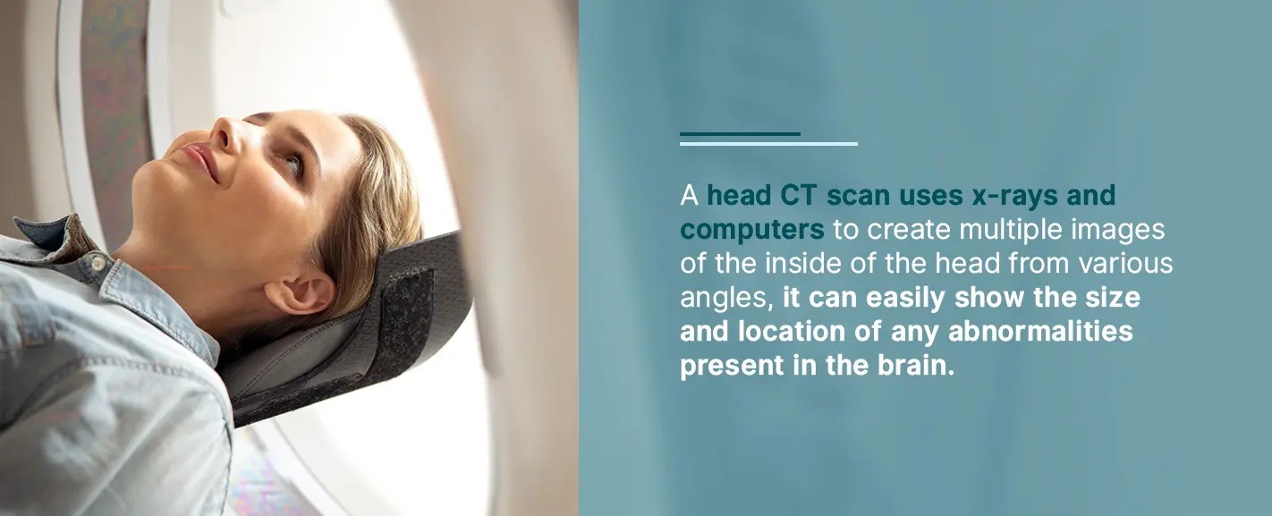
CT scans utilize X-rays to generate cross-sectional images of the brain. In the context of stroke diagnosis, CT scans are particularly valuable for their speed and ability to quickly identify hemorrhagic strokes.
Advantages of CT Scans in Stroke Diagnosis:
- Rapid Acquisition: CT scans are remarkably fast, typically taking only a few minutes to complete. This speed is critical in acute stroke settings where time is brain.
- Hemorrhage Detection: CT scans are highly sensitive in detecting blood within the skull. This makes them excellent for rapidly identifying hemorrhagic strokes, which require different management strategies than ischemic strokes. The presence of blood appears bright on a CT scan, making it easily distinguishable.
- Wide Availability: CT scanners are widely available in most hospitals and emergency departments, ensuring timely access to this diagnostic tool even in smaller healthcare facilities.
- Cost-Effective: Compared to MRIs, CT scans are generally less expensive, making them a more accessible initial diagnostic option.
- Exclusion of Other Conditions: CT scans can also help rule out other conditions that may mimic stroke symptoms, such as brain tumors or head trauma.
Limitations of CT Scans in Stroke Diagnosis:
- Limited Soft Tissue Detail: While excellent for detecting blood, CT scans provide less detailed images of soft tissues compared to MRIs. This can make it less sensitive in the very early stages of ischemic stroke when subtle tissue changes might be present.
- Ischemic Stroke Detection in Early Stages: In the hyperacute phase of ischemic stroke (within the first few hours), CT scans may not always show clear signs of ischemic damage. Subtle changes in brain tissue density related to early ischemia can be difficult to visualize on CT.
- Radiation Exposure: CT scans involve exposure to ionizing radiation, albeit at relatively low doses. While generally safe, radiation exposure is a consideration, especially for repeated scans.
Despite these limitations, CT scans remain the first-line imaging modality in many emergency stroke protocols due to their speed and effectiveness in ruling out hemorrhage, a critical initial step in stroke management.
MRI Scans for Stroke Diagnosis
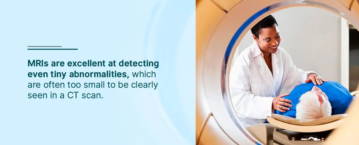
MRI utilizes strong magnetic fields and radio waves to generate detailed images of the brain. MRIs offer superior soft tissue contrast compared to CT scans, making them highly sensitive for detecting subtle brain changes associated with stroke, particularly ischemic stroke.
Advantages of MRI Scans in Stroke Diagnosis:
- Superior Soft Tissue Detail: MRI provides unparalleled detail of brain tissues, allowing for the detection of even subtle abnormalities. This is especially advantageous in identifying early ischemic changes that may be missed on CT scans.
- Early Ischemic Stroke Detection: MRI, particularly techniques like Diffusion-Weighted Imaging (DWI), is highly sensitive in detecting ischemic stroke within minutes of symptom onset, often earlier than CT scans. DWI detects the restricted water diffusion that occurs in acutely ischemic tissue.
- Accurate Assessment of Stroke Size and Location: The detailed images from MRI allow for a more precise assessment of the extent and location of brain damage caused by stroke.
- No Ionizing Radiation: MRI does not use ionizing radiation, making it a safer option in this regard, particularly for patients who may require repeated imaging.
- Detection of Lacunar Strokes: MRI is more sensitive in detecting small, deep ischemic strokes called lacunar strokes, which can be challenging to visualize on CT.
Limitations of MRI Scans in Stroke Diagnosis:
- Slower Acquisition Time: MRI scans take significantly longer than CT scans, often ranging from 30 minutes to an hour, depending on the specific protocols. This longer scan time can be a disadvantage in acute stroke management where rapid intervention is crucial.
- Limited Availability and Accessibility: MRI scanners are less widely available than CT scanners, particularly in smaller hospitals and emergency settings. Access to MRI in a timely manner can be a limiting factor in some locations.
- Higher Cost: MRI scans are generally more expensive than CT scans, which can impact healthcare costs.
- Contraindications: MRI is contraindicated in patients with certain metallic implants, such as pacemakers or some types of aneurysm clips. Screening for these contraindications adds to the preparation time.
- Claustrophobia: The enclosed nature of the MRI scanner can induce claustrophobia in some patients, potentially requiring sedation or making the procedure unsuitable.
Despite these limitations, MRI is considered the gold standard for detailed stroke imaging, especially for ischemic stroke. Its superior sensitivity and ability to detect early ischemic changes make it invaluable in comprehensive stroke evaluation and management when time allows.
CT vs MRI: A Comparative Summary for Stroke Diagnosis
| Feature | CT Scan | MRI Scan |
|---|---|---|
| Speed | Rapid (minutes) | Slower (30-60 minutes) |
| Hemorrhage Detection | Excellent | Good |
| Early Ischemic Detection | Less Sensitive (especially very early) | Highly Sensitive (especially with DWI) |
| Soft Tissue Detail | Less Detailed | Superior Detail |
| Availability | Widely Available | Less Widely Available |
| Cost | Lower | Higher |
| Radiation | Ionizing Radiation | No Ionizing Radiation |
| Contraindications | Fewer | More (metallic implants, claustrophobia) |
| Best For | Initial triage, hemorrhage exclusion, speed | Detailed assessment, early ischemic detection |


Conclusion: Choosing the Optimal Imaging Method for Stroke
Both CT scans and MRIs are indispensable tools in the diagnostic arsenal for stroke. The “best” imaging method is not absolute but rather depends on the clinical context, the suspected type of stroke, and the urgency of the situation.
In the acute setting, particularly in the first few hours after stroke onset, CT scan is often the preferred initial imaging modality due to its speed, widespread availability, and excellent ability to rapidly rule out hemorrhagic stroke. This quick assessment is critical for guiding immediate treatment decisions, such as the administration of thrombolytic therapy for ischemic stroke, which is contraindicated in hemorrhagic stroke.
MRI, with its superior soft tissue detail and sensitivity to early ischemia, is invaluable for a more comprehensive stroke evaluation. While MRI may take longer to perform and may not always be immediately available, it provides crucial information regarding the extent and location of ischemic damage, especially in the early stages when CT may be less conclusive for ischemic stroke. In many centers, MRI is used as a follow-up imaging modality after initial CT or as the primary imaging if time is less critical or if there is a high suspicion of ischemic stroke and hemorrhage has been ruled out.
Ultimately, the decision of whether to use CT or MRI, or both, for stroke diagnosis is a clinical one, guided by the individual patient’s presentation, the suspected stroke type, the available resources, and the need for rapid diagnosis and treatment initiation. Both imaging techniques play complementary roles in optimizing stroke care and improving patient outcomes.
Sources:
- How Are CT Scans Used in Diagnosing Strokes?: https://www.envrad.com/content/uploads/2020/07/How-are-CT-scans-used-in-diagnosing-strokes.jpg
- How Are MRI Scans Used in Diagnosing Strokes?: https://www.envrad.com/content/uploads/2020/07/How-are-MRI-scans-used-in-diagnosing-strokes.jpg
- National Heart, Lung, and Blood Institute (NHLBI) – Stroke: https://www.nhlbi.nih.gov/health-topics/stroke
- CT Scans Services – Envision Radiology: https://www.envrad.com/services/ct-scans/
- Thrombolytic Therapy for Stroke – MedlinePlus: https://medlineplus.gov/ency/article/007089.htm
- Stroke Diagnosis and Treatment – AgingCare.com: https://www.agingcare.com/articles/stroke-diagnosis-and-treatment-133116.htm
- MRI Scans Services – Envision Radiology: https://www.envrad.com/services/mri-scans/
- Colorado Springs Imaging – Envision Radiology: https://www.envrad.com/location/colorado-springs-imaging/
- Envision Radiology Locations: https://www.envrad.com/locations/
