A cerebrovascular accident (CVA), commonly known as a stroke, is a critical medical condition characterized by the disruption of blood flow to the brain. This interruption deprives brain tissue of essential oxygen and nutrients, leading to rapid cell damage and neurological deficits. Understanding stroke types, implementing timely nursing assessments, and developing effective care plans are crucial for optimal patient outcomes.
Types of Stroke: Ischemic and Hemorrhagic
Strokes are broadly classified into two primary categories: ischemic and hemorrhagic, each with distinct causes and management strategies.
Ischemic Stroke: This type, accounting for the majority of stroke cases, occurs due to a blockage in the arteries supplying blood to the brain. This blockage can be caused by:
- Thrombosis: Formation of a blood clot within a cerebral artery.
- Embolism: A blood clot or plaque debris traveling from another part of the body (often the heart) and lodging in a cerebral artery.
- Systemic Hypoperfusion: Reduced blood flow to all parts of the body, including the brain, often due to heart failure or severe hypotension.
The resulting lack of blood flow (ischemia) leads to oxygen and glucose deprivation, causing brain cells in the affected area to die within minutes.
Hemorrhagic Stroke: Hemorrhagic strokes result from the rupture of a blood vessel in the brain, leading to bleeding into brain tissue or the surrounding spaces. The two main types of hemorrhagic stroke are:
- Intracerebral Hemorrhage: Bleeding directly into the brain tissue, often caused by hypertension, arteriovenous malformations (AVMs), or amyloid angiopathy.
- Subarachnoid Hemorrhage: Bleeding into the space between the brain and the surrounding membrane (subarachnoid space), frequently caused by aneurysms or head trauma.
The blood accumulation increases intracranial pressure (ICP), compressing and damaging brain tissue.
Transient Ischemic Attack (TIA): Often referred to as a “mini-stroke,” a TIA involves a temporary disruption of blood flow to the brain. Symptoms are similar to a stroke but resolve within a short period, usually within an hour and almost always within 24 hours, without causing permanent brain damage. However, TIAs are significant warning signs, indicating an increased risk of future stroke and necessitating prompt medical evaluation and risk factor management.
The Nursing Process for Stroke Patients
Nursing care for stroke patients is multifaceted and requires a systematic approach based on the nursing process: assessment, diagnosis, planning, implementation, and evaluation. The severity of stroke, time to treatment initiation, and pre-existing conditions significantly influence patient disability levels. Initial care typically occurs in intensive care units (ICU) or step-down units, where nurses with specialized training, often NIH Stroke Scale (NIHSS) certified, closely monitor patients. Rapid and subtle changes in stroke symptoms demand critical thinking and swift interventions to prevent neurological deterioration.
Severe strokes can result in profound disabilities, requiring comprehensive care for basic needs such as feeding, bathing, and mobility. Long-term deficits can significantly impact patients and families, leading to emotional distress and depression. Nurses play a vital role in providing compassionate care, ensuring patient safety and dignity, and addressing both physical and psychosocial needs throughout the stroke recovery journey.
Nursing Assessment: Subjective and Objective Data Collection
The initial step in stroke nursing care is a comprehensive nursing assessment, gathering subjective and objective data to understand the patient’s condition and guide subsequent interventions.
Review of Health History: Subjective Data
1. Elicit General Symptoms: Sudden onset of specific symptoms is highly indicative of stroke. Nurses should inquire about:
- Motor Weakness: Hemiparesis (weakness on one side) or hemiplegia (paralysis on one side), typically affecting the face, arm, and leg on the same side of the body.
- Sensory Deficits: Numbness, tingling, or loss of sensation (paresthesias) in extremities.
- Swallowing Difficulty: Dysphagia, indicated by coughing, choking, or drooling when attempting to swallow.
- Visual Disturbances: Sudden vision loss in one or both eyes, double vision (diplopia), or visual field deficits (hemianopsia).
- Speech Impairment:
- Expressive Aphasia (Broca’s aphasia): Difficulty producing speech, understanding language remains relatively intact.
- Receptive Aphasia (Wernicke’s aphasia): Difficulty understanding spoken or written language, speech may be fluent but nonsensical.
- Dysarthria: Difficulty articulating words due to muscle weakness affecting speech.
- Balance and Coordination Issues: Ataxia (unsteady gait), dizziness, vertigo.
- Cognitive and Mental Status Changes: Confusion, disorientation, decreased level of consciousness, memory problems.
2. Determine Symptom Onset: The time of symptom onset or “last known well” time is crucial. This information is vital for determining eligibility for thrombolytic therapy (tPA) and other time-sensitive interventions.
3. Identify Stroke Risk Factors: Assess for modifiable and non-modifiable risk factors:
-
Non-Modifiable:
- Age: Risk increases with age, particularly after 55 years.
- Ethnicity: African Americans and Hispanics have a higher stroke incidence.
- Gender: Slightly higher incidence in males.
- Family History: Stroke in a first-degree relative, especially before age 65, increases risk.
- Prior Stroke or TIA: History of stroke or TIA significantly increases future stroke risk.
-
Modifiable:
- Hypertension: Uncontrolled high blood pressure is a major risk factor.
- Hyperlipidemia: High cholesterol levels contribute to atherosclerosis.
- Diabetes Mellitus: Diabetes increases the risk of blood clots and vascular damage.
- Obesity and Physical Inactivity: Contribute to other risk factors like hypertension and diabetes.
- Smoking: Damages blood vessels and increases clot formation.
- Excessive Alcohol Consumption: Can raise blood pressure and contribute to stroke risk.
- Atrial Fibrillation (Afib): Irregular heartbeat increases risk of clot formation and embolic stroke.
- Carotid Artery Stenosis: Narrowing of carotid arteries reduces blood flow to the brain.
- Obstructive Sleep Apnea: Associated with hypertension, Afib, and other stroke risk factors.
- COVID-19 Infection: Emerging evidence suggests increased stroke risk following COVID-19.
4. Review Medical History: Certain medical conditions significantly elevate stroke risk:
- Cardiovascular Disease: Coronary artery disease, heart failure.
- Peripheral Artery Disease: Indicates systemic atherosclerosis.
- Sickle Cell Disease: Can cause blood vessel blockage.
- Migraine with Aura: Slightly increased risk of ischemic stroke.
5. Family History: Inquire about family history of stroke, particularly in younger relatives, as genetic predispositions or conditions like CADASIL (Cerebral Autosomal Dominant Arteriopathy with Subcortical Infarcts and Leukoencephalopathy) can increase risk.
6. Medication Review: Assess current medications, including:
- Hormone Replacement Therapy and Oral Contraceptives: Estrogen-containing medications can increase stroke risk in some women.
- Antihypertensives: Non-adherence can lead to uncontrolled hypertension.
- Anticoagulants/Antiplatelets (Aspirin, Warfarin, Heparin, Enoxaparin): Usage indicates pre-existing conditions that increase stroke risk, or over-anticoagulation could increase hemorrhagic stroke risk.
7. Substance Use History: Illicit drug use, particularly stimulants like cocaine and amphetamines, is linked to increased stroke risk due to their effects on blood pressure and blood vessels.
8. Lifestyle Factors: Assess lifestyle habits that contribute to stroke risk:
- Diet: High in saturated and trans fats, cholesterol, and sodium.
- Physical Activity: Sedentary lifestyle.
- Alcohol Intake: Excessive alcohol consumption.
- Smoking: Current or past smoking history.
Physical Assessment: Objective Data
1. Utilize F.A.S.T. Recognition: Quickly assess for cardinal stroke signs using the F.A.S.T. acronym:
- F – Face: Check for facial drooping or asymmetry when smiling or showing teeth.
- A – Arms: Assess for arm weakness by asking the patient to raise both arms; observe for drifting or downward drift of one arm.
- S – Speech: Evaluate speech for slurring, difficulty forming words, or inability to speak.
- T – Time: Note the time of symptom onset and immediately activate emergency medical services (call 911 or local emergency number).
2. Assess ABCs: Prioritize airway, breathing, and circulation. Stroke patients are at risk for aspiration, airway obstruction, and respiratory compromise due to decreased consciousness or impaired swallowing.
3. Perform Neurological Examination: Conduct a comprehensive neurological assessment, often using the NIHSS, to quantify stroke severity and monitor neurological status. The NIHSS evaluates:
- Level of Consciousness (LOC): Alertness, orientation, response to stimuli.
- Visual Function: Visual fields, gaze, presence of hemianopsia.
- Facial Nerve Function: Facial symmetry, smile, brow raise.
- Motor Function (Arms and Legs): Strength and movement in upper and lower extremities.
- Cerebellar Function: Limb ataxia (coordination), finger-to-nose test, heel-to-shin test.
- Sensory Function: Sensation to touch and pain.
- Language and Speech: Aphasia (expressive and receptive), dysarthria.
- Neglect and Attention: Inattention to one side of the body or space (unilateral neglect).
4. Monitor Vital Signs: Closely monitor blood pressure, heart rate, respiratory rate, and oxygen saturation. Hypertension is common in acute stroke. However, rapid blood pressure reduction can be detrimental, especially in ischemic stroke, as it can compromise cerebral perfusion.
Diagnostic Procedures
1. Check Blood Glucose: Perform a point-of-care blood glucose test immediately to rule out hypoglycemia, which can mimic stroke symptoms.
2. Brain Imaging: Prompt neuroimaging is essential to confirm stroke diagnosis, differentiate between ischemic and hemorrhagic stroke, and guide treatment.
- Non-Contrast Head CT Scan: The initial imaging modality of choice, rapidly identifies hemorrhage and large ischemic strokes.
- CT Angiography (CTA) and CT Perfusion (CTP) Scan: Visualize blood vessels, identify clots, and assess brain tissue perfusion to determine the extent of ischemic damage and guide advanced treatments like thrombectomy.
- Magnetic Resonance Imaging (MRI): Provides more detailed images of brain tissue, particularly useful for detecting small ischemic strokes and assessing penumbra (potentially salvageable tissue).
- Carotid Duplex Ultrasound: Evaluates carotid artery stenosis, a risk factor for ischemic stroke.
- Digital Subtraction Angiography (DSA): Invasive procedure providing detailed visualization of cerebral blood vessels, used in select cases for diagnosis and intervention.
3. Laboratory Tests: Blood tests help identify underlying causes and contributing factors to stroke:
- Complete Blood Count (CBC): Evaluates for polycythemia (increased red blood cell count) or thrombocytopenia (low platelet count), which can contribute to stroke.
- Cardiac Biomarkers (Troponin, CK-MB): Assess for cardiac injury, as stroke can be associated with underlying cardiac conditions.
- Coagulation Studies (PT/INR, aPTT): Baseline coagulation status, important before administering anticoagulants or thrombolytics.
- Lipid Profile (Fasting): Assess cholesterol and triglyceride levels, risk factors for atherosclerosis.
- Electrolyte Panel and Renal Function Tests: Evaluate overall metabolic status and kidney function.
- Toxicology Screen: Rule out drug intoxication as a cause of stroke-like symptoms, especially in younger patients.
4. Pregnancy Test (for women of childbearing age): Fibrinolytic therapy (tPA) safety in pregnancy is not fully established, pregnancy testing is crucial before considering tPA in women of reproductive age.
5. Electrocardiogram (ECG): Detects cardiac arrhythmias, particularly atrial fibrillation, a major cause of cardioembolic stroke.
Nursing Interventions: Restoring Perfusion and Promoting Recovery
Nursing interventions are critical for minimizing brain damage, preventing complications, and facilitating stroke recovery.
Restoring Brain Perfusion: Acute Stroke Management
1. Rapid Initial Evaluation and Diagnostic Imaging: Within minutes of arrival in the emergency department, patients with suspected stroke should undergo a neurological assessment and STAT non-contrast head CT scan to confirm diagnosis and differentiate stroke type.
2. Restore Blood Flow in Ischemic Stroke: For confirmed ischemic stroke, initiate alteplase (tPA) administration as rapidly as possible within the eligible time window (typically within 3-4.5 hours of symptom onset, depending on guidelines and patient factors). tPA is a thrombolytic agent that dissolves blood clots. It is contraindicated in hemorrhagic stroke due to bleeding risk.
3. Continuous Neurological Monitoring: Frequent and meticulous neurological assessments are essential to detect changes in patient status and guide timely interventions. Monitor:
- Level of Consciousness (LOC): Using Glasgow Coma Scale (GCS) or NIHSS LOC component.
- Pupillary Response: Size, equality, and reactivity to light.
- Motor Strength and Sensation: Bilateral assessment of muscle strength and sensory function.
- Speech, Cognition, and Memory: Assess for changes in language, orientation, and cognitive abilities.
- Mood and Behavior: Observe for emotional lability, agitation, or depression.
4. Medication Administration: Manage secondary complications and optimize neurological recovery with prescribed medications:
- Antihypertensives: Carefully manage blood pressure within target ranges based on stroke type and treatment strategy. Avoid aggressive blood pressure lowering, especially in acute ischemic stroke, unless tPA is administered.
- Antiseizure Medications: Prophylactic anticonvulsants may be considered in hemorrhagic stroke or cortical ischemic stroke. Treat seizures promptly if they occur.
- Stool Softeners/Laxatives: Prevent constipation and straining during bowel movements, which can increase ICP.
5. Blood Pressure Management: Maintain blood pressure within recommended parameters:
- Ischemic Stroke (post-tPA): Maintain BP < 180/105 mmHg for the first 24 hours post-tPA.
- Ischemic Stroke (no tPA): Permissive hypertension may be allowed initially to maintain cerebral perfusion; avoid aggressive lowering unless extremely high.
- Hemorrhagic Stroke: Target systolic BP around 140 mmHg to minimize re-bleeding.
6. Prepare for Mechanical Thrombectomy: For large vessel occlusions in ischemic stroke, mechanical thrombectomy (surgical clot removal) may be indicated. Ensure informed consent is obtained and prepare the patient for the procedure.
7. Endovascular Procedures: For patients with carotid artery stenosis, prepare for potential interventions such as carotid endarterectomy (surgical removal of plaque) or carotid artery stenting to improve cerebral blood flow and prevent future stroke.
Stroke Recovery and Prevention: Rehabilitation and Education
1. Stroke Rehabilitation Referral: Initiate early referral to comprehensive stroke rehabilitation programs, including:
- Physical Therapy: Improve motor function, balance, gait, and mobility.
- Occupational Therapy: Enhance activities of daily living (ADLs), fine motor skills, and upper extremity function.
- Speech Therapy: Address communication deficits (aphasia, dysarthria), swallowing difficulties (dysphagia), and cognitive communication skills.
- Cognitive Therapy: Improve memory, attention, executive function, and address cognitive impairments.
2. Speech Deficit Management Education: For patients with aphasia or dysarthria, educate on:
- Communication Strategies: “Communication partner training” for family and caregivers, using visual aids, communication boards, apps, and written communication.
- Speech Practice: Encourage regular speech therapy exercises and home practice using flashcards, books, and computer programs.
3. Medication Education: Educate patients and families about prescribed medications upon discharge:
- Anticoagulants (Apixaban, Dabigatran, Rivaroxaban, Warfarin): For atrial fibrillation or other thromboembolic risks, prevent future clots.
- Antiplatelets (Aspirin, Clopidogrel): Prevent platelet aggregation and reduce secondary stroke risk.
- Antihypertensives (Diuretics, Beta-blockers, Calcium Channel Blockers, ACE Inhibitors): Manage hypertension to prevent recurrent stroke.
- Statins: Manage hyperlipidemia and reduce atherosclerotic risk.
4. Safety Strategies: Implement and educate on fall prevention strategies:
- Fall Precautions: Bed alarms, side rails, call light within reach, non-slip footwear, clear environment.
- Assistive Devices: Walkers, canes, wheelchairs, adaptive equipment for ADLs.
5. Support Group Encouragement: Recommend stroke support groups for patients and caregivers to provide emotional support, shared experiences, and peer encouragement.
6. Lifestyle Modification Promotion: Educate on modifiable risk factors and lifestyle changes to prevent secondary stroke:
- Healthy Diet: Low in saturated and trans fats, cholesterol, and sodium, rich in fruits, vegetables, and whole grains.
- Regular Exercise: Aim for at least 30 minutes of moderate-intensity exercise most days of the week.
- Smoking Cessation: Provide resources and support for quitting smoking.
- Moderate Alcohol Consumption: Limit alcohol intake or abstain altogether.
- Weight Management: Achieve and maintain a healthy weight.
7. Treatment Regimen Adherence: Emphasize the importance of medication adherence, follow-up appointments, and management of comorbidities (hypertension, diabetes, hyperlipidemia) to minimize future stroke risk.
Nursing Care Plans for CVA: Addressing Common Nursing Diagnoses
Nursing care plans provide a structured framework for addressing specific patient needs based on nursing diagnoses. Here are examples of care plans for common nursing diagnoses in stroke patients:
1. Impaired Verbal Communication
Nursing Diagnosis: Impaired Verbal Communication related to prolonged cerebral occlusion, dysarthria, and/or aphasia.
Defining Characteristics: Slurred speech, nonverbal communication, difficulty forming words, difficulty expressing thoughts, slow or delayed responses, extremity weakness or paralysis affecting writing/typing.
Expected Outcomes:
- Patient will establish an effective communication method to express needs and thoughts.
- Patient will actively participate in speech therapy to improve communication skills.
- Patient will utilize communication aids and resources to support communication.
Nursing Assessments:
- Identify type of aphasia: Differentiate between global, Wernicke’s, and Broca’s aphasia to tailor communication strategies.
- Observe patient’s communication methods: Identify gestures, signals, or sounds used by the patient and involve family in understanding these cues.
Nursing Interventions:
- Speak in short, direct sentences: Use clear, simple language, face the patient, and use visual cues. Ask “yes” or “no” questions.
- Utilize alternative communication methods: Employ writing, drawing, communication boards, gestures, and technology as needed.
- Encourage speech therapy: Emphasize the importance of speech-language therapy for language rehabilitation.
- Promote family participation: Involve family in therapy sessions and teach them communication techniques to support the patient.
2. Ineffective Cerebral Tissue Perfusion
Nursing Diagnosis: Ineffective Cerebral Tissue Perfusion related to interruption of blood flow, thrombus formation, artery occlusion, cerebral edema, or hemorrhage.
Defining Characteristics: Altered mental status, blurred vision, slurred speech, extremity weakness, changes in vital signs, neurological deficits on NIHSS.
Expected Outcomes:
- Patient will recognize stroke symptoms and seek immediate medical attention in the future.
- Patient will demonstrate improved cerebral perfusion as evidenced by stable vital signs and neurological status within acceptable parameters.
- Patient will show improvement in stroke deficits (speech, weakness, swallowing) by discharge.
Nursing Assessments:
- Establish baseline neurological status: Determine “last known well” time to guide treatment and assess for changes.
- Perform frequent neurological assessments: Use NIHSS or facility-specific stroke scales to monitor LOC, motor function, sensory function, and speech.
- Review brain imaging results (CT/MRI): Confirm stroke type (ischemic vs. hemorrhagic) and guide treatment.
Nursing Interventions:
- Maintain blood pressure within ordered parameters: Follow physician orders for target BP ranges based on stroke type and treatment.
- Administer thrombolytics (tPA) as indicated: For ischemic stroke within the time window, monitor for bleeding complications.
- Educate on stroke risk factors: Provide education on modifiable risk factors and lifestyle changes for secondary stroke prevention.
- Instruct on stroke symptom recognition using FAST: Emphasize the importance of timely recognition and activation of emergency services.
3. Risk for Injury
Nursing Diagnosis: Risk for Injury related to impaired judgment, spatial-perceptual deficits, weakness, poor motor coordination, poor balance, impaired sensory awareness, dysphagia, communication deficits, hemiplegia, and impulsivity.
Defining Characteristics: (Risk diagnosis, evidenced by risk factors, not actual injury)
Expected Outcomes:
- Patient will remain free from falls and injuries during hospitalization and rehabilitation.
- Patient will maintain intact skin integrity.
- Caregivers will create a safe environment to prevent patient injury post-discharge.
Nursing Assessments:
- Determine deficits related to brain area affected: Identify specific deficits like right-brain (spatial-perceptual) vs. left-brain (speech, swallowing) to anticipate injury risks.
- Assess sensory awareness: Evaluate ability to perceive pain, temperature, and pressure to prevent skin breakdown and injury.
- Note neglect or visual disturbances: Assess for unilateral neglect and hemianopia, which increase fall and injury risk.
Nursing Interventions:
- Use bed and chair alarms: Especially for patients with impulsivity or impaired judgment.
- Assist with eating and feeding: Implement dysphagia precautions, thickened liquids, and supervised meals to prevent aspiration.
- Teach scanning the environment: For patients with neglect or visual field deficits, train them to scan from left to right to improve awareness.
- Turn and assess skin frequently: Implement pressure ulcer prevention strategies, especially on paralyzed or insensate limbs.
4. Self-Care Deficit
Nursing Diagnosis: Self-Care Deficit related to neurobehavioral manifestations, weakness, musculoskeletal impairment, cognitive dysfunction, decreased motivation, impaired physical mobility, and unilateral neglect.
Defining Characteristics: Inability to independently perform cleansing, dressing, feeding, toileting activities.
Expected Outcomes:
- Patient will maintain skin integrity and be free from body odor.
- Patient will verbalize successful use of assistive devices for bathing and hygiene.
- Patient will express satisfaction with bathing and self-care, even with caregiver assistance.
- Patient will dress and perform ADLs to optimal potential.
- Patient will demonstrate competency in using assistive devices.
- Patient will feed self safely and effectively.
- Patient will maintain continence and skin integrity related to toileting.
Nursing Assessments:
- Assess functional abilities and limitations: Evaluate physical, cognitive, and emotional impairments affecting self-care.
- Assess patient preferences: Respect patient preferences for hygiene, food, and self-care to promote dignity and independence.
- Perform routine risk assessments: Fall risk (Morse Fall Scale), skin breakdown risk (Braden Scale), swallowing assessment.
Nursing Interventions:
- Establish a toileting schedule: Prevent constipation and promote bladder and bowel regularity.
- Encourage independence when possible: Promote self-care and ADL participation to maximize autonomy and self-esteem.
- Ensure adequate time for meals: Allow sufficient time for patients with dysphagia to eat safely.
- Assist with adaptive equipment: Provide and train on assistive devices for dressing, bathing, grooming, and feeding.
- Consult with physical and occupational therapy: Collaborate with rehabilitation team to optimize self-care skills and independence.
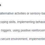
5. Unilateral Neglect
Nursing Diagnosis: Unilateral Neglect related to disease process, brain trauma or damage, ischemia of cerebral tissue.
Defining Characteristics: Altered safety behavior on neglected side, failure to move eyes or limbs on neglected side, difficulty grooming neglected side, unawareness of neglected limb position.
Expected Outcomes:
- Patient will demonstrate techniques to minimize unilateral neglect.
- Patient will care for both sides of the body appropriately and protect the neglected side from harm.
- Patient will achieve the highest possible level of functioning based on individual abilities and goals.
Nursing Assessments:
- Assess for signs of unilateral neglect: Observe for inattention to one side of the body, lack of self-care on one side, and unawareness of the affected side.
- Assess level of awareness of neglect: Determine patient’s understanding of their neglect and willingness to learn compensatory strategies.
- Assess skin integrity on neglected side: Monitor for skin breakdown due to lack of awareness and repositioning on the affected side.
Nursing Interventions:
- Initiate fall precautions: Implement measures to prevent falls due to one-sided weakness and neglect.
- Encourage assistive device use: Promote use of assistive devices to enhance safety and awareness of the neglected side.
- Instruct on neutral body positioning: Teach patient to maintain neutral alignment and regularly reposition to prevent pain and contractures.
- Position belongings on unaffected side: Place objects and approach patient from the unaffected side to encourage engagement with the neglected side.
- Coordinate rehabilitation program transfer: Ensure timely referral to rehabilitation services for ongoing management of unilateral neglect.
References
Original article likely used references. To enhance EEAT, include relevant, high-quality references here if available. Examples:
- American Heart Association/American Stroke Association (AHA/ASA) guidelines for stroke management.
- National Institute of Neurological Disorders and Stroke (NINDS) resources.
- Peer-reviewed nursing journals and textbooks on stroke care.
Alt Text: Diagram illustrating the two main types of stroke: Ischemic stroke caused by blockage and Hemorrhagic stroke caused by bleeding.
Alt Text: FAST acronym infographic for stroke recognition: Face drooping, Arm weakness, Speech difficulty, Time to call emergency services.
Alt Text: NIH Stroke Scale (NIHSS) assessment tool for quantifying neurological deficits in stroke patients, including categories like consciousness, vision, motor function, sensation, and language.