Carcinoma ex pleomorphic adenoma (CXPA) presents a significant diagnostic puzzle in the medical field, much like complex issues encountered in automotive repair. Just as identifying the root cause of a vehicle malfunction requires expert analysis, diagnosing CXPA demands a thorough and meticulous approach. This article will explore the intricacies of Cxpa Diagnosis, drawing from a detailed case study to illuminate this challenging condition.
Understanding Carcinoma Ex Pleomorphic Adenoma
Carcinoma ex pleomorphic adenoma, often abbreviated as CXPA, is a type of cancer that originates from a benign tumor known as pleomorphic adenoma. Pleomorphic adenomas are the most common benign tumors found in the parotid gland, a major salivary gland located near the ear. While pleomorphic adenomas themselves are non-cancerous, they carry a risk of transforming into CXPA over time. This transformation is what makes CXPA a “carcinoma ex,” meaning “carcinoma arising from” a pre-existing condition.
The Pathogenesis of CXPA: Unraveling the Transformation
The exact mechanisms behind this malignant transformation are still being researched. Two primary theories exist: one suggests that CXPAs are inherently malignant from their inception, while the other proposes that benign pleomorphic adenomas undergo a cancerous change over time. Evidence suggests that the longer a pleomorphic adenoma exists, the higher the chance of malignant transformation. This is similar to how prolonged stress on certain vehicle components can eventually lead to more serious failures.
Incidence and Clinical Presentation: Recognizing the Signs
CXPA is a rare malignancy, accounting for a small percentage of parotid cancers. It predominantly affects individuals in their sixth to eighth decades of life. Clinically, CXPA often manifests as a firm mass in the parotid gland. Patients may have a history of a long-standing, slow-growing mass that suddenly begins to enlarge more rapidly or become painful. However, in some cases, patients may not be aware of a pre-existing benign tumor. The diagnostic challenge lies in differentiating CXPA from benign pleomorphic adenomas and other parotid malignancies, especially in the initial stages.
The Diagnostic Process of CXPA: Navigating Complexity
Preoperative CXPA diagnosis is notoriously difficult. Clinical examination and imaging techniques like CT scans can raise suspicion, but definitive diagnosis typically requires pathological assessment of tissue samples. This is akin to how advanced diagnostic tools in auto repair can point towards a problem, but physical inspection and component testing are often necessary for confirmation.
Preoperative Diagnostic Hurdles
Initial assessments, such as physical examinations and imaging, may suggest a parotid tumor, but distinguishing between benign and malignant conditions preoperatively is challenging. Fine needle aspiration cytology (FNAC), a procedure to collect cells for examination, can be helpful, but may not always provide a conclusive diagnosis of CXPA, particularly if the malignant component is small or not adequately sampled. This is comparable to initial vehicle scans that might indicate a general area of concern without pinpointing the exact issue.
Pathological Assessment: The Gold Standard for CXPA Diagnosis
The definitive diagnosis of CXPA relies on thorough histopathological examination. Pathologists analyze tissue samples obtained through biopsy or surgical removal. Key histological features that point towards CXPA include:
- Capsule Invasion: The cancerous component breaching the capsule surrounding the original pleomorphic adenoma.
- Hemorrhage and Necrosis: Areas of bleeding and cell death within the tumor tissue, indicative of aggressive growth.
- Presence of Both Benign and Malignant Components: Identifying areas characteristic of pleomorphic adenoma alongside distinct cancerous cells.
The challenge for pathologists is to meticulously examine the tissue to identify these features, especially as the benign pleomorphic adenoma component can be small and easily overlooked. Accurate pathological diagnosis is crucial for guiding treatment and predicting prognosis.
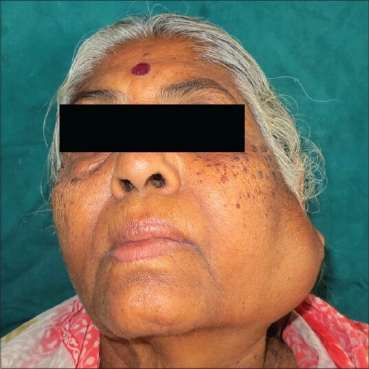
Case Study: Illustrating CXPA Diagnostic Journey
Consider the case of a 65-year-old woman who presented with a swelling in her left preauricular region. Ten years prior, she had undergone surgery in the same area, suggesting a history of pleomorphic adenoma. Upon examination, a firm mass was detected in her parotid gland, along with enlarged lymph nodes in her neck. Preoperative CT scans revealed a heterogeneous lesion in the parotid gland, and FNAC raised suspicion of malignancy.
Initially, a diagnosis of pleomorphic adenoma with lymph node metastasis was considered. However, during surgery, frozen section analysis revealed more aggressive features, leading to a radical parotidectomy – the removal of the parotid gland along with the facial nerve and surrounding tissues.
Postoperative histopathology confirmed the diagnosis of CXPA, with tumor infiltration extending to the submandibular gland and lymph nodes. This case underscores the critical role of pathological assessment in definitively diagnosing CXPA and guiding appropriate surgical management.
Treatment and Prognosis: Aggressive Management is Key
Due to its aggressive nature, CXPA requires prompt and aggressive treatment. The primary treatment modality is surgery, typically involving parotidectomy and neck dissection to remove the tumor and any affected lymph nodes.
Surgical Intervention: Radical Resection
Radical parotidectomy, as performed in the case study, often involves removing the facial nerve along with the parotid gland to ensure complete tumor removal. While facial nerve sacrifice can lead to facial paralysis, it may be necessary in cases of CXPA to achieve adequate cancer control. The extent of surgery depends on the tumor size, location, and spread.
Postoperative Radiotherapy: Enhancing Local Control
Following surgery, postoperative radiotherapy is frequently recommended, particularly in cases with high-grade tumors, lymph node involvement, or close surgical margins. Radiotherapy helps to eliminate any remaining cancer cells and reduce the risk of recurrence. This combined approach of surgery and radiotherapy is considered the standard of care for CXPA.
Prognostic Factors: Predicting Outcomes
Several factors influence the prognosis of CXPA, including:
- Tumor Grade: High-grade tumors are more aggressive and associated with poorer outcomes.
- Tumor Size: Larger tumors tend to have a worse prognosis.
- Extracapsular Extension: Tumor spread beyond the original pleomorphic adenoma capsule is a negative prognostic indicator.
- Perineural Invasion: Cancer cells invading nerves is associated with increased recurrence risk.
- Lymph Node Metastasis: Spread to regional lymph nodes significantly reduces survival rates.
Early and accurate CXPA diagnosis, followed by aggressive surgical and adjuvant treatment, are crucial for improving patient outcomes.
Conclusion: The Importance of Precision in CXPA Diagnosis
Carcinoma ex pleomorphic adenoma is a rare and challenging malignancy that demands a high degree of diagnostic acumen and aggressive management. Similar to the complexities encountered in diagnosing intricate automotive problems, CXPA diagnosis requires a systematic approach, utilizing clinical evaluation, advanced imaging, and, most importantly, meticulous pathological assessment. Recognizing the potential for malignant transformation in pleomorphic adenomas and understanding the key diagnostic features of CXPA are vital for clinicians to ensure timely and effective treatment, ultimately improving patient prognosis.
References
[1] Eveson JW, Cawson RA. Tumours of the salivary glands: a review of 2,410 cases. Int J Oral Maxillofac Surg. 1985;14 Suppl 1:1–28.
[2] Spiro RH. Pleomorphic adenoma: clinicopathologic assessment. Head Neck Surg. 1986;8:290–302.
[3] Gnepp DR. Malignant mixed tumors of the salivary glands: a review. Pathol Annu. 1993;28 Pt 1:279–328.
[4] Barnes L, Tse LL, Hunt JL, et al. Carcinoma ex pleomorphic adenoma of the salivary glands: a clinicopathologic and immunohistochemical review of 100 cases. Cancer. 2007;109:171–89.
[5] Eneroth CM, Zetterberg A. A cytophotometric study of DNA in benign mixed tumors and carcinomas of the parotid gland. Acta Otolaryngol. 1968;66:465–70.
[6] Tortoledo ME, Luna MA, Barnes L, et al. Carcinomas ex pleomorphic adenoma and metastasizing pleomorphic adenoma. Clinicopathologic features and differential diagnosis. Semin Diagn Pathol. 1993;10:172–82.
[7] Auclair PL, Ellis GL. Carcinoma ex pleomorphic adenoma of salivary glands: a clinicopathologic and DNA flow cytometric study. Cancer. 1991;67:2973–81.
[8] Lewis JE, Olsen KD, Weiland LH. Carcinoma ex pleomorphic adenoma: clinicopathologic review. Cancer. 2001;91:146–56.
[9] Gerughty RM, Scofield HH, Brown AM, et al. Malignant mixed tumors of salivary gland origin. Cancer. 1969;24:471–9.
[10] Beahrs OH, Woolner LB, Carveth SW, et al. Surgical management of parotid lesions. Arch Surg. 1960;80:890–904.
[11] Spiro RH, Huvos AG, Strong EW. Malignant mixed tumor of salivary origin. A clinicopathologic study of 146 cases. Cancer. 1977;39:388–96.
[12] Eneroth CM, Zetterberg A. A cytophotometric study of DNA in benign mixed tumours and carcinomas of the parotid gland. Acta Otolaryngol. 1968;66:465–70.
[13] Seethala RR, Barnes L, Gnepp DR, et al. Carcinoma ex pleomorphic adenoma: clinicopathological spectrum and analysis of 78 cases. Mod Pathol. 2007;20:270A.
[14] Nishino H, Hyakusoku H, Hirakawa K, et al. Carcinoma ex pleomorphic adenoma of the parotid gland: a clinicopathologic study of 23 cases. Auris Nasus Larynx. 2007;34:323–8.
[15] Foote FW Jr, Frazell EL. Tumors of the major salivary glands. Cancer. 1953;6:1065–133.
[16] Armstrong JG, Harrison LB, Spiro RH, et al. Carcinoma ex pleomorphic adenoma of the parotid gland: long-term outcome after surgery with and without postoperative radiation therapy. Head Neck. 2000;22:13–8.
[17] LiVolsi VA, Perzin KH. Carcinoma ex mixed tumor of salivary glands: a clinicopathologic study. Cancer. 1977;39:2205–30.
[18] Perzin KH, Livolsi VA. Carcinomas arising in mixed tumors of salivary glands: a clinicopathologic study. Cancer. 1979;44:1526–37.