Diagnostic imaging has revolutionized modern healthcare, providing clinicians with unprecedented insights into the human body. This technology enables earlier and more accurate diagnoses, reduces the need for invasive procedures, and ultimately leads to improved patient outcomes. Diagnosis Images are crucial tools in identifying a wide range of medical conditions and monitoring the effectiveness of treatments. Let’s delve into the world of diagnosis images to understand their significance and the various types available.
What are Diagnosis Images?
Diagnosis images, also known as medical imaging, encompass a range of techniques used to visualize the internal structures of the body. These techniques allow healthcare professionals to identify the underlying causes of illnesses or injuries and confirm diagnoses with greater precision. Furthermore, diagnosis images play a vital role in monitoring a patient’s response to medical treatments, such as the healing of fractures or the management of chronic conditions.
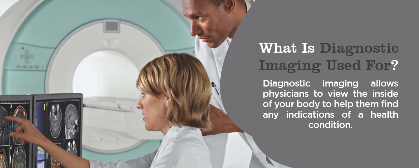
Diagnosis imaging empowers physicians to look inside your body and detect any indicators of a health issue. Various machines and methods are employed to generate detailed pictures of the body’s internal structures and activities. The selection of specific diagnosis imaging tests depends on the part of the body being examined and the patient’s presenting symptoms.
Many diagnosis imaging tests are non-invasive, straightforward, and painless. However, some may require patients to remain still within the imaging machine for extended periods, which can be slightly uncomfortable. Certain tests involve minimal exposure to radiation.
In other instances, diagnosis imaging may involve the insertion of a small camera attached to a flexible, thin tube, known as a “scope,” into the body. This scope is navigated through natural body openings or passageways to visualize the interior of specific organs, such as the lungs, heart, or colon. These procedures may necessitate anesthesia to ensure patient comfort and cooperation.
Types of Diagnosis Images Explained
There are numerous types of diagnosis imaging techniques, each with its unique capabilities and applications. Here are some of the most common and important types:
1. MRI (Magnetic Resonance Imaging) Scans
MRI scans are a powerful diagnosis imaging tool that utilizes strong magnetic fields and radio waves to create detailed images of the body’s organs and tissues without using radiation. Different types of MRI machines cater to varying patient needs and preferences:
- True open MRI: Designed to be open on all sides, true open MRI machines significantly reduce feelings of claustrophobia, making the experience more comfortable for patients who are anxious in enclosed spaces.
- Closed MRI: Traditional tube-like MRI machines, also known as closed MRIs, require the patient to lie down and be moved into the center of the machine for imaging.
- 3T MRI: Representing the cutting edge of MRI technology, 3T MRI machines utilize a stronger magnetic field (3 Tesla) to produce exceptionally detailed and high-resolution diagnosis images in a shorter amount of time. This advanced technology aids radiologists in distinguishing between benign and severe medical conditions more effectively. Like traditional MRIs, 3T MRIs are closed designs.
- Wide bore MRI: Often referred to as “open MRIs,” wide bore MRI machines resemble closed MRIs but feature a wider opening, offering a middle ground for patients who experience mild claustrophobia.
Doctors may recommend an MRI scan for a wide range of diagnostic purposes. MRIs provide exceptionally detailed views of internal structures, making them invaluable for examining:
- Brain and spinal cord abnormalities
- Cysts, tumors, and other irregularities throughout the body
- Joint injuries and abnormalities
- Breast tissue for cancer screening
- The female pelvic region to identify conditions like fibroids and endometriosis
- Suspected uterine anomalies
- Liver and abdominal diseases
- And many other conditions, as the applications of MRI technology continue to expand.
An MRI examination typically lasts between 30 to 60 minutes. In some cases, a contrast agent, a fluid injected into a vein to enhance image clarity, may be used, potentially extending the duration of the exam.
2. MRA (Magnetic Resonance Angiogram) Scans
MRA scans, or magnetic resonance angiograms, are a specialized type of MRI focused on producing highly detailed diagnosis images of blood vessels. MRAs employ radio wave energy pulses and magnetic fields to generate information about blood vessels that may not be readily obtainable through CT scans, ultrasounds, or X-rays.
MRA tests are commonly used to assess blood vessels in the legs, neck, brain, and kidneys, providing crucial information about blood flow and the condition of blood vessel walls. Doctors also utilize MRAs to detect aneurysms, blood clots, and calcium deposits within blood vessels. In certain situations, a contrast dye may be administered to further enhance the visualization of blood vessels in the MRA images.
MRA scans offer significant benefits for both patients and doctors in diagnosing health problems related to blood vessels:
- No radiation: Unlike CT scans and X-rays, MRAs do not involve radiation exposure.
- Non-invasive: MRAs are non-invasive procedures.
- Enhanced detection: MRAs can often detect critical information about blood vessels that may be missed by X-rays, ultrasounds, and CT scans.
- Blood flow assessment: MRAs are particularly effective in identifying issues within blood vessels that can lead to reduced blood flow.
MRA scans are valuable diagnostic tools primarily used to identify potential problems within blood vessels, including:
- Detecting aneurysms, calcium deposits, or blood clots in blood vessels
- Identifying narrowing of blood vessels
- Pinpointing abnormalities in brain blood vessels, such as congenital disabilities and inflammation
- Evaluating blood supply to vascular tumors in the brain
- Assessing patients who have experienced a stroke
- And many other vascular conditions.
3. CT (Computed Tomography) Scans
CT scans, sometimes referred to as “CAT scans,” are a diagnosis imaging technique that combines multiple X-ray images taken from various angles. Sophisticated computer software then processes these images to create cross-sectional views, or “slices,” of soft tissues and blood vessels within the body. CT scans provide a more comprehensive and detailed picture than standard X-rays. They are frequently employed in emergency situations to rapidly assess individuals with internal injuries resulting from trauma.
CT scans are versatile and can be used to evaluate the chest, neck, abdomen, brain, and spine. They produce clear diagnosis images of both soft and hard tissues, enabling doctors to make rapid medical decisions when necessary. This speed and clarity make CT scans a common procedure in hospitals and imaging centers. They assist physicians in identifying injuries and diseases that previously could only be discovered through surgery or autopsy. While CT scans utilize low doses of radiation, they are considered relatively safe and non-invasive.
CT scans are valuable in a wide array of medical scenarios where diagnosis imaging is required. They can detect subtle abnormalities in soft tissues, such as the brain and other organs. Doctors also use CT scans when patients present with symptoms like pain or dizziness. They are also useful in evaluating the extent of certain conditions, such as cancer. Depending on the specific area of the body being scanned, CT scans have diverse applications:
- Brain or head CT scans: To examine the skull and check for masses, bleeds, stroke, and other abnormalities.
- Chest CT scans: To provide further insights into abnormalities identified on standard chest X-rays.
- Neck CT scans: To investigate lumps and look for enlarged lymph nodes or glands.
- Spine CT scans: To detect spinal problems, including herniated discs, spinal canal narrowing, and fractures.
- Sinus CT scans: To diagnose sinus disease and identify obstructions.
- Pelvic or abdominal CT scans: To examine organs in these regions and diagnose unexplained abdominal pain.
4. Ultrasound
Ultrasound imaging, also known as sonography, is a safe and versatile diagnosis imaging method that generates real-time images of the body’s interior. Unlike X-rays and CT scans, ultrasound does not use radiation; instead, it utilizes high-frequency sound waves. This makes ultrasound a safe imaging option during pregnancy. Ultrasound images display the structure and movement of internal organs, as well as blood flow through vessels, in real-time.
During an ultrasound, a sonographer uses a handheld device called a transducer, which is moved over the skin, or sometimes placed internally. The transducer emits sound waves that travel through fluids and soft tissues. When these sound waves encounter denser surfaces, they echo or bounce back, creating the diagnosis images. Denser objects generate stronger echoes.
Ultrasound is a valuable tool for diagnosing a wide range of health conditions and assists physicians in developing effective treatment plans. If a patient presents with symptoms like pain, swelling, or infection, a doctor may recommend an ultrasound to determine the underlying cause. Ultrasounds are also used to guide anesthesiologists during surgical procedures when they need to precisely position needles near nerves.
Ultrasounds are frequently used to evaluate problems related to obstetrics, urology, circulation, abdominal issues, newborn care, and even musculoskeletal conditions. Common body parts and systems examined using ultrasound include:
- Heart
- Joints
- Uterus
- Blood vessels
- Muscles
- Bladder
- Kidneys
- And many others.
5. X-rays
X-rays are among the most well-known and commonly used diagnosis imaging tests. Doctors utilize X-rays to visualize the inside of the body, particularly bones. X-ray equipment generates a high-energy beam that passes through soft tissues but is absorbed by dense tissues like bone. This differential absorption creates an image, allowing doctors to identify bone injuries, such as fractures.
6. Mammography
Mammograms are a specialized type of X-ray diagnosis image of the breasts. They are crucial for the early detection of breast cancer, identifying small lumps or tissue changes that may be too subtle to be felt during a physical exam. Mammography utilizes low-dose X-rays to screen for early signs of breast cancer.
Digital mammography enhances the radiologist’s ability to detect and diagnose cancerous nodules that older mammography systems might miss. Mammograms are considered the most effective method for detecting breast cancer in its early stages, sometimes years before it becomes palpable. Regular mammograms offer significant benefits for women, including:
- Early detection and life-saving potential: Mammograms detect breast cancer at its earliest, most treatable stages, significantly increasing survival rates.
- Reduced risk of breast cancer mortality: Regular mammograms have been shown to reduce the risk of dying from breast cancer by approximately 30%.
- Less aggressive treatment options: Early detection often allows for less invasive treatment options, potentially avoiding mastectomy and preserving the breasts.
While some women may find mammograms uncomfortable or even painful due to breast compression, the discomfort is typically brief, lasting only a few minutes. The life-saving potential of mammography makes any temporary discomfort worthwhile.
7. Bone Density Scans
Bone density scans are an indirect diagnosis imaging test used to determine if a patient has osteoporosis. Also known as bone mineral density testing, this procedure measures the amount of bone mineral content per square centimeter in the bones.
Osteoporosis is a condition characterized by weakened and fragile bones that are prone to fractures. Bone density scans utilize X-ray equipment to measure the calcium and other mineral content packed into a small segment of bone, typically in the hip, forearm, or spine. Higher bone mineral content indicates denser, stronger bones that are less likely to fracture. Lower bone mineral content suggests reduced bone density and an increased risk of osteoporosis and fractures.
Before the advent of bone density scans, osteoporosis could often only be diagnosed after a fracture occurred, indicating that the bones were already significantly weakened. While older women are at higher risk, osteoporosis can affect individuals of any age or sex.
Doctors may recommend a bone density scan for individuals with risk factors for osteoporosis, such as:
- Fragile bones with increased fracture susceptibility
- Height loss of 1.5 inches or more
- Reduced levels of sex hormones
- Use of certain medications, like steroids, that can interfere with bone rebuilding
- Requirement for anti-rejection medications following organ transplantation.
8. Arthrogram
When joint function is impaired, limiting mobility and making everyday tasks challenging, an arthrogram may be used. An arthrogram, or arthrography, is a type of diagnosis imaging specifically designed to diagnose joint problems that may not be detectable by other imaging methods. Arthrograms involve taking a series of images of a joint using X-ray, fluoroscopy, CT scans, or MRI.
Prior to the arthrogram imaging, a radiologist injects a contrast dye, often iodine-based, into the joint. Fluoroscopy, a real-time X-ray technique, is used to guide the precise placement of the injection. The contrast dye coats the joint structures, making them appear white on the diagnosis images and highlighting any abnormalities. This allows the doctor to thoroughly evaluate joint function and arrive at an accurate diagnosis.
9. Myelogram
When detailed diagnosis imaging of the spinal canal is required, particularly of the spinal cord, spinal tissue, and surrounding nerves, a myelogram is often ordered. A myelogram is a procedure in which contrast dye is injected into the spinal cord space while fluoroscopy is used to capture moving X-ray images. As the dye flows through the spinal canal, the doctor carefully examines the area for any abnormalities, including tumors, inflammation, and infection.
A CT scan typically follows a myelogram to provide even greater detail and definition of any potential issues identified. Combined with CT technology, myelograms offer doctors more comprehensive and detailed information than X-rays alone.
Conclusion: The Power of Diagnosis Images in Healthcare
Diagnosis images are indispensable tools in modern medicine, providing a window into the human body that was previously unimaginable. From non-invasive techniques like ultrasound and MRI to detailed X-rays and CT scans, these technologies empower healthcare professionals to diagnose and monitor a vast array of medical conditions effectively. By enabling earlier detection, more accurate diagnoses, and less invasive procedures, diagnosis images play a critical role in improving patient care and outcomes. If you have health concerns, discuss with your doctor whether diagnosis images could be beneficial for your situation.
Sources
- https://mifimaging.com/2017/04/17/what-is-diagnostic-imaging/
- https://www.rasmussen.edu/degrees/health-sciences/blog/types-of-diagnostic-imaging/
- https://www.floridamedicalclinic.com/blog/what-is-diagnostic-radiology/
- https://www.envrad.com/services/x-ray/
- https://www.envrad.com/services/mammography/
- https://www.envrad.com/services/mra-scans/
- https://www.envrad.com/services/mri-scans/
- https://www.envrad.com/services/ultrasound-sonogram/
- https://www.healthimages.com/services/ct-scans/
- https://www.healthimages.com/services/bone-density/
- https://www.healthimages.com/services/arthrograms/
- https://www.healthimages.com/services/myelogram/
- https://www.healthimages.com/locations/
- https://www.healthimages.com/about-us/why-choose-us/