Mastoiditis, while seemingly unrelated to automotive repair, shares crucial diagnostic principles applicable across disciplines. For professionals at xentrydiagnosis.store, understanding complex diagnostic pathways is paramount. This article, tailored for our expert audience, delves into mastoiditis, a condition requiring meticulous diagnosis, drawing parallels to the intricate diagnostic processes in automotive repair.
Understanding Mastoiditis: Etiology and Types
Mastoiditis is fundamentally an inflammatory condition affecting the mastoid air cells, a section of the temporal bone connected to the middle ear cavity. It’s predominantly observed in children due to their higher susceptibility to middle ear infections. Acute mastoiditis typically arises as a complication of acute otitis media. However, it’s crucial to recognize other less common forms:
- Incipient Mastoiditis: This involves isolated infection within the mastoid air cells, without direct extension from the middle ear.
- Acute Coalescent Mastoiditis: The most prevalent type, characterized by inflammation that erodes the bony partitions between mastoid air cells. This erosion can lead to abscess formation and spread to adjacent structures.
- Subacute Mastoiditis: Develops from persistent or recurrent middle ear infections inadequately treated, leading to chronic infection and bone erosion in the mastoid.
While the incidence of mastoiditis has decreased significantly with antibiotics, prompt and accurate diagnosis remains critical to prevent severe complications.
Epidemiology of Mastoiditis: Risk Factors and Prevalence
Mastoiditis can affect individuals of any age, but it predominantly occurs in children under two years old, with a median age of around 12 months. Historically, in the pre-antibiotic era, mastoiditis was a frequent complication of acute otitis media, occurring in up to 20% of cases and often leading to serious intracranial complications.
The introduction of antibiotics, mastoidectomy procedures, and pneumococcal conjugate vaccines (PCV-7) has dramatically reduced the incidence. Before antibiotics, 5-10% of children with acute otitis media developed coalescent mastoiditis. Post-antibiotics and PCV-7, this figure has dropped to approximately 0.002% of children with acute otitis media progressing to acute coalescent mastoiditis. This highlights the significant impact of preventive and therapeutic medical advancements on disease epidemiology.
Pathophysiology: How Mastoiditis Develops
The majority of mastoiditis cases are a direct consequence of untreated or inadequately treated acute otitis media. The ear is anatomically divided into the outer, middle, and inner ear. The middle ear, extending from the tympanic membrane to the cochlea, houses vital structures like the ossicles (malleus, incus, stapes) and the Eustachian tube. Critically, the middle ear cavity is continuous with the mastoid air cells lining within the temporal bone.
The Eustachian tube’s function is to drain fluid and regulate air pressure in the middle ear, connecting it to the oral cavity. When this tube becomes blocked due to inflammation or debris, it creates a conducive environment for bacterial proliferation. In acute mastoiditis, infection extends from the middle ear into the mastoid air cells. This can cause erosion of the bony septa separating these air cells, leading to coalescence and the formation of pus-filled cavities – characteristic of acute coalescent mastoiditis. The accumulated pus can then spread through direct extension, thrombophlebitis, or bony pathways, resulting in serious sequelae such as subperiosteal abscess, sigmoid sinus thrombosis, meningitis, and intracranial abscess.
Streptococcus pneumoniae is the most common bacterial pathogen implicated in mastoiditis. Other frequently involved pathogens include Group A beta-hemolytic streptococci, Staphylococcus aureus, Streptococcus pyogenes, and Haemophilus influenzae. Risk factors increasing susceptibility to mastoiditis include young age (under two years), immunocompromised conditions, recurrent acute otitis media, and incomplete pneumatization of the mastoid process.
Clinical Presentation: Recognizing Mastoiditis Symptoms
Diagnosis of mastoiditis relies heavily on recognizing its characteristic clinical presentation. In children, mastoiditis often manifests as irritability, unexplained fussiness, lethargy, fever, ear pulling, and ear pain, particularly in the postauricular region. Adult patients typically report severe ear pain, fever, and headache.
Physical examination findings are crucial. Key indicators include postauricular erythema (redness), tenderness to palpation, warmth, and fluctuance (a sign of fluid collection) behind the ear. Protrusion of the auricle (ear lobe) is also a significant sign. Otoscopic examination frequently reveals bulging of the posterosuperior wall of the external auditory canal and a bulging tympanic membrane, often with pus visible behind it. In some cases, the tympanic membrane may have ruptured, with purulent drainage.
It’s important to note:
- A normal tympanic membrane, while less common, does not always exclude acute mastoiditis.
- While a history of recent or concurrent otitis media is typical, mastoiditis can develop rapidly, even during the initial stages of acute otitis media.
Diagnostic Evaluation: Confirming Mastoiditis
Mastoiditis diagnosis is primarily clinical, based on history and physical examination. However, laboratory and imaging studies play a vital role in confirming the diagnosis, assessing severity, and identifying potential complications. These are analogous to advanced diagnostic tools used in automotive repair, such as OBD-II scanners and pressure testing equipment, which complement visual inspection.
Initial laboratory investigations often include:
- Complete Blood Count (CBC) with differential: Typically shows leukocytosis (elevated white blood cell count) with a left shift, indicating bacterial infection.
- Erythrocyte Sedimentation Rate (ESR) and C-reactive protein (CRP): These inflammatory markers are usually elevated, supporting the presence of an inflammatory process.
Radiologic Evaluation: Computed Tomography (CT) scanning is the imaging modality of choice for evaluating mastoiditis. CT scans provide detailed visualization of the mastoid bone and surrounding structures, revealing:
- Fluid and/or mucosal thickening in the middle ear and mastoid air cells, indicating infection and inflammation.
- Loss of definition of the bony septae within the mastoid air cells, a hallmark of coalescent mastoiditis.
- Destruction or irregularity of the mastoid cortex (outer layer of the mastoid bone).
- Periosteal thickening, disruption, or the presence of a subperiosteal abscess, indicating extra-mastoid spread of infection.
Image showing a CT scan of the head in axial plane demonstrating mastoiditis in the left mastoid bone.
This image illustrates the diagnostic power of CT scanning in visualizing mastoiditis, similar to how diagnostic imaging in automotive repair, like thermal imaging, can reveal hidden issues.
Mastoiditis Treatment and Management Strategies
Antibiotics are the cornerstone of mastoiditis treatment. However, antibiotics alone are associated with a complication rate of around 8.5%. Depending on the severity and presence of complications, additional interventions may be necessary, including myringotomy (surgical incision of the tympanic membrane), tympanostomy tube placement, and mastoidectomy (surgical removal of infected mastoid air cells).
Hospital admission is typically required for patients with acute mastoiditis. Uncomplicated cases, in patients without significant underlying medical conditions, may be managed on an outpatient basis with daily intravenous (IV) ceftriaxone, demonstrating low complication rates. “Uncomplicated” mastoiditis is defined by minimal physical examination findings and no significant past medical history.
Factors that complicate mastoiditis cases include:
- Large postauricular swelling or abscess.
- Radiographic evidence of bone erosion on CT scan.
- High fever.
- Neurological signs suggesting intracranial involvement.
Inpatient management of uncomplicated cases usually involves:
- IV antibiotics: Vancomycin is often the initial antibiotic of choice to cover common pathogens like Streptococcus pneumoniae.
- High-dose IV steroids: To reduce inflammation.
- Myringotomy with tympanostomy tube placement: To drain middle ear fluid and pus.
Close monitoring with serial physical examinations is crucial, as a patient’s condition can deteriorate rapidly. If there is no clinical improvement within 48 hours of initiating IV antibiotics, mastoidectomy is generally indicated. For patients with a history of chronic ear infections, an anti-pseudomonal antibiotic is often added to vancomycin to broaden coverage. Studies have shown that Streptococcus pyogenes is associated with a higher rate of complications at initial presentation, while Staphylococcus aureus is linked to increased complications during hospitalization.
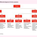
Differential Diagnosis: Distinguishing Mastoiditis from Mimics
Accurate diagnosis requires considering conditions that can mimic mastoiditis. Common differential diagnoses include:
- Cellulitis: Skin infection that can cause redness and swelling, but typically lacks the ear-specific findings of mastoiditis.
- Otitis externa: Infection of the external ear canal; while ear pain is present, postauricular signs are less prominent.
- Lymphadenopathy: Swollen lymph nodes behind the ear, which can be tender but lack the inflammatory signs specific to mastoiditis.
- Trauma: History of injury to the mastoid area.
- Tumors: Rare, but tumors like rhabdomyosarcoma, Ewing sarcoma, and myofibroblastic tumors can sometimes present similarly to mastoiditis. Tumors are more likely to be bilateral, involve cranial nerves, and less likely to present with fever.
Differentiating these conditions is essential to avoid misdiagnosis and treatment delays, especially in children, who are susceptible to both mastoiditis and certain types of tumors.
Prognosis and Potential Complications
The prognosis for uncomplicated acute mastoiditis is generally good with prompt and appropriate treatment. Most patients recover fully with conservative measures like antibiotics, steroids, and myringotomy, without needing mastoidectomy. However, research comparing conservative versus invasive treatment approaches is still ongoing.
Close monitoring is critical, especially in the first 48 hours of treatment. If a patient’s condition does not improve or worsens, mastoidectomy becomes necessary. Untreated or complicated mastoiditis can lead to severe consequences. Complications are categorized as extracranial and intracranial:
Extracranial Complications:
- Subperiosteal abscess: Abscess formation outside the mastoid bone, near the skull’s surface.
- Facial nerve palsy: Compression of the facial nerve leading to weakness or paralysis of facial muscles.
- Labyrinthitis: Infection spreading to the inner ear, causing tinnitus, vertigo, and hearing loss.
- Petrous apicitis: Osteomyelitis of the petrous part of the temporal bone, potentially causing Gradenigo syndrome (otorrhea, retro-orbital pain, ipsilateral abducens nerve palsy).
- Bezold abscess: Abscess extending into the sternocleidomastoid muscle in the neck.
Intracranial Complications (6-23% of cases):
- Temporal lobe or cerebellar abscess: Brain abscess formation.
- Epidural or subdural abscess: Abscess between the skull and brain coverings.
- Venous sinus thrombosis: Blood clot in the venous sinuses of the brain, the least common but most severe intracranial complication.
Patients with intracranial complications often present with neurological signs such as seizures, nuchal rigidity, severe headaches, and altered mental status, necessitating urgent and aggressive intervention.
Interdisciplinary Consultation and Patient Education
Effective mastoiditis management often involves collaboration between various medical specialties. Emergency medicine, family medicine, and otolaryngology (ENT) providers are frequently the first point of contact for patients with ear pain. While emergency medicine and family medicine practitioners manage routine otitis media, recognizing mastoiditis is crucial for timely referral.
Patients exhibiting signs and symptoms of acute mastoiditis require immediate evaluation in an emergency department with ENT consultation. These patients typically require hospital admission for IV antibiotics and possible surgical interventions. Patients with recurrent or chronic otitis media, without acute mastoiditis symptoms, can be referred to an ENT specialist on an outpatient basis for further evaluation and preventive strategies.
Deterrence and Patient Education:
Vaccination, particularly pneumococcal vaccination, is the most effective preventive measure against mastoiditis. Vaccination reduces the risk of pneumococcal infections, a leading cause of otitis media and subsequent mastoiditis. Early and appropriate treatment of acute otitis media is also crucial to prevent progression to mastoiditis. Patient education should emphasize the importance of vaccination and seeking prompt medical attention for ear infections, especially in children.
Key Clinical Pearls and Emerging Diagnostic Tools
While CT scanning remains the gold standard for mastoiditis diagnosis, research is exploring adjunctive diagnostic tools. One promising area is the use of ultrasound to detect complications of mastoiditis, particularly subperiosteal abscesses. A study involving a small patient cohort demonstrated ultrasound’s ability to identify complications in a significant number of cases. Although further research is needed, ultrasound could potentially serve as a valuable screening tool, especially in pediatric patients, to reduce unnecessary CT scan radiation exposure and healthcare costs.
Enhancing Healthcare Team Outcomes through Collaborative Management
Effective management of mastoiditis, from initial diagnosis to treatment and follow-up, requires a collaborative approach among healthcare professionals. This includes accurate initial assessment, timely referral to specialists when needed, and coordinated inpatient and outpatient care. Continued research into optimal treatment strategies, including outpatient management of uncomplicated mastoiditis, is essential to improve patient outcomes, reduce hospitalizations, and enhance healthcare efficiency. Just as in automotive repair, where a team of specialists might be needed for complex diagnostics and repairs, a multidisciplinary approach in medicine ensures the best possible outcomes for patients with conditions like mastoiditis.
References
- Cassano P, Ciprandi G, Passali D. Acute mastoiditis in children. Acta Biomed. 2020 Feb 17;91(1-S):54-59. [PMC free article: PMC7947742] [PubMed: 32073562]
- Schilder AG, Chonmaitree T, Cripps AW, Rosenfeld RM, Casselbrant ML, Haggard MP, Venekamp RP. Otitis media. Nat Rev Dis Primers. 2016 Sep 08;2(1):16063. [PMC free article: PMC7097351] [PubMed: 27604644]
- Atkinson H, Wallis S, Coatesworth AP. Acute otitis media. Postgrad Med. 2015 May;127(4):386-90. [PubMed: 25913598]
- Mansour T, Yehudai N, Tobia A, Shihada R, Brodsky A, Khnifies R, Barzilai R, Srugo I, Luntz M. Acute mastoiditis: 20 years of experience with a uniform management protocol. Int J Pediatr Otorhinolaryngol. 2019 Oct;125:187-191. [PubMed: 31369930]
- Hogan CJ, Tadi P. StatPearls [Internet]. StatPearls Publishing; Treasure Island (FL): Oct 31, 2022. Ear Examination. [PubMed: 32310474]
- Siddiq S, Grainger J. The diagnosis and management of acute otitis media: American Academy of Pediatrics Guidelines 2013. Arch Dis Child Educ Pract Ed. 2015 Aug;100(4):193-7. [PubMed: 25395494]
- Alkhateeb A, Morin F, Aziz H, Manogaran M, Guertin W, Duval M. Outpatient management of pediatric acute mastoiditis. Int J Pediatr Otorhinolaryngol. 2017 Nov;102:98-102. [PubMed: 29106885]
- Do TN, Linabery AM, Patterson RJ, Tu A. Cranial Rhabdomyosarcoma Masquerading as Infectious Mastoiditis: Case Report and Literature Review. Pediatr Neurosurg. 2018;53(5):317-321. [PubMed: 30145587]
- Egan G, Pierro J, Madhusoodhan PP, Ilyas G, Cohen B, Bhatla T. Primary Ewing Sarcoma of the Mastoid: A Novel Case Mimicking Acute Mastoiditis. J Pediatr Hematol Oncol. 2018 Mar;40(2):148-151. [PubMed: 29176463]
- Rodgers B, Bhalla V, Zhang D, El Atrouni W, Wang F, Sundararajan J, Lin J. Bilateral inflammatory myofibroblastic tumor mastoiditis. Head Neck. 2015 Nov;37(11):E142-5. [PubMed: 25546323]
- Loh R, Phua M, Shaw CL. Management of paediatric acute mastoiditis: systematic review. J Laryngol Otol. 2018 Feb;132(2):96-104. [PubMed: 28879826]
- Mantsopoulos K, Wurm J, Iro H, Zenk J. Role of ultrasonography in the detection of a subperiosteal abscess secondary to mastoiditis in pediatric patients. Ultrasound Med Biol. 2015 Jun;41(6):1612-5. [PubMed: 25796413]
- Mustafa A, Toçi B, Thaçi H, Gjikolli B, Baftiu N. Acute Mastoiditis Complicated with Concomitant Bezold’s Abscess and Lateral Sinus Thrombosis. Case Rep Otolaryngol. 2018;2018:8702532. [PMC free article: PMC5884287] [PubMed: 29755803]
- Tawfik KO, Ishman SL, Tabangin ME, Altaye M, Meinzen-Derr J, Choo DI. Pediatric acute mastoiditis in the era of pneumococcal vaccination. Laryngoscope. 2018 Jun;128(6):1480-1485. [PubMed: 29105776]
- Dagan R, Pelton S, Bakaletz L, Cohen R. Prevention of early episodes of otitis media by pneumococcal vaccines might reduce progression to complex disease. Lancet Infect Dis. 2016 Apr;16(4):480-92. [PubMed: 27036355]