Chronic diarrhea, defined as diarrhea lasting for more than four weeks, is a prevalent condition affecting approximately 5% of the global population, irrespective of age, gender, ethnicity, or socioeconomic background. This condition significantly impacts patients’ well-being and daily life, posing substantial economic burdens on healthcare systems due to diagnostic and management complexities. The range of potential causes for chronic diarrhea is extensive, encompassing infections, endocrine disorders, malabsorption syndromes, and gut-brain interaction disorders. The symptomatic overlap among these conditions complicates accurate diagnosis, potentially leading to diagnostic delays or errors. This review aims to provide a detailed overview of the differential diagnosis of chronic diarrhea, with a particular emphasis on differentiating irritable bowel syndrome with diarrhea (IBS-D) and exocrine pancreatic insufficiency (EPI)—two conditions with similar presentations but distinct etiologies and management strategies. We will outline a structured, four-step diagnostic approach and present a practical algorithm to aid clinicians in effectively distinguishing IBS-D from EPI and other causes of chronic diarrhea. Our goal is to enhance diagnostic precision, ultimately improving patient outcomes, quality of life, and reducing healthcare costs.
Diarrhea, when persistent, becomes a significant health concern. Global surveys highlight the widespread nature of functional diarrhea, with nearly 5% of individuals reporting symptoms. The impact is substantial, affecting daily activities, diminishing health-related quality of life, and increasing the demand for healthcare resources. In the United States alone, gastrointestinal issues, with diarrhea as a leading symptom, account for millions of ambulatory care visits annually, demonstrating the considerable clinical and economic burden.
The landscape of chronic diarrhea’s differential diagnosis is broad and diverse. It includes infectious agents (bacterial, parasitic, viral), endocrine disorders (hyperthyroidism, diabetes), maldigestive and malabsorptive conditions (celiac disease, lactose intolerance, exocrine pancreatic insufficiency [EPI]), gut-brain interaction disorders (irritable bowel syndrome [IBS]), inflammatory conditions (Crohn’s disease, ulcerative colitis), medication side effects (laxatives), and toxic ingestions (alcohol abuse). The similarity in symptoms across these diverse conditions often makes definitive diagnosis challenging. This diagnostic ambiguity can lead to delayed or incorrect diagnoses, prolonging patient discomfort and potentially worsening outcomes. Therefore, a systematic and efficient approach to diagnosis is crucial.
This article offers a strategic overview for differentiating and accurately diagnosing the various causes of diarrhea encountered in clinical practice. We focus on IBS-D and EPI, two conditions that frequently require careful distinction. By elucidating the key differences between IBS-D, EPI, and other conditions presenting with similar symptoms, we aim to facilitate earlier and more accurate diagnoses, guide appropriate treatment strategies, improve patient quality of life, and optimize healthcare resource utilization.
IRRITABLE BOWEL SYNDROME (IBS)
Irritable bowel syndrome stands as the most common cause of diarrhea in developed countries, with prevalence rates ranging from 4% to 9% in the United States, based on Rome III/IV criteria. While IBS can manifest at any age, it is most frequently diagnosed in women between 20 and 40 years old. Women are about twice as likely as men to receive an IBS diagnosis (14% versus 8.9%).
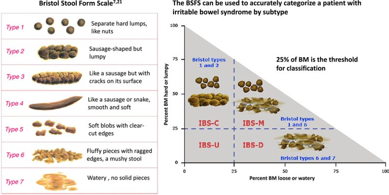
The Rome IV criteria define IBS based on recurrent abdominal pain occurring at least once a week, associated with changes in bowel habits related to defecation and/or alterations in stool form or frequency. These symptoms must also negatively impact the patient’s quality of life and daily functioning. While the Rome IV clinical criteria do not specify a symptom duration, clinicians must ensure other potential diagnoses are thoroughly considered and excluded. The research-oriented Rome IV criteria are more stringent, requiring symptoms to be present for the last three months, with symptom onset at least six months prior to diagnosis (Table 1). Commonly reported symptoms in IBS-D, though not part of the core definition, include abdominal bloating and distension, fecal urgency, a sensation of incomplete evacuation, and mucus in stools. Stool consistency is typically watery, classified as Bristol Stool Form Scale 6-7 (Figure 1), and bowel movements usually occur during waking hours. Stress is a recognized exacerbating factor. Crucially, certain “red flag” symptoms necessitate further investigation to rule out organic diseases beyond IBS. These alarm features include symptom onset after age 50, unintentional weight loss, sudden unexplained changes in symptoms, recurrent rectal bleeding or anemia, and a family history of inflammatory bowel disease, celiac disease, or colorectal cancer (Figures 2 and 3).
TABLE 1. Rome IV Diagnostic Criteria for Irritable Bowel Syndrome
| Clinical Diagnostic Criteria | Research Diagnostic Criteria (Clinical Trials, Epidemiological Studies) |
|---|---|
| Recurrent abdominal pain on average at least 1 day/week, associated with two or more of the following*: Related to defecation Associated with a change in frequency of stool Associated with a change in form (appearance) of stool Bothersome symptoms: Interfere with daily activities Require attention Cause worry or interfere with the quality of life | Recurrent abdominal pain on average at least 1 day/week in the last 3 months, associated with two or more of the following†: Related to defecation Associated with a change in frequency of stool * Associated with a change in the form (appearance) of stool |
*For the last 8 weeks.
†For the last 3 months with symptom onset at least 6 months before diagnosis.
FIGURE 1. Bristol Stool Form Scale and IBS Subtypes
Image alt text: Bristol Stool Chart illustrating stool types 1 through 7, with types 6 and 7 representing diarrhea, associated with IBS-D. Chart also shows IBS subtypes: IBS-C (constipation), IBS-M (mixed), and IBS-U (unspecified).
FIGURE 2. General Sequence for Differential Diagnosis of Chronic Diarrhea
Image alt text: Flowchart outlining a 4-step diagnostic approach for chronic diarrhea, starting with history and physical exam, progressing to risk factor identification, alarm feature assessment, and initial lab workup, including tests for celiac disease, IBD, and EPI.
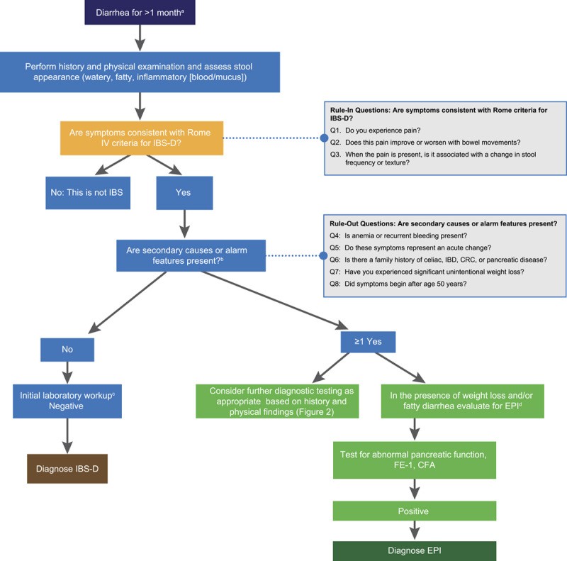
FIGURE 3. A General Strategy for the Differential Diagnosis of Patients with Chronic Diarrhea: EPI or IBS-D?
Image alt text: Algorithm specifically designed to differentiate between EPI and IBS-D in patients with chronic diarrhea, incorporating Rome IV criteria, alarm features, and diagnostic tests like fecal elastase-1 and fecal fat measurement.
While chronic abdominal pain is a defining characteristic of IBS, distinguishing it from functional diarrhea can be challenging due to symptom overlap and diagnostic fluidity. Patients with IBS, particularly those experiencing frequent pain, often report higher levels of psychological distress and co-existing somatic conditions compared to those with functional diarrhea. Early and accurate diagnosis is crucial for effective management of these overlapping conditions.
Currently, there is no universally accepted biomarker for IBS. Extensive diagnostic testing to exclude organic causes is generally discouraged due to its high cost, inefficiency, and low diagnostic yield. Accurate IBS diagnosis can often be achieved through a detailed patient history alone. Studies have shown that Rome criteria, in the absence of alarm symptoms, exhibit high specificity and positive predictive values for IBS diagnosis. Therefore, current guidelines emphasize a positive diagnostic strategy, minimizing unnecessary investigations. The American College of Gastroenterology (ACG) and the American Gastroenterological Association (AGA) recommend against routine colonoscopy for IBS diagnosis, except in patients over 45 years old (for age-appropriate screening) or those presenting with alarm signs suggestive of more serious conditions.
Recommended initial diagnostic tests are limited and targeted. These include serologic testing for celiac disease (serum IgA and tissue transglutaminase IgA), fecal calprotectin (or lactoferrin) and C-reactive protein (CRP) to rule out inflammatory bowel disease in patients without alarm signs, and stool antigen testing for Giardia in individuals with risk factors such as travel to endemic areas, exposure to untreated water, or daycare attendance.
Irritable Bowel Syndrome Subtypes
IBS is further categorized into four subtypes based on predominant stool patterns: IBS-D (diarrhea-predominant), IBS-C (constipation-predominant), IBS-M (mixed), and IBS-U (unclassified). Among these, IBS-D is the most prevalent, affecting up to 40% of adults diagnosed with IBS. Globally, IBS-D affects approximately 1.2% of the population, with a slight female predominance, mirroring the overall IBS prevalence.
IBS-D is characterized by Bristol Stool Form Scale type 6 or 7 stools (loose, mushy, watery) in more than 25% of bowel movements, and type 1 or 2 stools (hard, lumpy, pellet-like) in less than 25% of bowel movements (Figure 1). Rome IV criteria emphasize assessing stool texture on days with abdominal pain to enhance the accuracy of IBS subtype classification.
The symptomatic overlap between IBS-D and other conditions, such as EPI, celiac disease, small intestinal bacterial overgrowth (SIBO), disaccharidase deficiencies, Crohn’s disease, ulcerative colitis, and infections, makes accurate diagnosis challenging. However, initial stool characterization as watery (suggesting IBS), fatty/greasy (suggesting EPI), or inflammatory (suggesting inflammatory bowel disease) can help narrow the differential diagnosis (Table 2).
TABLE 2. Narrowing the Diagnosis According to Stool Characteristics
| Watery | Fatty/Greasy | Inflammatory |
|---|---|---|
| Osmotic Carbohydrate malabsorption Celiac disease Osmotic laxatives Secretory Bile acid malabsorption Microscopic colitis Endocrinopathies (e.g., diabetes, hyperthyroidism) Medications (e.g., metformin) Functional Functional diarrhea * Irritable bowel syndrome | Malabsorption or Maldigestion Celiac disease Small intestinal bacterial overgrowth Giardiasis Whipple disease Inadequate luminal bile acid concentration Exocrine pancreatic insufficiency | Inflammatory Bowel Disease Segmental colitis associated with diverticulosis (SCAD) Infectious disease Clostridium difficile Invasive bacterial infections Invasive parasitic infections Ischemic colitis Radiation colitis Lymphoma |
EXOCRINE PANCREATIC INSUFFICIENCY (EPI)
Exocrine pancreatic insufficiency (EPI) is most commonly associated with pancreatic diseases but can also arise from extrapancreatic conditions. Approximately 80% of children with cystic fibrosis develop EPI within the first two years of life. EPI prevalence varies in chronic pancreatitis, affecting 10% to 30% in mild cases and up to 85% in severe cases. Among pancreatic cancer patients, about 72% develop EPI, with higher frequency (3.36 times) when tumors are located in the pancreatic head. In pancreatic diseases, EPI results from reduced pancreatic enzyme and bicarbonate secretion due to parenchymal damage or pancreatic duct obstruction.
EPI is characterized by a deficiency in the quantity or activity of pancreatic enzymes in the small intestine, leading to impaired food digestion. Fat maldigestion is a primary clinical feature of EPI. Diarrhea in EPI occurs when the digestive capacity is overwhelmed by the quantity and quality of ingested food. Symptom prevalence in EPI is highly variable, influenced by dietary habits and restrictions. Clinical studies in confirmed EPI patients report steatorrhea (fatty/oily stools) in 23% to 70% of chronic pancreatitis cases, 46% in preoperative pancreatic cancer, and 15% in cystic fibrosis. A detailed patient history is essential, as dietary modifications, such as low-fat diets adopted to manage symptoms, can mask EPI and complicate diagnosis. Patients on low-fat diets might be asymptomatic, obscuring the underlying condition.
EPI typically presents with symptoms of malabsorption syndrome, including diarrhea, abdominal distension and cramps, flatulence, and weight loss, as well as nutritional deficiencies (fat-soluble vitamins, micronutrients, and proteins). Symptom severity varies based on the underlying cause, enzyme deficiency, and dietary fat intake. Common patient reports include foul-smelling, fatty, loose stools, flatulence, and weight loss. Long-term consequences of EPI can include sarcopenia, osteoporosis, low-trauma fractures, metabolic bone disease (especially in chronic pancreatitis), increased infection risk, and cardiovascular disease.
EPI should be considered in patients with chronic diarrhea and a history of pancreatic disease (acute, relapsing, or chronic pancreatitis; cystic fibrosis; pancreatic cancer; acute necrotizing pancreatitis; and type 1, 2, or 3c diabetes mellitus), risk factors for pancreatic disease (alcohol abuse and/or smoking), family history of pancreatic diseases (especially chronic pancreatitis or pancreatic cancer), or prior pancreatic or gastric surgery. EPI testing is warranted in patients with chronic diarrhea and high clinical suspicion, even without a prior pancreatic disease diagnosis.
Diagnosing EPI definitively is often challenging due to the lack of highly accurate tests, yet it is crucial to prevent complications. Diagnosis typically relies on a combination of symptoms, nutritional markers, and non-invasive pancreatic function tests, such as the coefficient of fat absorption (CFA) and fecal elastase-1 (FE-1). While other pancreatic function tests exist, they are often invasive (e.g., endoscopic pancreatic function test [ePFT]) or not readily accessible (e.g., 13C-labeled breath tests) in routine clinical practice. The direct secretin-cholecystokinin (CCK) test, though highly sensitive for detecting reduced pancreatic secretion, is invasive, expensive, and cumbersome, limiting its clinical utility. ePFT with intravenous secretin has been developed as an alternative but faces similar limitations. These more complex tests are primarily used for functional diagnosis of chronic pancreatitis in cases with inconclusive imaging, not for routine EPI diagnosis.
The CFA test is considered the gold standard for EPI diagnosis. However, it requires patients to adhere to a high-fat diet (100 g of fat/day for 5 days) and collect total fecal output for 3 days (days 3-5), making it cumbersome, unpleasant, and poorly compliant in clinical practice. 13C-labeled breath tests are accurate and standardized alternatives to CFA for EPI diagnosis but are not yet widely available. Fecal elastase-1 (FE-1) testing measures the concentration of this pancreatic-specific enzyme in stool, reflecting pancreatic secretion. FE-1 testing is simple, widely available, and thus the most frequently used pancreatic function test. However, the optimal cutoff and accuracy of FE-1 for EPI diagnosis, using CFA as the gold standard, vary across studies. Sensitivity ranges from 68% to 94%, and specificity from 48% to 82%, depending on the cutoff value (84 to 200 µg/g) used in different studies.
In patients with chronic diarrhea and high EPI probability (e.g., pancreatic head cancer, advanced chronic calcifying pancreatitis, pancreaticoduodenectomy, gastrectomy), pancreatic function tests may be less critical for diagnosis. Conversely, in patients with chronic diarrhea and low EPI probability (no prior pancreatic disease, no risk factors, no weight loss or nutritional deficiencies), normal FE-1 levels can effectively exclude EPI. Low FE-1 levels may suggest EPI, prompting further pancreatic evaluation, but false-positive FE-1 results can occur, especially in watery diarrhea.
EPI is one of several organic gastrointestinal diseases that can mimic IBS. Studies have shown that EPI, defined by low FE-1, is present in 5% to 6% of patients meeting Rome criteria for IBS-D and in 4.6% of patients with unexplained abdominal pain, diarrhea, or IBS-D. However, due to the possibility of false-positive FE-1 results in watery diarrhea, low FE-1 levels do not definitively rule out IBS-D.
DIFFERENTIAL DIAGNOSIS OF CHRONIC DIARRHEA: A STEP-WISE APPROACH
Early and accurate diagnosis is paramount in managing chronic diarrhea. Patients may present with a range of symptoms indicative of various disorders, including IBS-D, EPI, celiac disease, SIBO, inflammatory bowel disease, and infections (e.g., giardiasis). These conditions share overlapping symptoms like diarrhea, abdominal pain, bloating, and flatulence. To facilitate accurate diagnosis, we propose a four-step diagnostic process (Figure 2).
Step 1: Comprehensive History and Physical Examination
The diagnostic process should begin with a thorough patient history and physical examination. Patients with chronic diarrhea (lasting ≥4 weeks) should be questioned in detail about their symptoms and diarrhea history. While clinical diarrhea is defined as loose or watery stools ≥3 times in 24 hours, patient definitions vary (e.g., loose stools, increased frequency, urgency), highlighting the importance of a detailed history. Abnormal stool form is often more critical than frequency, as patients with functional constipation may also report “diarrhea” due to increased defecation attempts, while further questioning reveals straining, incomplete evacuation, and hard stools.
Initial assessment should categorize diarrhea as watery (suggestive of IBS, celiac disease, endocrinopathy, or laxative abuse), fatty/greasy (suggestive of malabsorption/maldigestion like celiac disease or EPI), or inflammatory (suggestive of infectious or inflammatory bowel disease). However, overlap exists (Table 2). Further history should include: diarrhea pattern (continuous, intermittent, meal-related – differentiating secretory from osmotic), onset, precipitating events, stool volume, presence of blood, mucus, or fat, nocturnal diarrhea, and fecal urgency or incontinence. Other gastrointestinal and extraintestinal symptoms, aggravating factors (diet, stress, medications), and alleviating factors should also be explored.
Step 2: Identification of Risk Factors, Iatrogenic Causes, and Pre-existing Conditions
Ruling out extrinsic causes of chronic diarrhea requires assessing recent travel (to regions with specific diarrhea-related pathogens like Giardia), prior gastrointestinal surgery (gallbladder removal, ileocecal resection, Roux-en-Y gastric bypass), radiation therapy, and diarrhea-inducing medications. Pre-existing mucosal (celiac disease), hepatic, pancreaticobiliary, neoplastic, or systemic (endocrine, vascular, immunologic) diseases can also increase diarrhea risk.
Step 3: Exclusion of Alarm Features (“Red Flags”)
Certain clinical features suggest more serious underlying pathology and must be excluded. These alarm features include: recent onset, especially in older individuals; nocturnal diarrhea; severe or worsening symptoms; unexplained weight loss; family history of gastrointestinal or systemic diseases like celiac disease, inflammatory bowel disease, or colorectal cancer; rectal bleeding; and unexplained iron deficiency anemia.
Step 4: Initial Laboratory Workup
History and physical examination guide subsequent diagnostic testing. In the presence of alarm signs, testing should target the most likely etiologies. For example, meal-related fatty-greasy diarrhea with weight loss and fat-soluble vitamin deficiencies warrants EPI investigation. FE-1 measurement is the most common initial test for exocrine pancreatic function; levels <200 µg/g suggest EPI. In patients without alarm features, presenting with symptoms consistent with functional diarrhea or IBS-D, current AGA and ACG guidelines recommend screening for celiac disease (anti-tissue transglutaminase IgA and total IgA), inflammatory bowel disease (fecal calprotectin or lactoferrin and CRP [ACG only]), and Giardia (in at-risk populations). The AGA also suggests considering bile acid diarrhea testing (48-hour fecal bile acid assay or serum fibroblast growth factor 19 level).
DIAGNOSTIC ALGORITHM FOR CHRONIC DIARRHEA: FOCUSING ON IBS-D AND EPI
Building on the four-step approach, we present a streamlined algorithm to aid clinicians in differentiating IBS-D and EPI from other diarrhea syndromes (Figure 3). This algorithm aims to minimize unnecessary testing and healthcare costs by guiding efficient and appropriate diagnoses, particularly for IBS-D. The algorithm begins with three “rule-in” questions aligned with Rome IV criteria for IBS: (1) Do you experience abdominal pain? (2) Does pain improve or worsen with defecation? (3) Is pain associated with changes in stool frequency or form? Positive answers to these questions, in the absence of alarm symptoms (Figure 3), suggest IBS-D with high accuracy (approximately 97%). In such cases, minimal testing is needed. However, if alarm symptoms are present (e.g., weight loss) and EPI is suspected, nutritional markers and FE-1 testing are indicated. Abnormal results necessitate pancreatic imaging. If pancreatic imaging is normal, a false-positive FE-1 result is likely, EPI can be excluded, and other causes of chronic diarrhea should be investigated.
DISCUSSION
Diarrhea is a common and complex condition with diverse causes and mechanisms. Differentiating these causes, especially when symptoms overlap, is challenging. This is particularly evident in IBS-D, where up to 75% of affected individuals in the United States remain undiagnosed. This is concerning because IBS is a prevalent cause of diarrhea, diagnosable with high accuracy using simple criteria and limited testing.
Exocrine pancreatic insufficiency (EPI) is a significant condition that often mimics IBS-D, leading to misdiagnosis. To address this, we have developed an algorithm specifically designed to distinguish between IBS-D and EPI (Figure 3) and differentiate them from other causes of chronic diarrhea. Importantly, this algorithm incorporates recommendations from recent ACG and AGA IBS guidelines, emphasizing a positive diagnostic strategy. This simple algorithm can assist clinicians in making timely and accurate diagnoses, reducing unnecessary testing, minimizing treatment delays, and improving patient health and quality of life.
ACKNOWLEDGMENTS
The authors acknowledge Moira A. Hudson, PhD, and Janet E. Matsuura, PhD, of ICON plc, for medical writing and editing assistance in the development of this manuscript, funded by AbbVie.
Footnotes
Funding for this manuscript was provided by AbbVie. AbbVie provided medical review, with content decisions made by the authors. No honoraria or payments were made for authorship.
D.M.B. has consulting, advisory, or speaking roles with Alnylam, Alfasigma, Anji, Ardelyx, Arena, Bayer, AbbVie, Mahana, Owlstone, Ironwood, Salix, Takeda, Redhill, QoL Medical, Gemelli Biotech, and Vibrant. He is a Board member of the International Foundation for Gastrointestinal Disorders (IFFGD) and has received grants from the IDP Foundation. J.E.D.-M. has received honoraria for lectures and advisory roles from AbbVie, Viatris, and Abbott Pharmaceuticals and research grants from AbbVie.
Contributor Information
Darren M. Brenner, Email: [email protected].
J. Enrique Domínguez-Muñoz, Email: [email protected].