Facial swelling presents a diagnostic challenge in emergency medicine, demanding a systematic approach to differentiate between benign conditions and life-threatening emergencies. As frontline providers, emergency clinicians must be adept at rapidly identifying and managing critical causes of facial edema to ensure optimal patient outcomes. This article provides an in-depth exploration of the differential diagnosis of facial edema, emphasizing critical conditions that require immediate recognition and intervention.
Facial edema, characterized by abnormal fluid accumulation in the facial tissues, can stem from a diverse range of etiologies. These range from localized infections to systemic diseases, each with varying degrees of severity and requiring distinct management strategies. The key to effective emergency management lies in a rapid, accurate differential diagnosis to promptly address potentially life-threatening conditions.
Categorizing the Differential Diagnosis of Facial Edema
To streamline the diagnostic process, facial edema can be categorized based on the anatomical location of swelling. This anatomical approach aids in narrowing the differential and guiding targeted investigations and interventions. While the spectrum of causes is extensive, certain diagnoses are critical to rule out due to their immediate life-threatening potential. These “can’t miss” diagnoses are highlighted below for emphasis.
- Orbital/Periorbital Edema: Swelling around the eyes and eyelids.
- Critical Diagnoses:
- Cavernous Sinus Thrombosis (CST)
- Myxedema Coma
- Orbital Cellulitis
- Critical Diagnoses:
- Subcutaneous Facial Edema: Swelling in the skin and tissues beneath the skin of the face.
- Facial Cellulitis
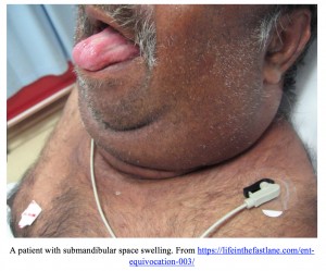
- Submandibular Space Edema: Swelling beneath the jaw.
- Critical Diagnoses:
- Ludwig’s Angina
- Salivary Gland Pathology
- Critical Diagnoses:
- Intra-oral Edema: Swelling inside the mouth.
- Critical Diagnoses:
- Angioedema
- Odontogenic Infection
- Critical Diagnoses:
- Miscellaneous Causes of Facial Edema:
- Trauma
- Superior Vena Cava Syndrome (SVCS)
- Malignancy
Immediate Life-Threatening Causes of Facial Edema: A Detailed Review
Prompt recognition of life-threatening causes of facial edema is paramount in the emergency setting. These conditions necessitate immediate intervention to prevent severe morbidity and mortality.
Angioedema: A Critical Cause of Facial Swelling
Angioedema stands out as a particularly critical “can’t miss” diagnosis in patients presenting with facial swelling. It is defined by rapid subcutaneous or submucosal swelling resulting from extravasation of fluid into interstitial tissues. The onset is typically rapid, often within minutes of exposure to a trigger. For emergency physicians, the crucial etiologies of angioedema to consider are:
- Anaphylaxis-related Angioedema: Triggered by an IgE-mediated allergic reaction.
- ACE Inhibitor-Induced Angioedema: A side effect of Angiotensin Converting Enzyme (ACE) inhibitors.
- Hereditary Angioedema (HAE): Caused by a deficiency in C1 inhibitor.
Anaphylactic angioedema is driven by an IgE-mediated immune response. This response activates mast cells, leading to the release of inflammatory mediators such as histamine, which dramatically increase vascular permeability, resulting in rapid edema formation [1]. In contrast, both ACE inhibitor-induced angioedema and hereditary angioedema involve the bradykinin pathway. ACE inhibitors block the degradation of bradykinin, while C1 inhibitor deficiency leads to increased bradykinin production. Both mechanisms result in an excess of bradykinin, a potent vasodilator that increases vascular permeability and causes angioedema [2].
Clinically, angioedema often presents with a rapid onset of swelling affecting the face, lips, tongue, larynx, and sometimes the bowel. Facial involvement frequently manifests as lip, tongue, and uvular swelling, particularly prominent in ACE inhibitor-induced cases [3]. Identifying the underlying etiology is crucial as it guides treatment strategies. A thorough medication history, specifically noting ACE inhibitor use, is essential.
Diagnosis is primarily clinical, based on the characteristic presentation of rapid swelling. Patients may exhibit varying degrees of facial angioedema, affecting the lips, tongue, uvula, or larynx. Anaphylaxis-induced angioedema may be accompanied by urticaria, skin flushing, bronchospasm, and pruritus [4].
While many angioedema cases are benign and self-limiting, airway compromise due to tongue, upper airway, or laryngeal swelling poses a significant life threat. Intubation should be considered in any patient showing signs of airway obstruction. Fiberoptic nasal intubation may be necessary in cases of severe tongue swelling. Given the potential for difficult intubation, preparation for surgical cricothyrotomy as a rescue airway is crucial.
Treatment of angioedema, beyond airway management, depends on the etiology. For anaphylaxis-related angioedema, immediate intramuscular epinephrine (0.3 mg for adults) is the cornerstone of treatment and may need to be repeated based on symptom severity. Glucocorticosteroids and antihistamines are adjunctive therapies, but epinephrine is the primary life-saving intervention.
For ACE inhibitor-induced angioedema, the long-term management involves discontinuing the offending ACE inhibitor. Acute treatment options are more complex and less definitively effective. Icatibant (a bradykinin receptor antagonist), C1 inhibitor concentrate (to reduce bradykinin production), and Fresh Frozen Plasma (FFP) have been considered, but their efficacy is debated [5-7]. Current evidence suggests that early administration of these therapies may be beneficial. In most cases, discontinuing the ACE inhibitor and supportive airway management remain the primary strategies. ACE inhibitor-induced angioedema typically resolves within 1-3 days [8].
Hereditary angioedema management, in addition to airway support, includes specific therapies such as C1 inhibitor concentrate, icatibant, and ecallantide (a kallikrein inhibitor). These therapies are significantly more effective in HAE compared to ACE inhibitor-induced angioedema.
Ludwig’s Angina: Submandibular Space Infection
Ludwig’s angina, a submandibular space infection, represents a rapidly progressing and potentially lethal condition. This infection of the floor of the mouth can spread aggressively, leading to significant airway compromise and systemic sepsis. While historically associated with high mortality rates (up to 50%), early diagnosis and aggressive antibiotic therapy have dramatically reduced mortality to 0-4% [9]. Odontogenic infections, particularly involving the second and third mandibular molars, are implicated in approximately two-thirds of Ludwig’s angina cases [10]. The infection can rapidly spread, causing substantial tongue and submandibular swelling, potentially extending to the epiglottis.
Diagnosis relies on clinical assessment, supported by imaging. Patients typically present with fever, chills, and oral pain. As the infection progresses, symptoms can include neck stiffness, difficulty speaking, drooling, and stridor. Examination findings include symmetric swelling and tenderness in the submandibular region, sometimes with palpable crepitus, and potential tongue protrusion. CT imaging is the preferred modality to evaluate for Ludwig’s angina, allowing for visualization of submandibular space infection and localization for potential surgical drainage [11, 12]. However, obtaining CT should not delay airway management in unstable patients.
The primary goals in managing Ludwig’s angina are securing the airway and initiating early antibiotic therapy. While not all cases necessitate immediate intubation, a low threshold for early airway intervention is crucial. Airway control is best achieved before signs of compromise (stridor, hypoxia) manifest. Nasal fiberoptic intubation is often favored if feasible, but video laryngoscopy can also be considered [13]. Anesthesia consultation should be sought if available for airway management assistance.
Once the airway is addressed, prompt antibiotic administration is critical. Antibiotic selection depends on the patient’s immune status. For immunocompetent patients, recommended regimens [14] include:
- Ampicillin-Sulbactam (3 grams IV every 6 hours)
- Penicillin G (2 to 4 million units IV every 4-6 hours) plus Metronidazole (500 mg IV every 6-8 hours)
- Clindamycin (600 mg IV every 6-8 hours)
In immunocompromised patients, broader coverage is necessary:
- Cefepime (2 g IV every 12 hours) plus Metronidazole (500 mg IV every 6-8 hours)
- Imipenem (500 mg IV every 6 hours)
- Meropenem (1 g IV every 8 hours)
- Piperacillin-Tazobactam (4.5 g IV every 6 hours)
Early consultation with otolaryngology (ENT) is essential. Surgical intervention (incision and drainage – I&D) may be required, or the ENT team may opt for observation with intravenous antibiotics. They can also provide backup for surgical cricothyrotomy if needed. Depending on the severity, ICU admission for airway monitoring may be necessary. While steroids have been suggested to reduce swelling and inflammation, potentially decreasing the need for surgical airway, their effectiveness remains unproven in randomized controlled trials [15].
Myxedema Coma: Decompensated Hypothyroidism
Myxedema coma, although not primarily presenting with facial swelling, is a critical consideration in the differential diagnosis of facial edema, especially in patients with a history of hypothyroidism and suggestive symptoms. This condition carries a high mortality rate even with appropriate treatment.
Myxedema coma represents severe hypothyroidism leading to altered mental status, hypothermia, and other hypothyroidism-related symptoms such as bradycardia, hypotension, and hypoglycemia [16]. It can be triggered by uncontrolled hypothyroidism or precipitating events like myocardial infarction, sepsis, or certain medications [17].
Facial swelling in myxedema coma is characterized by a diffuse “puffiness,” often with bilateral periorbital edema and lip and/or tongue swelling. This swelling is due to abnormal deposition of proteins and mucopolysaccharides in the tissues, resulting in non-pitting edema.
Diagnosis is primarily clinical, supported by laboratory findings. T4 levels are typically very low, while TSH levels may be high or low depending on the underlying cause [19]. However, in suspected myxedema coma, treatment should not be delayed for lab confirmation. Immediate treatment involves intravenous thyroid hormone supplementation. Adrenal insufficiency should also be considered and managed with hydrocortisone.
The mortality rate of myxedema coma remains high (around 30%) [20], emphasizing the need for a high index of suspicion in patients with facial swelling and other signs of hypothyroidism. Death often results from multi-organ failure.
Cavernous Sinus Thrombosis (CST)
Cavernous sinus thrombosis (CST), while rare, is a life-threatening condition to consider in patients with periorbital facial swelling. Untreated CST carries a high mortality [21]. Before antibiotics, mortality approached 100%, but with modern management, it is now less than 30%.
CST involves thrombus formation within the cavernous sinus, a dural venous sinus located centrally, lateral to the sphenoid sinus. The cavernous sinuses receive blood from facial veins, middle cerebral veins, and sphenoid veins, draining into the internal jugular veins. The absence of valves in these sinuses allows for retrograde spread of infection from the face or sinuses, commonly leading to CST, with Staphylococcus aureus being the most frequent causative organism.
Initial symptoms can be variable. Headache, often localized to the trigeminal nerve (CN V) distribution, is common. Fever, eye pain, and vision changes may also occur. As severity increases, patients can develop confusion, somnolence, and coma. Examination findings may include chemosis, ptosis, proptosis, and ophthalmoplegia due to cranial nerve involvement (CN III, IV, VI) in proximity to the cavernous sinus [22].
Non-contrast CT head scan, often the initial imaging, can be normal in up to 30% of cases [23]. Abnormal CT findings may include filling defects in the cavernous sinus and dilation of the superior ophthalmic vein.
While CT is readily available, MRI with venography (MRV) is the most accurate imaging modality, demonstrating reduced or absent flow in the affected cavernous sinus [24]. MRI/MRV sensitivity can reach 100% [25].
Immediate ED treatment includes empiric antibiotics. While S. aureus is common, CST can be polymicrobial, involving gram-positive, gram-negative, and anaerobic bacteria. Empiric antibiotic choices should include a penicillinase-resistant penicillin and a 3rd or 4th generation cephalosporin. Vancomycin should be added if MRSA is suspected. Anticoagulation for CST is controversial due to limited data. However, current evidence suggests heparin or low molecular weight heparin (LMWH) may be safe to prevent thrombus progression [26]. Corticosteroids may be considered as adjunctive therapy to reduce inflammation and edema, but antibiotics are the mainstay of treatment. ICU admission is necessary for close monitoring and further management due to the high morbidity and mortality associated with CST.
Orbital Cellulitis
Orbital cellulitis should be considered when facial swelling is primarily localized to the orbital/periorbital region. Untreated orbital cellulitis can lead to severe complications, including permanent vision loss, abscess formation, cavernous sinus thrombophlebitis, and death [27]. Orbital cellulitis is an infection involving the orbital tissues, distinct from periorbital cellulitis, which is limited to the eyelid. Differentiating these is critical due to the serious complications associated with orbital cellulitis.
Orbital cellulitis is more common in young children but can affect adults. Rhinosinusitis is the most frequent cause [28]. Other causes include recent eye surgery, trauma, and dental infections. Staphylococcus aureus and Streptococci are the most common causative organisms [29]. Less common causes include anaerobes, Pseudomonas aeruginosa, Haemophilus influenzae, and fungal infections (mucormycosis in diabetic ketoacidosis).
Patients present with eye pain, eyelid swelling, and double vision. Examination reveals pain with eye movements, proptosis, and ophthalmoplegia due to inflammation of extraocular muscles (EOM). Periorbital cellulitis, in contrast, does not typically present with pain on EOM or ophthalmoplegia [30].
Diagnosis combines clinical judgment and imaging. CT scan of the orbits is a reasonable first-line imaging modality, revealing EOM inflammation, fat stranding, and anterior globe displacement [31].
Empiric antibiotic therapy is the mainstay of treatment. Recommended regimens include:
- Vancomycin (15-20 mg/kg IV every 8-12 hours) AND
- Ceftriaxone (2 g IV every 24 hours) OR
- Cefotaxime (2 g IV every 24 hours) OR
- Ampicillin-Sulbactam (3 g IV every 6 hours) OR
- Piperacillin-Tazobactam (4.5 g IV every 6 hours)
While surgery is not always required, ophthalmology and otolaryngology consultation is essential for further management and close monitoring for infection progression.
Other Differential Diagnoses of Facial Edema
Beyond the life-threatening conditions, several other etiologies can cause facial edema and require consideration in the differential diagnosis.
Dental Infections
Dental infections are a frequent cause of facial swelling. Periodontal disease affects a significant portion of the adult population [32]. While localized odontogenic infections are rarely life-threatening with antibiotics, untreated infections can spread and lead to serious conditions like Ludwig’s angina. Dental infections arise from bacterial colonization on teeth, forming plaque. Infection spread depends on plaque location. Supragingival plaque leads to dental caries, pulpitis, and periapical abscesses [33]. Subgingival plaque causes gingivitis and periodontal infections, which can extend to deeper facial and neck spaces [34]. Odontogenic infections can also involve the jaw bone, causing osteomyelitis.
Dental caries, particularly in patients under 35, is a common cause of odontogenic infections, primarily caused by Streptococcus mutans [35]. Progression can lead to pulp exposure, pulpitis, and periapical abscesses. Periodontal infections are more common in patients over 35 [36]. While often slow-progressing, rapid periodontitis can occur, leading to periodontal abscesses and deep fascial infections.
Thorough examination for odontogenic infections is essential. Assess dentition, periapical areas, and gingivae. Evaluate the submandibular space for signs of Ludwig’s angina.
Imaging is often unnecessary for uncomplicated odontogenic infections, but CT can be useful to evaluate for osteomyelitis and deep fascial space involvement [37].
Treatment depends on severity. Dental caries referral to a dentist is appropriate. Uncomplicated gingivitis may respond to chlorhexidine 0.12% oral rinse [38]. More severe cases or abscesses require systemic antibiotics:
- Amoxicillin 500 mg PO every 8 hours
- Amoxicillin-Clavulanate 875 mg PO every 12 hours
- Clindamycin 450 mg PO every 6-8 hours
Simple abscesses can be drained in the ED and referred to a dentist. Complex abscesses may require specialist surgical drainage, especially if spreading to fascial planes.
Facial Cellulitis
Facial cellulitis, infection of the dermis and subcutaneous tissue, can present with facial swelling, erythema, and warmth. Streptococcus pyogenes and Staphylococcus species, including MRSA, are common pathogens [39]. Differentiating simple facial cellulitis from more severe infections like orbital cellulitis or deep space infections (e.g., Ludwig’s angina) is critical. Imaging is usually not needed unless there is concern for deeper infections. Treatment involves antibiotics targeting the suspected organisms.
Superior Vena Cava Syndrome (SVCS)
Superior vena cava syndrome (SVCS) is an important cause of facial swelling to consider. SVCS results from obstruction of blood flow in the superior vena cava. Malignancy, particularly non-small cell lung cancer (NSCLC), is the most common cause [40]. Obstruction occurs via external compression or tumor invasion. Other causes include thrombosis (especially with intravascular devices), infection, and radiation therapy. Clinical presentation includes dyspnea, facial swelling, and head fullness [41]. Arm swelling, cough, and chest pain may also be present. Severity depends on the rate of SVC obstruction development. Examination reveals facial edema and venous distention in the neck and chest. Severe cases can present with stridor due to acute compression.
Chest X-ray may show a mediastinal mass but is nonspecific. CT chest with IV contrast is the preferred imaging modality to define vascular anatomy, blockage location, and etiology [42].
Treatment is primarily supportive, addressing the underlying cause. Elevate the head of the bed. Avoid IM injections in the upper extremities. Corticosteroids and diuretics are sometimes used, though their effectiveness is debated [43, 44]. Radiation therapy is often indicated for malignancy-related SVCS. Anticoagulation is recommended for thrombus-related SVCS.
Malignancy
Head and neck malignancies, the 6th most common malignancy, are frequently squamous cell carcinoma (SCC) [45]. Malignancies can involve various structures, including the oral cavity, pharynx, soft tissue, bone, and larynx. Facial malignancies typically present as chronic, unilateral facial swelling [46]. Patients often present with a slow-growing facial mass. Advanced imaging (CT/MRI) is necessary to evaluate anatomy and tumor extent. Rule out other etiologies, such as cellulitis, and assess for airway involvement.
Trauma
Facial trauma should always be considered, especially when history is limited. The face’s complex structures (bones, nerves, vasculature, glands, muscles) make it vulnerable to injuries that can compromise breathing, vision, speech, and the central nervous system. Blunt trauma from violence or motor vehicle collisions (MVCs) is the most common cause [47]. Gunshot wounds (GSW) to the face carry higher mortality.
Prioritize primary trauma survey to identify life-threatening injuries, particularly airway compromise. Secondary survey includes a thorough facial exam. Key points include:
- Ocular trauma: Assess pupils, EOMs, visual acuity. Evaluate for proptosis, retrobulbar hematoma, open globe injuries – ophthalmologic emergencies.
- Medial eyelid lacerations: Suspect nasolacrimal duct injury requiring specialist repair.
- Halo test for CSF leak: Sensitive but not specific for CSF.
- Facial fractures requiring urgent specialist evaluation and admission:
- LeFort midface fractures
- Facial fractures in multi-trauma patients
- Zygomatic arch fractures with trismus (airway risk)
- Nasoethmoid fractures (CSF leak risk)
Salivary Gland Pathology
Salivary gland pathology (stones or infection) can cause facial swelling, though rarely life-threatening. Sialolithiasis (salivary stones) is common. Gland inflammation (sialoadenitis) can result from primary infection (e.g., viral parotitis/mumps, bacterial sialoadenitis) or secondary infection due to ductal obstruction (e.g., sialolithiasis) [49].
Sialolithiasis is more common in men over 30. Risk factors include trauma, smoking, diuretics, anticholinergics, and dehydration [50]. The submandibular gland is most commonly affected (up to 90%) [51].
Patients present with localized gland pain and swelling, worsened by eating or salivating. Palpable ductal stones may be present. Inflamed glands may be tender and erythematous [52].
Imaging is not always needed but can help locate stones and identify complications (tumors, abscesses). Ultrasound detects 90% of stones >2 mm. CT has high sensitivity for stone detection [53].
ED treatment is typically conservative: hydration, gland/duct massage, and sialogogues (e.g., lemon drops). Antibiotics are indicated for secondary infection.
Conclusion
Facial edema presents a broad differential diagnosis in emergency medicine, requiring clinicians to efficiently differentiate between benign and life-threatening conditions. Rapid and accurate diagnosis is crucial for timely intervention and improved patient outcomes.
Take Home Points for Facial Edema Differential Diagnosis
- Recognize the critical “can’t miss” diagnoses of facial swelling: Angioedema, Ludwig’s Angina, Myxedema coma, Cavernous Sinus Thrombosis, and Orbital Cellulitis.
- Location of swelling is a key factor in narrowing the differential diagnosis.
- Conduct a thorough examination, focusing on airway assessment and identifying potential airway compromise.
- In cases of potential airway compromise, secure the airway promptly. Early intervention is crucial.
- Develop an airway management algorithm for patients with significant facial swelling, and be proficient in various intubation techniques (Video Laryngoscopy, Fiberoptic Intubation, Cricothyrotomy).
- For critically ill patients with facial swelling and suspected infection, initiate antibiotics expeditiously.
References
- Bernstein A, Cremonesi P, Hoffmann TK, et al. Angioedema in the emergency department: a practical guide to differential diagnosis and management. International Journal of Emergency Medicine. 2017;10:15.
- Bas M, Adams V, Suvorava T, et al. Nonallergic angioedema: role of bradykinin. Allergy. 2007 Aug. 62(8):842-56.
- Alcoceba E, Gonzalez M, Gaig P, et al. Edema of the uvula: etiology, risk factors, diagnosis, and treatment. J Investig Allergol Clin Immunol 2010; 20:80.
- Greaves MW, Sabroe RA. ABC of allergies. Allergy and the skin. I–Urticaria. BMJ 1998; 316:1147.
- Baş M, Greve J, Stelter K, et al. A randomized trial of icatibant in ACE-inhibitor-induced angioedema. N Engl J Med 2015; 372:418.
- Straka BT, Ramirez CE, Byrd JB, et al. Effect of bradykinin receptor antagonism on ACE inhibitor-associated angioedema. J Allergy Clin Immunol 2017; 140:242.
- Sinert R, Levy P, Bernstein JA, et al. Randomized Trial of Icatibant for Angiotensin-Converting Enzyme Inhibitor-Induced Upper Airway Angioedema. J Allergy Clin Immunol Pract 2017; 5:1402.
- Banerji A, Clark S, Blanda M, et al. Multicenter study of patients with angiotensin-converting enzyme inhibitor-induced angioedema who present to the emergency department. Ann Allergy Asthma Immunol 2008; 100:327.
- Furst IM, Ersil P, Caminiti M. A rare complication of tooth abscess–Ludwig’s angina and mediastinitis. J Can Dent Assoc 2001; 67:324.
- Boscolo-Rizzo P, Da Mosto MC. Submandibular space infection: a potentially lethal infection. Int J Infect Dis 2009; 13:327.
- Hurley MC, Heran MK. Imaging studies for head and neck infections. Infect Dis Clin North Am 2007; 21:305.
- Kulkarni AH, Pai SD, Bhattarai B, et al. Ludwig’s angina and airway considerations: a case report. Cases Journal. 2008;1:19.
- Ovassapian A, Tuncbilek M, Weitzel EK, et al. Airway management in adult patients with deep neck infections: a case series and review of the literature. Anesth Analg 2005; 100:585.
- Brook I. Microbiology and principles of antimicrobial therapy for head and neck infections. Infect Dis Clin North Am 2007; 21:355.
- Hasan W, Leonard D, Russell J, “Ludwig’s Angina—A Controversial Surgical Emergency: How We Do It,” International Journal of Otolaryngology, vol. 2011, Article ID 231816, 4 pages, 2011
- Kwaku MP, Burman KD. Myxedema coma. J Intensive Care Med 2007; 22:224.
- Ono Y, Ono S, Yasunaga H, et al. Clinical characteristics and outcomes of myxedema coma: Analysis of a national inpatient database in Japan. J Epidemiol 2017; 27:117.
- Berger T, James WD, Elston D. Andrews’ Diseases of the skin: clinical dermatology (11th ed.)
- Garber J, Cobin R, Gharib H, et al. Clinical Practice Guidelines for Hypothyroidism in Adults: Cosponsored by the American Association of Clinical Endocrinologists and the American Thyroid Association. Endocrine Practice: November 2012, Vol. 18, No. 6, pp. 988-1028.
- Ono Y, Ono S, Yasunaga H, et al. Clinical characteristics and outcomes of myxedema coma: Analysis of a national inpatient database in Japan. J Epidemiol 2017; 27:117.
- Laupland KB. Vascular and parameningeal infections of the head and neck. Infect Dis Clin North Am. 2007 Jun. 21(2):577-90, viii.
- Duong DK, Leo MM, Mitchell EL. Neuro-ophthalmology. Emerg Med Clin North Am. 2008 Feb. 26(1):137-80, vii.
- Bousser MG, Russell RR. Cerebral venous thrombosis. In: Major Problems in Neurology, Warlow CP, Van Gijn J (Eds), WB Saunders, London 1997. p.27, 104.
- Coutinho JM, Ferro JM, Canhão P, et al. Unfractionated or low-molecular weight heparin for the treatment of cerebral venous thrombosis. Stroke. 2010 Nov. 41(11):2575-80
- Sadigh G, Mullins M, Saindane A, Diagnostic Performance Of MRI Sequences For Evaluation Of Dural Venous Sinus Thrombosis. American Journal of Roentgenology2016 206:6, 1298-1306
- Durand, ML. Periocular infections. In: Principles and Practice of Infectious Diseases, 7th ed, Mandell, GL, Bennett, JE, Dolin, R (Eds), Churchill Livingstone Elsevier, Philadelphia 2010. p.1569.
- Sobol SE, Marchand J, Tewfik TL, et al. Orbital complications of sinusitis in children. J Otolaryngol 2002; 31:131.
- McKinley SH, Yen MT, Miller AM, et al. Microbiology of pediatric orbital cellulitis. Am J Ophthalmol 2007; 144:497.
- Seltz LB, Smith J, Durairaj VD, et al. Microbiology and antibiotic management of orbital cellulitis. Pediatrics 2011; 127:e566.
- Botting AM, McIntosh D, Mahadevan M. Paediatric pre- and post-septal peri-orbital infections are different diseases. A retrospective review of 262 cases. Int J Pediatr Otorhinolaryngol 2008; 72:377.
- Eustis HS, Mafee MF, Walton C, et al. MR imaging and CT of orbital infections and complications in acute rhinosinusitis. Radiol Clin North Am 1998; 36:1165.
- Eke PI, Dye BA, Wei L, et al. Prevalence of periodontitis in adults in the United States: 2009 and 2010. J Dent Res 2012; 91:914.
- Selwitz RH, Ismail AI, Pitts NB. Dental caries. Lancet 2007; 369:51.
- Loesche W. Dental caries and periodontitis: contrasting two infections that have medical implications. Infect Dis Clin North Am 2007; 21:471.
- Chow AW. Infections of the oral cavity, neck and head. In: Principles and Practice of Infectious Diseases, 6th ed, Mandell GL, Bennett JE, Dolin R (Eds), Churchill Livingstone, Philadelphia 2005. p.787.
- Albandar JM, Brunelle JA, Kingman A. Destructive periodontal disease in adults 30 years of age and older in the United States, 1988-1994. J Periodontol 1999; 70:13.
- Hurley MC, Heran MK. Imaging studies for head and neck infections. Infect Dis Clin North Am 2007; 21:305.
- Krayer JW, Leite RS, Kirkwood KL. Non-surgical chemotherapeutic treatment strategies for the management of periodontal diseases. Dent Clin North Am 2010; 54:13.
- Rath E, Skrede S, Mylvaganam H, et al. Aetiology and clinical features of facial cellulitis: a prospective study,Infectious Diseases, 2018; 50:1, 27-34.
- Rice TW, Rodriguez RM, Light RW. The superior vena cava syndrome: clinical characteristics and evolving etiology. Medicine (Baltimore) 2006; 85:37.
- Markman M. Diagnosis and management of superior vena cava syndrome. Cleve Clin J Med 1999; 66:59.
- Kim HJ, Kim HS, Chung SH. CT diagnosis of superior vena cava syndrome: importance of collateral vessels. AJR Am J Roentgenol 1993; 161:539.
- Rowell NP, Gleeson FV. Steroids, radiotherapy, chemotherapy and stents for superior vena caval obstruction in carcinoma of the bronchus: a systematic review. Clin Oncol (R Coll Radiol) 2002; 14:338.
- Wilson LD, Detterbeck FC, Yahalom J. Clinical practice. Superior vena cava syndrome with malignant causes. N Engl J Med 2007; 356:1862.
- Siegel RL, Miller KD, Jemal A. Cancer Statistics, 2017. CA Cancer J Clin 2017; 67:7.
- Farhood AI, Hajdu SI, Shiu MH, et al. Soft tissue sarcomas of the head and neck in adults. Am J Surg 1990; 160:365.
- Erdmann D, Follmar KE, Debruijn M, et al. A retrospective analysis of facial fracture etiologies. Ann Plast Surg 2008; 60:398.
- Dula DJ, Fales W. The ‘ring sign’: is it a reliable indicator for cerebral spinal fluid? Ann Emerg Med 1993; 22:718.
- Huoh KC, Eisele DW. Etiologic factors in sialolithiasis. Otolaryngol Head Neck Surg 2011; 145:935.
- Williams MF. Sialolithiasis. Otolaryngol Clin North Am 1999; 32:819.
- Mandel L. Salivary gland disorders. Med Clin North Am 2014; 98:1407.
- Paterson JR, Murphy MJ. Bones, groans, moans… and salivary stones? J Clin Pathol 2001; 54:412.
- Thomas WW, Douglas JE, Rassekh CH. Accuracy of Ultrasonography and Computed Tomography in the Evaluation of Patients Undergoing Sialendoscopy for Sialolithiasis. Otolaryngol Head Neck Surg 2017; 156:834.