Hip pain can significantly impact daily life, and a labral tear is a common culprit. The labrum, a ring of cartilage surrounding the hip socket, provides stability and cushioning to the hip joint. Diagnosing a labral tear accurately is the first crucial step towards effective treatment and pain relief. This article delves into the diagnosis of labral tears, exploring the symptoms, examination methods, and imaging techniques used to identify this condition.
Recognizing the Symptoms of a Labral Tear
Patients with a labral tear often experience a range of symptoms that can vary in intensity. Common indicators include:
- Hip or Groin Pain: This is often the primary symptom, frequently described as a deep ache in the hip or groin area. The pain may worsen with activity, particularly hip flexion, rotation, or prolonged sitting.
- Clicking, Catching, or Locking Sensation: Many individuals report a clicking, popping, or snapping feeling within the hip joint. Sometimes, the hip may feel like it is catching or locking up during movement.
- Stiffness and Limited Range of Motion: Hip stiffness, especially after periods of rest, is another frequent complaint. Patients may notice a decreased ability to move their hip through its full range of motion.
- Pain Radiating to the Buttock or Thigh: In some cases, pain can extend beyond the hip and groin, radiating into the buttock or down the thigh.
- Pain with Specific Activities: Activities like pivoting, twisting, running, or squatting can exacerbate pain associated with a labral tear.
It’s important to note that these symptoms can overlap with other hip conditions, making accurate diagnosis crucial.
Physical Examination in Labral Tear Diagnosis
A thorough physical examination by an experienced orthopedic specialist is a vital component of Labral Tear Diagnosis. The examination typically involves:
- Medical History Review: The doctor will ask about the patient’s symptoms, when they started, what activities aggravate the pain, and any prior hip injuries.
- Observation: The physician will observe the patient’s posture, gait (walking pattern), and any visible signs of hip dysfunction.
- Palpation: Careful touching of the hip area to identify specific points of tenderness.
- Range of Motion Assessment: Evaluating the hip’s flexibility by moving the leg in different directions to assess for limitations and pain.
- Provocative Maneuvers: Specific hip movements and tests are performed to reproduce symptoms and assess for labral pathology. Common tests include the FABER test (Flexion, ABduction, External Rotation), the FADDIR test (Flexion, ADduction, Internal Rotation), and the hip scour test. These maneuvers stress the hip joint and labrum, helping to pinpoint the source of pain.
While physical examination findings can suggest a labral tear, imaging studies are usually necessary to confirm the diagnosis and rule out other conditions.
Imaging Techniques for Labral Tear Diagnosis
Advancements in medical imaging have significantly improved the accuracy of labral tear diagnosis. Several imaging modalities are utilized:
-
X-rays: While X-rays do not directly visualize the labrum (a soft tissue structure), they are crucial for ruling out other causes of hip pain, such as fractures, dislocations, or arthritis. X-rays assess the bony structures of the hip joint and can identify conditions that may mimic labral tear symptoms.
-
Magnetic Resonance Imaging (MRI): MRI is considered the gold standard imaging technique for diagnosing labral tears. MRI provides detailed images of soft tissues, including the labrum, cartilage, ligaments, and muscles around the hip joint. A standard MRI can often detect labral tears.
-
Magnetic Resonance Arthrography (MR Arthrography): For enhanced visualization of labral tears, MR arthrography is often employed. This technique involves injecting contrast dye into the hip joint before the MRI scan. The contrast agent distends the joint and highlights labral tears, improving diagnostic accuracy, especially for subtle tears.
-
Computed Tomography (CT) Arthrography: CT arthrography is an alternative imaging method, particularly useful when MRI is contraindicated (e.g., patients with pacemakers). Similar to MR arthrography, contrast dye is injected into the hip joint before a CT scan. CT arthrography provides detailed bony and soft tissue images, aiding in labral tear diagnosis.
Arthroscopic view highlighting a fragmented labral tear and the result after debridement.
Arthroscopic Diagnosis: A Direct Look
In some complex cases, or when non-surgical treatments have failed and surgery is being considered, diagnostic hip arthroscopy may be performed. Hip arthroscopy is a minimally invasive surgical procedure that allows the surgeon to directly visualize the inside of the hip joint using a small camera and instruments.
During diagnostic arthroscopy, the surgeon can:
- Directly Inspect the Labrum: Arthroscopy provides a clear, magnified view of the labrum, allowing for definitive confirmation of a tear, its location, and severity.
- Assess Other Intra-articular Pathology: Beyond the labrum, arthroscopy allows for evaluation of the articular cartilage, ligaments, and other structures within the hip joint, identifying any coexisting conditions.
- Perform Therapeutic Procedures: If a labral tear is confirmed during arthroscopy, the surgeon can often proceed with surgical treatment, such as labral debridement or repair, in the same procedure.
While arthroscopy is a more invasive diagnostic approach compared to imaging, it offers the advantage of direct visualization and the potential for immediate treatment.
Differential Diagnosis: Ruling Out Other Conditions
Accurate labral tear diagnosis also involves considering and ruling out other conditions that can cause similar hip pain symptoms. These include:
- Hip Osteoarthritis: Degenerative joint disease of the hip.
- Hip Bursitis: Inflammation of the fluid-filled sacs (bursae) around the hip joint.
- Iliopsoas Tendonitis: Inflammation of the iliopsoas tendon, a major hip flexor.
- Piriformis Syndrome: A condition affecting the piriformis muscle in the buttock, which can compress the sciatic nerve and cause hip and leg pain.
- Stress Fractures of the Hip or Pelvis: Small cracks in the bone due to repetitive stress.
- Referred Pain from the Back: Pain originating in the lower back can sometimes be felt in the hip region.
A comprehensive diagnostic approach, including medical history, physical examination, and appropriate imaging, helps to differentiate labral tears from these other conditions.
The Importance of Accurate Diagnosis for Effective Treatment
Accurate labral tear diagnosis is paramount because it guides treatment decisions. Once a labral tear is confirmed, the appropriate treatment strategy can be determined based on factors such as the tear’s severity, location, patient activity level, and presence of other hip conditions.
Treatment options for labral tears range from conservative management (rest, physical therapy, pain medication) to surgical interventions like:
-
Labral Debridement: Removal of the damaged portion of the labrum. As noted in research, selective debridement may offer favorable outcomes in specific cases with small tears and sufficient remaining labral tissue. However, studies also suggest that debridement might lead to inferior long-term outcomes compared to repair due to the loss of the labrum’s suction seal effect.
-
Labral Repair: Suturing the torn labrum back to its anatomical position. Labral repair is increasingly favored to preserve the labrum’s function in joint stability and fluid pressurization. Research indicates that labral repair outperforms partial resection in maintaining the hip joint’s fluid seal.
-
Labral Reconstruction: Replacing the damaged labrum with a graft (autograft or allograft) when repair is not feasible due to extensive damage or previous debridement. While labral reconstruction aims to restore the labrum’s biomechanical function, long-term studies are still needed to fully evaluate its effectiveness in preventing osteoarthritis progression.
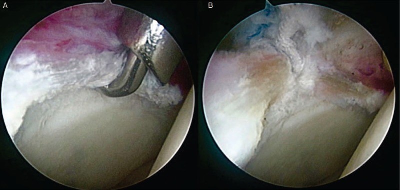 Figure 7
Anterior labral repair being performed, showing anchor placement and suture passage through the labrum.
Conclusion: Seeking Expert Evaluation for Labral Tear Diagnosis
If you are experiencing persistent hip pain, especially with symptoms suggestive of a labral tear, seeking evaluation from an orthopedic specialist is essential. Accurate labral tear diagnosis is a multi-faceted process involving symptom recognition, thorough physical examination, and advanced imaging techniques. A precise diagnosis ensures that you receive the most appropriate and effective treatment plan to address your hip pain, restore function, and improve your quality of life.