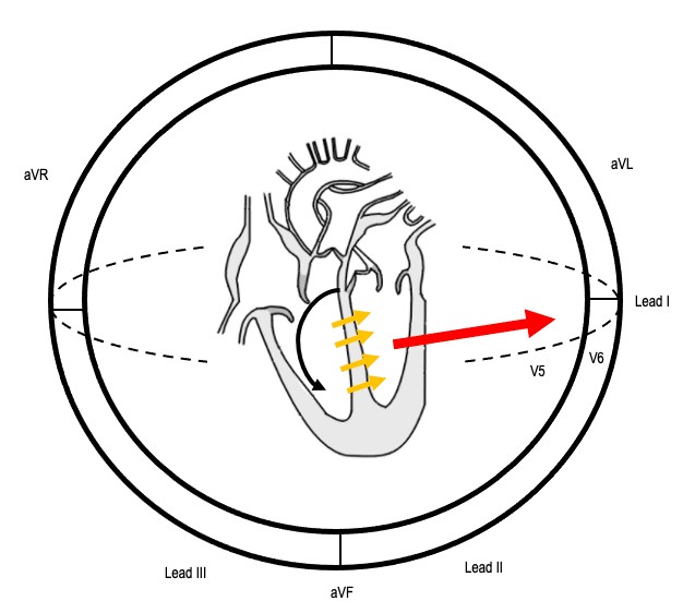
Left Bundle Branch Block (LBBB) is a critical electrocardiogram (ECG) finding that indicates a delay or blockage in the electrical conduction pathway to the left ventricle of the heart. Accurate Lbbb Diagnosis is essential as it can signify underlying cardiac conditions and influence patient management. This guide provides an in-depth exploration of LBBB diagnosis, covering ECG criteria, electrophysiology, morphological characteristics, and clinical implications.
Understanding LBBB: ECG Diagnostic Criteria
The cornerstone of LBBB diagnosis lies in recognizing specific patterns on a 12-lead ECG. The primary diagnostic criteria include:
- QRS duration ≥ 120ms: This signifies prolonged ventricular depolarization due to delayed conduction through the left bundle branch.
- Dominant S wave in V1: Lead V1, positioned over the right ventricle, records a deep S wave as the electrical impulse moves away from it, towards the delayed left ventricle activation.
- Broad monophasic R wave in lateral leads (I, aVL, V5-6): Lateral leads, viewing the left ventricle, exhibit tall, broad R waves, often notched or slurred, reflecting the slow and asynchronous depolarization of the left ventricle.
- Absence of Q waves in lateral leads: Normal septal depolarization from left to right produces small Q waves in lateral leads. In LBBB, this septal activation is reversed or absent, eliminating these Q waves.
- Prolonged R wave peak time > 60ms in leads V5-6: The time from the beginning of the QRS complex to the peak of the R wave is prolonged in lateral leads, further indicating delayed left ventricular activation.
Sequence of conduction in LBBB: illustrating the delayed electrical pathway and altered ventricular activation.
Electrophysiological Basis of LBBB
To fully grasp LBBB diagnosis, understanding the underlying electrophysiology is crucial. In a healthy heart, electrical impulses travel simultaneously down the left and right bundle branches, resulting in coordinated ventricular contraction. Septal activation occurs from left to right, producing small Q waves in lateral leads.
In LBBB, a block in the left bundle branch forces the electrical impulse to initially travel through the right bundle branch, activating the right ventricle first. The impulse then spreads to the left ventricle via slower cell-to-cell conduction through the septum. This altered sequence leads to:
- Reversed Septal Activation: The septum depolarizes from right to left, abolishing the normal Q waves in lateral leads.
- Delayed Left Ventricular Depolarization: The slow, circuitous route of electrical activation to the left ventricle significantly prolongs the QRS duration.
- Altered Depolarization Vector: The overall depolarization vector shifts, resulting in tall R waves in lateral leads and deep S waves in right precordial leads (V1-3). The delay between right and left ventricular activation causes the characteristic “M-shaped” or notched R wave morphology in lateral leads.
ECG Morphology in LBBB: Recognizing Key Features
Beyond the diagnostic criteria, specific QRS morphologies in different ECG leads are vital for accurate LBBB diagnosis.
Lateral Leads (I, aVL, V5-V6)
The R wave morphology in lateral leads can vary, presenting as:
- “M-shaped” or Notched R wave: This is a classic finding in LBBB, reflecting the delayed and asynchronous left ventricular depolarization.
- Monophasic R wave: A broad, single peaked R wave.
- RS complex: In some cases, an RS complex may be observed, though the R wave remains broad and dominant.
Lead V1 Morphology
In lead V1, the QRS complex in LBBB diagnosis typically presents as:
- rS complex: A small initial R wave followed by a deep and wide S wave is the most common morphology.
- QS complex: A completely negative complex, where the R wave is absent, and only a deep Q or S wave is present.
It’s important to note the “appropriate discordance” in LBBB, particularly in lead V1. Due to the altered depolarization, the ST segment and T wave should be discordant to the QRS complex. In leads with deep S waves (like V1 in LBBB), a degree of ST elevation can be normal and part of this expected discordance. However, concordant ST elevation in LBBB is highly concerning for myocardial ischemia and warrants further investigation, potentially utilizing Sgarbossa criteria for STEMI detection in LBBB.
Clinical Significance and Causes of LBBB
While LBBB diagnosis is made via ECG, it’s crucial to understand its clinical implications. LBBB is rarely found in healthy individuals and often indicates underlying structural heart disease. Common causes include:
- Ischemic Heart Disease: Myocardial infarction, particularly anterior MI, can damage the left bundle branch.
- Hypertension: Chronic hypertension can lead to left ventricular hypertrophy and conduction system disease.
- Dilated Cardiomyopathy: Enlargement and weakening of the heart muscle can disrupt the electrical conduction system.
- Aortic Stenosis: Obstruction of the aortic valve can cause left ventricular hypertrophy and LBBB.
- Lenègre-Lev Disease: A primary degenerative disease affecting the cardiac conduction system.
- Hyperkalemia: Elevated potassium levels can impair electrical conduction in the heart.
- Digoxin Toxicity: Excessive digoxin levels can also induce LBBB.
Historically, new-onset LBBB in the setting of chest pain was considered a STEMI equivalent. However, current guidelines emphasize assessing for concordant ST changes or excessive discordance, as per Sgarbossa criteria, to accurately identify acute myocardial infarction in patients with LBBB.
LBBB with Atrial Fibrillation: Demonstrating appropriate ST-segment discordance in the context of LBBB.
Incomplete LBBB and Differential Diagnosis
Incomplete LBBB shares morphological features with complete LBBB but with a QRS duration less than 120ms. It represents a less severe conduction delay and may have similar clinical implications.
The differential diagnosis for LBBB includes:
- Right Ventricular Paced Rhythm: Paced rhythms originating from the right ventricle can mimic LBBB morphology. However, pacing spikes preceding the QRS complexes will be present.
- Left Ventricular Hypertrophy (LVH): LVH can cause QRS widening and ST-T wave changes in lateral leads, potentially resembling LBBB. However, LVH typically lacks the dominant S wave in V1 and broad R wave in lateral leads characteristic of LBBB.
Conclusion: Mastering LBBB Diagnosis
Accurate LBBB diagnosis is a critical skill in ECG interpretation. By understanding the ECG criteria, electrophysiological mechanisms, and morphological variations, clinicians can confidently identify LBBB and appreciate its clinical significance. Recognizing LBBB is not just about ECG interpretation; it’s about understanding the potential underlying cardiac pathology and guiding appropriate patient management. This comprehensive guide serves as a valuable resource for healthcare professionals seeking to enhance their expertise in LBBB diagnosis.
References:
- Life in the Fast Lane ECG Library: https://litfl.com/ecg-library/
- Sgarbossa Criteria: https://litfl.com/sgarbossa-criteria-ecg-library/
- Dilated Cardiomyopathy: https://litfl.com/dilated-cardiomyopathy-dcm-ecg-library/
- Anterior MI: https://litfl.com/anterior-myocardial-infarction-ecg-library/
- Lenègre-Lev disease: https://litfl.com/lenegre-lev-disease/
- Hyperkalaemia: https://litfl.com/hyperkalaemia-ecg-library/
- Digoxin toxicity: https://litfl.com/digoxin-toxicity-ecg-library/
- Left ventricular hypertrophy: https://litfl.com/left-ventricular-hypertrophy-lvh-ecg-library/
