Wound care is a critical aspect of healthcare, addressing injuries that compromise the skin’s integrity and underlying tissues. A wound, defined as any damage to the skin, tissues, or organs, can range from minor abrasions to severe injuries resulting from trauma, surgery, or chronic conditions. Effective wound management is paramount to prevent infection, promote healing, and improve patient outcomes. This article delves into the essential nursing diagnoses and interventions for wound care, providing a comprehensive guide for healthcare professionals.
Understanding Wound Infection and Healing
Wound Infection
Wound infection occurs when pathogens breach the body’s primary defense mechanism – the skin – and overwhelm the immune system. This often results from inadequate aseptic techniques or contamination. Patients with chronic conditions such as diabetes mellitus or HIV, which weaken the immune system or impede healing, are at a heightened risk of wound infections.
Untreated wound infections can escalate into serious, even life-threatening conditions, including cellulitis, osteomyelitis (bone infection), and sepsis. Recognizing the signs of wound infection and implementing timely interventions are crucial in preventing these complications.
Common signs and symptoms of a local wound infection include:
- Purulent drainage (pus) from the wound
- Erythema (redness) and increased warmth around the wound
- Edema (swelling)
- Malodor (foul smell)
- Increased pain or tenderness
Systemic signs of infection, indicating a more severe condition, may include:
- Fever
- Chills
- Lymphadenopathy (swollen lymph nodes)
Wound Healing Process
Wound healing, the body’s natural process of tissue restoration, commences immediately after injury. Any disruption or delay in this intricate process can increase the vulnerability to infection and impede recovery. Wound healing progresses through four distinct phases:
-
Hemostasis (Immediate): This initial phase is characterized by immediate vasoconstriction of blood vessels to minimize blood loss. Coagulation occurs, forming a fibrin clot that acts as a temporary barrier and matrix for cellular migration. Platelets aggregate at the injury site, further contributing to clot formation and releasing growth factors essential for subsequent healing stages.
-
Inflammation (0-4 days): The inflammatory phase is a crucial defense mechanism. Vasodilation increases blood flow to the wound, delivering neutrophils and macrophages to combat potential infection and clear debris. Neutrophils are the first responders, followed by macrophages that phagocytize bacteria and necrotic tissue. Clinically, this phase manifests as erythema, warmth, edema, and pain. While inflammation is a natural and necessary part of healing, prolonged or excessive inflammation can be detrimental.
-
Proliferation (2-24 days): This phase is marked by the formation of granulation tissue, a critical step in wound repair. Fibroblasts proliferate and synthesize collagen, providing structural support to the new tissue. Angiogenesis, the formation of new blood vessels, ensures adequate oxygen and nutrient supply to the healing tissue. Epithelialization occurs as epithelial cells migrate from the wound edges to resurface the wound bed. The wound appears red and beefy due to the rich capillary network in the granulation tissue.
-
Maturation (24+ days to years): Also known as the remodeling phase, maturation involves the reorganization of collagen fibers, increasing tensile strength of the wound. Collagen synthesis and breakdown continue, remodeling the scar tissue. Scar tissue is initially weaker than original skin, reaching approximately 80% of the original tensile strength over time. The scar gradually fades and becomes more pliable.
Types of Wound Healing
Understanding the different types of wound healing is essential for guiding appropriate nursing interventions. Wounds heal through three primary intentions:
-
Primary Intention: This type of healing occurs when wound edges are approximated or closed together, typically using sutures, staples, adhesives, or tapes like steri-strips. Primary intention healing is common in surgical incisions or clean lacerations. It results in minimal scarring and a faster healing time.
-
Secondary Intention: Secondary intention healing is employed when wound edges cannot be easily approximated, often due to tissue loss or infection. The wound is left open to heal from the bottom up. Granulation tissue gradually fills the wound cavity, followed by epithelialization. This method is common in pressure ulcers and wounds with significant tissue loss. Secondary intention healing takes longer and results in more significant scarring and a higher risk of infection compared to primary intention.
-
Tertiary Intention (Delayed Primary Closure): Tertiary intention, or delayed primary closure, is used when a wound is initially left open, typically to manage infection or edema. Once the infection is controlled and the wound bed is clean and granulating, the wound edges are approximated and closed, usually with sutures. This approach combines the benefits of secondary intention (infection control) and primary intention (wound closure).
Factors Affecting Wound Healing
Numerous factors can influence the body’s ability to heal effectively. Nurses must consider these factors when assessing and planning care for patients with wounds:
-
Nutritional Status: Adequate nutrition, particularly protein, vitamins (especially Vitamin C and Zinc), and minerals, is crucial for tissue repair and regeneration. Malnutrition can significantly impair wound healing.
-
Stress: Psychological and physiological stress can negatively impact wound healing by releasing cortisol, which can suppress the immune system and delay the inflammatory response.
-
Comorbidities: Underlying health conditions like diabetes mellitus, peripheral vascular disease, and autoimmune disorders can compromise circulation, immune function, and overall healing capacity.
-
Age: Both very young and older individuals may have impaired healing due to immature or declining immune and physiological functions.
-
Obesity: Adipose tissue has poor vascularity and can contribute to chronic inflammation, both of which hinder wound healing. Obesity is also associated with increased risk of wound dehiscence and infection.
-
Medications: Certain medications, such as corticosteroids, immunosuppressants, and chemotherapy drugs, can suppress the immune system or interfere with the inflammatory and proliferative phases of healing.
-
Lifestyle Factors: Alcohol consumption and smoking impair wound healing. Smoking, in particular, reduces tissue oxygenation due to vasoconstriction and carbon monoxide, significantly delaying healing and increasing infection risk.
-
Mechanical Factors: Friction, shear forces, and pressure can cause further tissue damage and disrupt the healing process. Poor mobility increases the risk of pressure injuries.
-
Moisture Balance: Both excessive and insufficient wound moisture can impede healing. Maceration (excessive moisture) can damage periwound skin, while a dry wound bed can hinder cell migration and epithelialization.
-
Knowledge Deficits: Lack of patient and caregiver knowledge about proper wound care techniques can lead to suboptimal wound management and delayed healing.
Nursing Process for Wound Care
The nursing process provides a systematic framework for wound care, ensuring comprehensive and individualized patient care. It involves assessment, diagnosis, planning, implementation, and evaluation.
Nursing Assessment
A thorough nursing assessment is the cornerstone of effective wound care. It involves gathering subjective and objective data to understand the patient’s condition comprehensively.
Review of Health History
-
General Symptoms: Assess for symptoms indicative of wound infection, such as purulent discharge, skin discoloration, swelling, foul odor, warmth, tenderness, pain, or inflammation. Inquire about systemic symptoms like fever, chills, and lymphadenopathy, which may suggest a more serious infection.
-
Underlying Cause: Determine the etiology of the wound. Was it caused by trauma, surgery, pressure, or a chronic condition? Understanding the cause helps guide appropriate treatment strategies. For example, pressure ulcers require pressure relief measures, while surgical wounds necessitate aseptic care.
-
Medical Risk Factors: Identify pre-existing conditions that may impair wound healing or increase infection risk. Diabetes mellitus, immunosuppression, renal failure, obesity, advanced age, neuropathy, and peripheral vascular disease are significant risk factors.
-
Surgical History: For surgical wounds, review surgical records to note factors that could contribute to infection, such as poor surgical technique, prolonged hospital stay, intraoperative contamination, or hypothermia.
-
Medication Review: Document all medications, including prescription, over-the-counter, and herbal supplements. Pay particular attention to immunosuppressants, steroids, NSAIDs, DMARDs, and chemotherapeutic agents, as these can delay wound healing.
-
Nutritional Status: Evaluate the patient’s nutritional intake, focusing on protein, vitamin, and fluid intake. Protein deficiency and inadequate micronutrients can significantly impair wound healing. Assess dietary habits and water intake.
Physical Assessment
-
Wound Type Determination: Accurately classify the wound type, as this dictates the specific wound care approach. Common wound types include skin tears, diabetic foot ulcers, arterial ulcers, venous stasis ulcers, pressure ulcers, surgical wounds, and traumatic wounds.
-
Wound Characteristics Assessment and Documentation: At each dressing change, meticulously assess and document wound characteristics, including:
- Location: Anatomical site of the wound.
- Size: Measure length, width, and depth in centimeters. Consistent measurement techniques are essential for tracking wound progression.
- Drainage: Note the type (serous, serosanguineous, sanguinous, purulent), color, odor, and amount of exudate.
- Wound Bed: Describe the tissue types present: granulation tissue (red, beefy, indicates healing), slough (yellow or tan, stringy or mucinous, nonviable tissue), eschar (black or brown, hard, necrotic tissue).
- Wound Edges and Periwound Skin: Assess wound edges for approximation, rolled edges (epibole), or undermining. Examine periwound skin for erythema, edema, maceration, or induration.
- Tunneling or Undermining: Probe for the presence and extent of tunneling (linear channels) or undermining (tissue destruction under intact skin at wound edges).
- Signs of Infection: Reassess for local and systemic signs of infection at each dressing change.
-
T.I.M.E. Assessment Framework: Utilize the T.I.M.E. acronym as a structured approach to wound assessment:
- T (Tissue): Assess tissue type in the wound bed. Healthy granulation tissue (red) or epithelial tissue (pink) indicates healing. Slough (yellow/gray) and necrotic tissue (black) are nonviable and impede healing.
- I (Infection/Inflammation): Differentiate between normal inflammation (part of healing) and infection (abnormal). Infection delays healing and can spread systemically.
- M (Moisture): Evaluate wound moisture balance. A moist wound environment promotes healing, but excessive moisture can cause maceration. Assess the type and amount of wound exudate.
- E (Edges): Monitor wound edges for signs of non-advancing edges (rolled edges), undermining, or tunneling, which indicate impaired healing. Assess periwound skin condition.
-
Pain Assessment: Pain is a common symptom associated with wounds. Use a validated pain scale (e.g., numeric rating scale, visual analog scale) to quantify pain intensity. Assess pain characteristics (location, quality, radiation, aggravating/alleviating factors). Regular pain assessment is essential to evaluate treatment effectiveness and patient comfort.
Diagnostic Procedures
-
Wound Culture: Obtain a wound swab for culture and sensitivity testing, especially if infection is suspected. This helps identify the causative pathogens and guide antibiotic therapy.
-
Biopsy or Aspiration: In cases of deep or chronic wounds, a tissue biopsy may be necessary for accurate pathogen identification and to rule out other conditions. Aspiration of fluid is used to assess for deeper infections, such as abscesses.
-
Laboratory Values: Blood tests can provide valuable information about systemic infection and overall health status. Monitor:
- White Blood Cell (WBC) Count: Elevated WBC count indicates systemic inflammation or infection.
- C-Reactive Protein (CRP): An acute-phase reactant, elevated CRP levels suggest inflammation.
- Procalcitonin (PCT): PCT is a more specific marker for bacterial infection than CRP.
- Presepsin: Another marker for bacterial infection, particularly sepsis.
- Microbial DNA: PCR-based tests can identify specific pathogens in wound samples.
- Bacterial Protease Activity (BPA): Elevated BPA levels may indicate bacterial burden in chronic wounds.
-
Imaging Studies: Imaging techniques can aid in assessing the extent and depth of infection, particularly in deeper tissues:
- Plain X-rays: Useful for detecting soft-tissue gas or foreign bodies.
- Computed Tomography (CT): Provides detailed images of soft tissues and can identify abscesses or deep infections.
- Magnetic Resonance Imaging (MRI): Offers superior soft-tissue resolution and is excellent for evaluating osteomyelitis or deep tissue infections.
- Ultrasound: Can be used to assess superficial soft-tissue infections and fluid collections.
-
Peripheral Perfusion Assessment: Evaluate peripheral perfusion, especially in patients with lower extremity wounds or suspected peripheral artery disease.
- Ankle-Brachial Index (ABI) and Toe-Brachial Index (TBI): These non-invasive tests assess arterial blood flow to the lower extremities.
- Monofilament Testing: Evaluates protective sensation in the feet, particularly important for diabetic neuropathy and preventing foot ulcers.
Nursing Interventions
Nursing interventions are crucial for promoting wound healing, preventing infection, and managing associated symptoms.
Provide Appropriate Wound Care
-
Debridement of Nonviable Tissue: Debridement, the removal of necrotic tissue, is often essential for wound healing. Nonviable tissue can harbor bacteria and impede granulation tissue formation. Debridement methods include:
- Autolytic Debridement: Utilizes the body’s own enzymes to break down necrotic tissue under moist dressings. Suitable for wounds with minimal necrotic tissue.
- Enzymatic Debridement: Topical application of enzymatic ointments to digest necrotic tissue.
- Sharp Debridement: Surgical removal of necrotic tissue using sharp instruments (scalpel, scissors) by a trained healthcare professional. This method is faster and more effective for larger amounts of necrotic tissue.
-
Moisture Management: Maintain optimal wound moisture balance.
- Moist Wound Dressings: Use dressings that maintain a moist wound environment, promoting cell migration and preventing desiccation. Hydrogels, hydrocolloids, and foams are examples.
- Exudate Management: For wounds with excessive exudate, use highly absorbent dressings like alginates or hydrofibers to prevent maceration of periwound skin. Frequent dressing changes may be necessary.
-
Wound Packing: For deep wounds with tunneling or undermining, pack the wound loosely with moistened sterile gauze or hydrogel-impregnated dressings to maintain wound bed contact and prevent premature closure of the wound opening, which could lead to abscess formation.
-
Periwound Skin Protection: Protect the skin surrounding the wound (periwound skin) from maceration and excoriation. Apply barrier creams, powders, protective wipes, or barrier wafers to create a protective layer.
-
Wound Vac Therapy: Vacuum-assisted closure (VAC) therapy is an option for complex, open wounds, skin grafts, flaps, and pressure ulcers. It applies negative pressure to the wound bed, removing exudate, reducing edema, promoting granulation tissue formation, and enhancing wound closure.
-
Pain Management: Wound care procedures, particularly dressing changes and debridement, can be painful. Premedicate with prescribed analgesics (oral or topical) prior to wound care procedures to improve patient comfort and cooperation.
Prevent or Manage Infection
-
Aseptic or Clean Technique: Employ appropriate techniques to minimize infection risk. Aseptic technique (sterile gloves and equipment) is essential for high-risk wounds like burns, surgical wounds, and when dealing with immunocompromised patients. Clean technique (non-sterile gloves and clean equipment) is often sufficient for chronic wounds like pressure ulcers and simple wounds like skin tears in non-immunocompromised individuals.
-
Antibiotic Administration: For wounds exhibiting signs of infection, administer antibiotics as prescribed. Topical antibiotics or silver dressings may be used for local infections. Systemic infections require oral or intravenous antibiotic therapy. Culture and sensitivity results guide targeted antimicrobial therapy.
-
Immediate Wound Cleaning: Emphasize the importance of immediate wound cleaning after skin injury. Washing the wound with mild soap and water is crucial to remove contaminants and reduce infection risk, especially for “dirty” wounds (animal bites, contaminated objects).
-
Avoid Harsh Cleansing Agents: Educate patients to avoid using hydrogen peroxide or rubbing alcohol to clean wounds. These agents are cytotoxic to healthy tissue and can impair wound healing. Use gentle wound cleansers or sterile saline.
-
Hand Hygiene: Strict hand hygiene is paramount. Wash hands thoroughly with soap and water or use an alcohol-based hand sanitizer before and after wound care procedures, and whenever touching a wound or dressing.
-
Wound Dressings: Keep wounds covered with appropriate dressings. Dressings protect the wound from external contamination, maintain a moist healing environment, and absorb exudate. Educate patients that wounds should be covered to promote healing and prevent infection.
Promote Wound Healing
-
Nutritional Support: Optimize nutritional status to support tissue repair. Encourage a high-protein diet rich in vitamins and minerals. Nutritional supplements, such as protein-enriched drinks or vitamin supplements, may be necessary for patients with nutritional deficits. Ensure adequate hydration to promote circulation and tissue oxygenation.
-
Education on Skin Breakdown Prevention: Educate patients and caregivers on strategies to prevent further skin injury and promote wound healing:
- Pressure Relief: Implement pressure relief measures, such as frequent repositioning (at least every 2 hours for bedridden patients), pressure-redistributing support surfaces, and offloading devices for pressure ulcers.
- Foot Protection: Advise patients to always wear appropriate footwear (shoes or socks) to protect feet, especially for diabetic patients.
- Edema Management: For patients with venous insufficiency, promote leg elevation and use compression stockings to control edema and improve circulation.
- Skin Hygiene: Maintain clean and dry skin, especially in patients with incontinence. Use gentle cleansers and barrier creams as needed.
- Smoking Cessation: Strongly advise smokers to quit smoking, as smoking significantly impairs wound healing.
-
Wound Documentation: Document wound assessment findings and wound care provided at each dressing change. Include wound size, drainage, wound bed characteristics, periwound skin condition, treatments administered, and patient response. Wound photography can be a valuable tool for tracking wound progression over time.
-
Referral to Wound Care Specialist: For chronic or non-healing wounds, or complex wounds, refer the patient to a wound care specialist or wound care clinic. Specialists can provide advanced wound care modalities, such as specialized dressings, negative pressure wound therapy, bioengineered skin substitutes, or hyperbaric oxygen therapy.
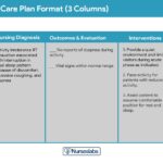
Nursing Care Plans
Nursing care plans provide a structured approach to addressing patient needs based on identified nursing diagnoses. Common nursing diagnoses related to wound care include:
Acute Pain
Nursing Diagnosis: Acute Pain related to wound infection and tissue damage.
Related to:
- Tissue injury
- Inflammation
- Nerve irritation
As evidenced by:
- Verbal report of pain
- Guarding behavior
- Restlessness
- Facial grimacing
- Changes in vital signs (increased heart rate, blood pressure)
Expected Outcomes:
- Patient will report pain at a manageable level (e.g., using a pain scale).
- Patient will demonstrate relaxed body posture.
- Patient will participate in wound care without excessive distress.
Assessments:
- Pain Assessment: Use a pain scale (numeric, visual) to quantify pain intensity. Assess pain location, quality, onset, duration, aggravating/alleviating factors.
- Type of Pain: Differentiate between nociceptive pain (tissue damage) and neuropathic pain (nerve damage).
- Palpation: Gently palpate periwound skin for tenderness, warmth, and swelling.
Interventions:
- Premedication: Administer prescribed analgesics (oral, IV, topical) prior to painful procedures like dressing changes or debridement. Allow adequate time for medication to take effect.
- Pain Management Education: Educate patient about pain medication regimen, including dosage, frequency, and potential side effects. Discuss non-pharmacological pain management strategies (e.g., positioning, relaxation techniques, distraction).
- Symptom Management: Address contributing factors to pain, such as edema (elevation), wound drainage (appropriate dressings), and skin maceration (barrier creams).
- Splinting/Support: Splint or support the wounded extremity to minimize movement and protect from further injury.
Impaired Skin Integrity
Nursing Diagnosis: Impaired Skin Integrity related to physical injury, pressure, or impaired healing.
Related to:
- Trauma
- Pressure
- Shear
- Friction
- Moisture
- Surgical incision
- Impaired circulation
- Delayed healing process (e.g., diabetes)
As evidenced by:
- Disruption of skin surface
- Wound drainage
- Erythema
- Edema
- Delayed wound healing
- Presence of necrotic tissue
Expected Outcomes:
- Patient will demonstrate progressive wound healing.
- Wound will exhibit reduced drainage and inflammation.
- Patient will verbalize understanding of wound care regimen.
- Patient will participate in wound care activities.
Assessments:
- Wound Assessment: Assess wound characteristics at each dressing change (size, depth, drainage, wound bed, periwound skin).
- Wound Classification: Identify the type of wound (pressure ulcer stage, surgical wound, etc.).
- Risk Assessment: Use risk assessment tools (e.g., Braden Scale for pressure ulcer risk) to identify patients at risk for skin breakdown.
- Wound Culture: Obtain wound culture if signs of infection are present.
Interventions:
- Wound Cleansing: Cleanse wound with appropriate antiseptic or wound cleanser as prescribed. Avoid harsh agents like alcohol or hydrogen peroxide.
- Decontamination: Remove foreign material and debris from the wound.
- Debridement: Implement debridement methods as needed to remove necrotic tissue.
- Appropriate Dressings: Apply appropriate wound dressings based on wound type, drainage, and stage of healing. Use non-adherent dressings to minimize trauma during dressing changes.
- Wound Healing Stages Management: Adjust wound care plan based on the phase of wound healing.
- Moist Wound Environment: Maintain a moist wound environment with appropriate dressings.
- Topical Agents: Apply topical antibiotics or antiseptics as prescribed for infected wounds.
- Suture/Staple Removal: Remove sutures or staples as per protocol once wound is sufficiently healed.
- Referral to Specialist: Refer to wound care specialist for complex or non-healing wounds.
Ineffective Protection
Nursing Diagnosis: Ineffective Protection related to compromised skin integrity and impaired defenses.
Related to:
- Skin breakdown
- Tissue trauma
- Impaired immune function
- Older age
- Malnutrition
- Immobility
- Incontinence
As evidenced by:
- Wound development
- Delayed healing
- Signs of infection
- Pressure ulcer formation
Expected Outcomes:
- Patient will remain free from wound infection.
- Patient will demonstrate protective measures to prevent skin breakdown and wounds.
Assessments:
- Vital Signs Monitoring: Monitor vital signs (temperature, heart rate, blood pressure) for signs of systemic infection.
- Nutritional Assessment: Assess nutritional status, including protein and fluid intake.
- ADL Assessment: Evaluate patient’s ability to perform activities of daily living (mobility, hygiene) to assess risk for skin breakdown.
Interventions:
- Antibiotic Therapy: Administer antibiotics as prescribed for existing infections or as prophylaxis in high-risk situations.
- Dietary Referral: Refer to dietitian for nutritional assessment and optimization of dietary intake to support immune function and wound healing.
- Infection Control Education: Educate patient and caregivers on infection control measures, including hand hygiene and proper wound care techniques.
- Wound Care Provision: Provide or ensure proper wound care according to wound type and patient needs.
Ineffective Tissue Perfusion
Nursing Diagnosis: Ineffective Tissue Perfusion (peripheral) related to compromised circulation.
Related to:
- Vascular insufficiency
- Edema
- Smoking
- Chronic conditions (diabetes, PVD)
- Sedentary lifestyle
As evidenced by:
- Delayed wound healing
- Skin discoloration (pallor, cyanosis)
- Cool extremities
- Decreased peripheral pulses
- Pain (intermittent claudication)
- Edema
Expected Outcomes:
- Patient will demonstrate improved tissue perfusion as evidenced by wound healing.
- Patient will verbalize understanding of factors affecting tissue perfusion.
Assessments:
- Diagnostic Test Review: Monitor results of peripheral perfusion tests (ABI, TBI, skin perfusion pressure).
- Perfusion Signs: Assess for signs of ineffective tissue perfusion in extremities and wound area (skin color, temperature, pulses, edema, pain).
- Comorbidity Consideration: Consider comorbidities (diabetes, vascular disease) that affect perfusion.
Interventions:
- Foot Care Education: Educate on proper foot care, especially for diabetic patients (daily inspection, proper footwear).
- Exercise Encouragement: Encourage walking or exercise to improve circulation (if appropriate and not contraindicated by wound location).
- Leg Elevation/Compression: Elevate lower extremities for venous insufficiency. Apply compression stockings as prescribed for venous disease, but avoid compression in arterial insufficiency.
- Smoking Cessation: Emphasize smoking cessation due to its detrimental effects on blood vessels and tissue perfusion.
- Referral for HBOT: Consider referral for hyperbaric oxygen therapy (HBOT) for wounds with severely compromised perfusion and delayed healing.
Knowledge Deficit
Nursing Diagnosis: Knowledge Deficit related to wound care management.
Related to:
- Lack of information
- Misinformation
- Unfamiliarity with wound care procedures
As evidenced by:
- Verbalization of lack of knowledge
- Questions about wound care
- Non-adherence to wound care regimen
- Wound complications
Expected Outcomes:
- Patient will verbalize understanding of wound care management.
- Patient will demonstrate proper wound care techniques.
- Patient will adhere to wound care treatment plan.
Assessments:
- Knowledge Assessment: Assess patient’s current knowledge about wound care and healing process.
- Demonstration Assessment: Ask patient or caregiver to demonstrate wound care techniques to assess competency.
- Misunderstanding Identification: Identify any cultural beliefs or misconceptions about wound care practices.
- Resource Assessment: Assess patient’s access to wound care resources (financial, social support).
Interventions:
- Wound Care Education: Provide comprehensive education on wound care plan, including dressing changes, wound cleansing, infection prevention, and medication regimen.
- Question Time: Allow ample time for patient and caregiver to ask questions and clarify doubts.
- Caregiver Involvement: Involve caregivers in education and training to ensure consistent wound care at home.
- Infection Control Emphasis: Emphasize infection control measures, especially hand hygiene and aseptic/clean techniques.
- Resource Referral: Refer to social worker or case manager for assistance with resources (home health, equipment, financial aid).
- Dietitian Referral: Refer to dietitian for nutritional counseling to support wound healing.
References
(References would be listed here, mirroring those present in the original article or updated with more recent sources if aiming for enhanced content.)