The skin, the body’s largest organ, acts as a crucial protective barrier against the external environment, preventing pathogens from entering and causing infections. When this barrier is compromised due to various factors such as cuts, abrasions, ulcers, surgical incisions, and wounds, it becomes vulnerable to bacterial invasion, leading to infections and other complications. Therefore, a thorough understanding of assessing, preventing, treating, and educating patients about impaired skin integrity is paramount for nurses and healthcare professionals.
In this article, we will delve into a comprehensive Nursing Diagnosis Care Plan For Impaired Skin Integrity, covering its causes, signs and symptoms, assessment strategies, nursing interventions, and expected outcomes. This guide aims to equip healthcare providers with the knowledge and tools necessary to effectively manage and prevent skin integrity issues in their patients.
Causes of Impaired Skin Integrity
Impaired skin integrity can arise from a multitude of internal and external factors that compromise the skin’s structure and function. Understanding these causes is crucial for identifying at-risk patients and implementing preventive measures.
Internal Factors:
- Extremes in Age: Both very young and elderly individuals have more fragile skin. Newborns have delicate skin that is easily irritated, while aging skin becomes thinner, less elastic, and has reduced moisture, making it more susceptible to injury and slower to heal.
- Cognitive Impairment: Patients with cognitive impairments may be unable to recognize or communicate discomfort or injury, increasing their risk of prolonged pressure and skin breakdown. They might also be less likely to reposition themselves or maintain hygiene, contributing to skin issues.
- Physical Immobilization/Bedrest: Prolonged pressure on bony prominences due to immobility restricts blood flow and oxygen supply to the skin, leading to tissue ischemia and necrosis, ultimately resulting in pressure ulcers. Conditions requiring bed rest, such as paralysis or severe illness, significantly elevate this risk.
- Paralysis: Loss of motor and sensory function due to paralysis prevents individuals from independently repositioning or feeling pressure, making them highly vulnerable to pressure injuries.
- Underlying Health Conditions: Certain medical conditions can compromise skin integrity. For example, diabetes mellitus can lead to poor circulation and neuropathy, increasing the risk of foot ulcers. Peripheral vascular disease also impairs blood flow, making the skin more susceptible to damage. Malnutrition and dehydration weaken the skin and impede healing.
External Factors:
- Hyperthermia: Elevated body temperature can lead to increased perspiration, causing skin maceration and breakdown, especially in skin folds and areas under pressure.
- Hypothermia: Conversely, low body temperature can reduce blood flow to the skin, making it more susceptible to damage and slowing down the healing process.
- Radiation: Radiation therapy, used in cancer treatment, can cause radiation dermatitis, leading to skin redness, dryness, peeling, and breakdown in the treated area.
- Chemicals: Exposure to harsh chemicals, irritants, and allergens can cause contact dermatitis, leading to skin inflammation, itching, and breakdown. This can include cleaning products, certain soaps, and industrial chemicals.
- Moisture/Secretions: Excessive moisture from perspiration, urine, stool, wound drainage, or saliva can macerate the skin, weakening its barrier function and making it prone to breakdown and infection. Incontinence is a major risk factor.
- Shearing/Friction/Pressure: These mechanical forces are major contributors to pressure ulcer development. Friction occurs when skin rubs against surfaces, like bed sheets, causing superficial damage. Shearing happens when skin layers slide over each other, damaging blood vessels and deep tissues. Pressure, especially sustained pressure over bony prominences, restricts blood flow and leads to tissue ischemia.
- Surgery: Surgical incisions disrupt skin integrity, creating a direct pathway for infection. Wound healing complications, such as dehiscence or infection, can further impair skin integrity.
Image: Close-up of a stage 2 pressure ulcer on a sacrum, illustrating partial thickness skin loss affecting the epidermis and dermis.
Signs and Symptoms of Impaired Skin Integrity
Recognizing the signs and symptoms of impaired skin integrity is crucial for early intervention and preventing further complications. These manifestations can be both subjective, reported by the patient, and objective, observed by the nurse during assessment.
Subjective Symptoms (Patient Reports):
- Pain: Pain is a common symptom, ranging from mild discomfort to severe, localized pain at the site of skin breakdown.
- Itching (Pruritus): Itching can be a sign of skin irritation, dryness, or inflammation, often preceding visible skin damage.
- Numbness: Numbness or altered sensation in the affected or surrounding skin may indicate nerve damage or impaired circulation, increasing the risk of unnoticed injury and delayed healing.
Objective Signs (Nurse Assesses):
- Changes to Skin Color:
- Erythema: Redness of the skin, indicating inflammation or increased blood flow to the area.
- Bruising (Ecchymosis): Discoloration due to blood leakage into the subcutaneous tissues, suggesting trauma or pressure damage.
- Blanching: Paleness or whitening of the skin when pressure is applied, indicating impaired blood flow. Non-blanchable erythema is a key sign of a pressure ulcer.
- Warmth to Skin: Increased skin temperature around an area of breakdown may indicate inflammation or infection.
- Swelling to Tissues (Edema): Swelling can be a sign of fluid accumulation in the tissues due to injury, inflammation, or impaired lymphatic drainage.
- Observed Open Areas or Breakdown: This includes:
- Excoriation: Superficial damage to the skin, often caused by scratching or rubbing.
- Abrasions: Scrapes or superficial wounds involving the epidermis.
- Lacerations: Cuts or tears in the skin.
- Ulcers: Deeper wounds involving tissue loss, such as pressure ulcers, venous ulcers, or arterial ulcers.
- Blisters: Fluid-filled pockets on the skin, often caused by friction or burns.
Image: Illustration of skin layers showing an abrasion, a superficial wound limited to the epidermis.
Expected Outcomes for Impaired Skin Integrity
Setting realistic and measurable goals is essential in the nursing care plan for impaired skin integrity. The expected outcomes focus on promoting healing, preventing complications, and empowering patients in self-care. Common goals include:
- Patient will maintain intact skin integrity: This is a preventative goal for patients at risk of skin breakdown.
- Patient will experience timely healing of wounds without complications: This outcome focuses on the resolution of existing skin integrity issues.
- Patient will demonstrate effective wound care: This goal ensures the patient or caregiver can perform necessary wound care procedures correctly.
- Patient will verbalize proper prevention of pressure injuries: Patient education is vital for long-term prevention and management of skin integrity.
Nursing Assessment for Impaired Skin Integrity
A comprehensive nursing assessment is the cornerstone of effective care for impaired skin integrity. It involves gathering both subjective and objective data to identify risk factors, existing skin problems, and guide appropriate interventions.
1. Conduct a Thorough Skin Assessment:
A systematic head-to-toe skin examination should be performed upon admission, during unit transfers, and regularly (at least once per shift, or more frequently based on risk and facility policy). This routine assessment is crucial for early detection and prevention of skin breakdown during hospitalization. Special attention should be given to high-risk areas prone to pressure, such as the heels, sacrum, elbows, hips, and scapulae.
2. Utilize Braden Scale for Pressure Ulcer Risk Assessment:
The Braden Scale is an evidence-based tool widely used to assess a patient’s risk for developing pressure ulcers. It evaluates six key risk factors:
- Sensory Perception: Ability to feel and respond to discomfort or pressure.
- Moisture: Degree of skin exposure to moisture.
- Activity: Level of physical activity.
- Mobility: Ability to change and control body position.
- Nutrition: Usual food intake pattern.
- Friction and Shear: Resistance to movement (friction) and forces causing skin layers to slide over each other (shear).
A score is calculated based on these criteria, ranging from 6 to 23. Lower scores indicate a higher risk of pressure ulcer development. Institutional policies dictate the frequency of Braden Scale assessments, but nurses should also use it whenever they suspect a patient is at risk of skin breakdown.
3. Assess Circulatory Status:
Evaluate skin circulation, sensation, and turgor. Poor skin turgor (decreased elasticity), diminished sensation (indicating nerve damage, especially in conditions like diabetic neuropathy), and poor circulation (manifested by reddish or purple skin discoloration of lower legs and weak or absent palpable pulses) all increase the risk of tissue injury.
4. Assess Activity Level and Mobility:
Observe the patient’s ability to ambulate and reposition themselves in bed or chair. Immobility or limited mobility significantly increases the risk of pressure ulcers. Patients using restraints are also at higher risk due to restricted movement and potential for increased pressure.
5. Determine Risk of Skin Breakdown Related to Moisture:
Assess for factors contributing to excessive skin moisture, such as increased perspiration (diaphoresis) and incontinence (urinary or fecal). Evaluate the nature and amount of body secretions, including sweat, urine, and stool, as these can irritate and macerate the skin.
6. Evaluate Patient’s Ability for Self-Care:
Assess the patient’s capacity to manage hygiene and incontinence independently. Patients who are incontinent or unable to request assistance for toileting require vigilant monitoring and care to maintain clean, dry skin and prevent breakdown.
7. Describe and Document Wound Characteristics:
Accurate and detailed documentation of observed wounds and skin breakdown is essential for tracking healing progress and evaluating treatment effectiveness. Wound assessment should include:
- Location and Stage (if a pressure ulcer): Use standardized staging systems for pressure ulcers.
- Size: Measure length, width, and depth in centimeters.
- Wound Bed: Describe tissue type (e.g., granulation, slough, eschar).
- Periwound Area: Assess the skin surrounding the wound for color, temperature, and signs of maceration or erythema.
- Drainage: Note the amount, color, consistency, and odor of any wound drainage.
- Odor: Document any foul odor, which may indicate infection.
- Tunneling or Undermining: Assess for extensions of the wound under the skin edges.
Photography of the wound is recommended for comparative purposes and objective tracking of healing.
8. Assess Patient’s Nutrition and Hydration Status:
Monitor the patient’s nutritional intake and fluid balance. Adequate hydration and nutrition, particularly protein intake, are crucial for tissue repair and wound healing. Deficiencies can impair the healing process and increase susceptibility to skin breakdown.
9. Assess Stoma and Ostomy Site (if applicable):
For patients with new ostomies, assess the stoma site for proper healing, color (should be pink to red and moist), edema, and any signs of complications. Evaluate the appropriateness and fit of the ostomy appliance and skin barrier. Collaboration with a wound care specialist is essential in these cases.
Image: Depiction of a healthy stoma, characterized by its moist, pink appearance and protrusion from the abdominal wall.
Nursing Interventions for Impaired Skin Integrity
Nursing interventions are crucial for both preventing and treating impaired skin integrity. These interventions aim to address the identified risk factors, promote skin healing, and prevent complications.
1. Implement Wound Care Protocols as Prescribed:
Apply appropriate wound care protocols based on the type, location, and severity of the wound, as well as facility policies and wound care specialist recommendations. Wound care may include cleansing, debridement, application of topical medications or dressings, and infection management.
2. Position and Reposition Patient Frequently and Comfortably:
Reposition patients at high risk for pressure ulcers at least every two hours, or more frequently as indicated by their condition and facility protocols. Use a turn clock to aid in consistent repositioning. Protect bony prominences by relieving pressure. Encourage patients who are able to assist with repositioning themselves.
3. Ensure Adequate Skin Perfusion:
Use pressure-redistributing support surfaces, such as specialty mattresses and cushions. Utilize positioning devices, such as pillows, wedges, and heel protectors, to support bony prominences (elbows, knees, hips, heels) and offload pressure.
4. Maintain Skin Hygiene and Manage Moisture:
Keep the skin clean and dry. Gently cleanse the skin regularly with mild soap and water. Avoid harsh soaps or excessive scrubbing. Promptly cleanse and dry the skin after episodes of incontinence or excessive perspiration. Use moisture barriers to protect skin exposed to excessive moisture.
5. Alleviate Pressure and Friction:
Utilize pressure-reducing mattresses, such as low-air loss mattresses that cycle inflation and deflation to mimic patient movement. Employ support surfaces like air mattresses, gel overlays for chairs and beds, waffle boots, and wedge pillows to offload pressure from bony prominences. Educate patients and caregivers on proper transfer techniques to minimize friction and shear forces.
6. Promote Proper Nutrition and Hydration:
Collaborate with a dietitian to ensure patients receive adequate nutrition and hydration, particularly protein, vitamins, and minerals essential for wound healing. Encourage sufficient fluid intake to maintain skin hydration and support tissue perfusion.
7. Protect Skin from Further Injury:
Implement measures to protect fragile skin from further injury. This includes:
- Padding and protecting vulnerable areas.
- Avoiding adhesive tape directly on fragile skin.
- Using lift sheets for repositioning instead of dragging patients across surfaces.
- Ensuring patients wear appropriate footwear, such as socks and non-slip shoes, especially those with compromised neurovascular status (e.g., diabetic patients).
8. Coordinate with Wound/Ostomy Specialist:
Consult with a wound care or ostomy specialist for complex wounds, stoma care issues, or when standard interventions are ineffective. Specialists can provide expert recommendations, evaluate wound progress, and educate staff and patients on advanced wound care techniques.
9. Minimize Skin Irritation:
Use barrier creams, pastes, or powders to protect skin exposed to moisture or irritants, such as around stomas or incontinent areas. Avoid products containing alcohol or fragrances, which can dry or irritate the skin. Use adhesive removers to facilitate ostomy pouch changes or dressing removal without causing skin trauma.
10. Manage Ostomy Pouch System (if applicable):
Educate patients on proper ostomy pouch application, emptying techniques, and skin care around the stoma. Ensure proper sizing of the adhesive wafer to create a secure seal and prevent leakage. Instruct patients to empty ostomy pouches when they are ⅓ to ½ full to prevent excessive weight and potential detachment.
Image: Diagram showing the correct application of an ostomy pouch system, emphasizing skin barrier placement and secure attachment around the stoma.
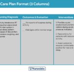
Nursing Care Plans for Impaired Skin Integrity
Nursing care plans are structured frameworks that guide nursing care by prioritizing assessments and interventions to achieve specific patient outcomes. Here are examples of nursing care plans for impaired skin integrity, addressing various underlying causes:
Care Plan #1: Impaired Skin Integrity related to Immobility
Diagnostic Statement: Impaired skin integrity related to immobility as evidenced by a stage 2 pressure ulcer on the sacrum.
Expected Outcomes:
- Patient will experience improvement of pressure ulcer from stage 2 to stage 1 within 2 weeks of nursing interventions.
- Patient will maintain skin integrity by keeping the skin dry and clean at the end of each shift.
- Patient will verbalize cooperation and compliance during wound care within 1 hour of nursing intervention.
- Patient will enumerate three ways to prevent pressure ulcers within 1 hour of nursing education.
Assessment:
- Assess the skin and wounds regularly: Monitor skin at risk for breakdown at least once per shift, paying close attention to pressure points. Observe wounds for signs of worsening or infection, such as increased redness, drainage, or odor. Measure wound dimensions weekly to track healing.
- Determine patient’s mobility level: Assess the patient’s ability to move and reposition independently. Identify any limitations in mobility that contribute to prolonged pressure.
- Assess the need for positioning devices: Evaluate the patient’s need for and use of positioning aids (pillows, wedges, specialty mattresses) to offload bony prominences and maintain proper body alignment.
Interventions:
- Perform wound care per guidelines and orders: Implement prescribed wound care protocols, which may include cleansing, dressing changes, and topical treatments. Ensure aseptic technique to prevent infection.
- Reposition patient and support bony prominences: Implement a turning schedule (at least every 2 hours) for bedridden patients. Use pillows and positioning devices to support bony prominences (hips, knees, heels, elbows) and promote pressure redistribution.
- Keep the skin clean and dry: Provide meticulous skin hygiene, especially after episodes of incontinence or perspiration. Use moisture barriers as needed. Ensure bed linens and clothing remain clean and dry.
- Utilize appropriate support surfaces: Employ pressure-reducing support surfaces like air mattresses, gel overlays, and waffle boots to minimize pressure on bony prominences.
- Encourage nutrition and hydration: Promote adequate fluid intake and a diet rich in protein and essential nutrients to support wound healing and skin integrity. Consult with a dietitian as needed.
Care Plan #2: Impaired Skin Integrity related to Diabetic Neuropathy
Diagnostic Statement: Impaired skin integrity related to decreased skin sensation secondary to diabetic neuropathy as evidenced by redness and an open area on the left lower leg.
Expected Outcomes:
- Patient will demonstrate intact skin on the lower extremities at the end of the shift.
- Patient will enumerate three ways to prevent skin infection within 1 hour of nursing education.
- Patient will maintain skin perfusion by managing blood glucose within target range at the end of the shift.
- Patient will verbalize understanding of the importance of daily skin inspection within 1 hour of patient education.
Assessment:
- Assess skin for signs of infection: Monitor the open area and surrounding skin for signs of infection, such as redness, warmth, swelling, purulent drainage, foul odor, and increased pain.
- Determine skin perfusion: Assess circulation to the lower extremities, noting skin color, temperature, and pulses. Evaluate sensation in the feet and lower legs to identify areas of neuropathy.
- Monitor patient’s blood glucose levels: Regularly monitor blood glucose levels to assess glycemic control, as hyperglycemia impairs wound healing and increases infection risk.
Interventions:
- Control blood glucose levels: Implement strategies to maintain blood glucose within the target range, including medication administration, dietary management, and patient education on diabetes management.
- Educate on diabetic neuropathy and skin care: Educate the patient about diabetic neuropathy, its impact on sensation and skin integrity, and the importance of daily foot and skin inspections. Teach proper foot care practices, including gentle washing, drying, and moisturizing (avoiding between toes).
- Ensure foot protection at all times: Emphasize the importance of wearing protective footwear, such as well-fitting shoes and socks, at all times to prevent injury to the feet. Advise against walking barefoot.
Care Plan #3: Impaired Skin Integrity related to Surgical Incision and Stoma Creation
Diagnostic Statement: Impaired skin integrity related to surgical incision and stoma creation to the abdomen.
Expected Outcomes:
- Patient will verbalize understanding of preventing skin irritation around the stoma within 1 hour of nursing education.
- Patient will manifest a moist and pinkish stoma at the end of the shift.
- Patient will enumerate three ways to protect the stoma from skin breakdown within 1 hour of nursing education.
- Patient will verbalize the proper technique for fitting and emptying the ostomy pouch within 1 hour of demonstration.
Assessment:
- Determine indication for surgery/stoma: Understand the underlying medical condition necessitating stoma creation to provide context for care.
- Assess incision and stoma: Evaluate the surgical incision for signs of infection or dehiscence. Assess the stoma for color (pink to red and moist), edema (initially expected), and protrusion. Report any signs of stoma necrosis (dark or dusky color).
- Determine patient’s dietary intake: Assess the patient’s nutritional status and dietary habits, as nutrition plays a vital role in surgical recovery and wound healing.
Interventions:
- Collaborate with wound/ostomy specialist: Consult with a wound/ostomy specialist for ongoing stoma care management, appliance selection, and patient education.
- Create meal plans with the patient: Educate the patient on dietary recommendations for ostomy management, including strategies to manage stool consistency and minimize odor. A low-residue diet may be initially recommended.
- Minimize skin irritation around the stoma: Apply skin barrier pastes or powders around the stoma to protect the peristomal skin from irritation and moisture. Ensure proper pouch fit to prevent leakage.
- Educate patient on ostomy care: Provide comprehensive education on proper ostomy pouch application, emptying techniques, skin care around the stoma, and troubleshooting common problems.
Care Plan #4: Impaired Skin Integrity related to Burn Wounds
Diagnostic Statement: Impaired skin integrity related to burn wounds.
Expected Outcomes:
- Patient will demonstrate skin restoration as evidenced by tissue regeneration within the expected healing timeframe for the burn severity.
- Patient will maintain intact wound dressing at the end of each dressing change.
- Patient will verbalize cooperation and compliance during wound care procedures at the end of each session.
- Patient will enumerate three ways to prevent skin infection in burn wounds within 1 hour of patient education.
Assessment:
- Assess the severity of the burn wound: Determine the extent and depth of the burn to guide treatment and predict prognosis.
- Determine the degree of burn: Classify the burn according to depth (first, second, third, or fourth degree) to understand the level of tissue damage.
- Assess patient’s knowledge about wound dressing and care: Evaluate the patient’s understanding of burn wound care procedures and their attitude towards dressing changes, which can be painful and anxiety-provoking.
Interventions:
- Apply wound dressing: Apply appropriate burn wound dressings according to established protocols and physician orders. Maintain aseptic technique during dressing changes to prevent infection.
- Cleanse the wound regularly: Cleanse the burn wound as prescribed to remove debris, exudate, and nonviable tissue. Debridement may be necessary to promote healing.
- Promote new skin growth: Implement strategies to promote tissue regeneration and wound closure. Skin tissue engineering and grafting may be considered for extensive burns.
- Encourage patient compliance in wound dressing: Provide adequate pain management prior to dressing changes. Educate the patient on the importance of wound care compliance for optimal healing and infection prevention. Address patient anxieties and concerns related to burn wound care.
Care Plan #5: Impaired Skin Integrity related to Radiation Therapy
Diagnostic Statement: Impaired skin integrity related to radiation therapy as evidenced by erythema and reports of irritation to the axillary area.
Expected Outcomes:
- Patient will verbalize two interventions to prevent skin irritation during radiation therapy.
- Patient will alert the nurse to signs of worsening skin breakdown, such as peeling, open areas, or drainage.
Assessment:
- Assess the skin prior to each treatment: Examine the skin in the radiation treatment area before each session to monitor for early signs of skin reactions and breakdown.
- Assess patient’s understanding of skin reactions: Evaluate the patient’s knowledge about expected skin reactions during radiation therapy and when to report concerns to the healthcare team.
Interventions:
- Moisturize after treatments: Instruct the patient to apply a non-irritating, fragrance-free moisturizer to the treated area after each radiation session, as recommended by the radiation oncology team. Avoid application immediately before treatment.
- Keep the skin clean and dry: Advise the patient to maintain hygiene in the treated area using warm water and mild soap. Pat the skin dry gently; avoid rubbing.
- Avoid abrasive cleaners: Instruct the patient to avoid harsh soaps, antibacterial cleansers, alcohol-based products, and scrubbing the treated skin.
- Wear loose clothing and comfortable bra: Recommend loose-fitting, breathable cotton clothing to minimize friction. Advise women receiving radiation to the chest area to wear a soft, non-underwire bra.
- Avoid direct sun exposure: Educate the patient to protect the treated skin from direct sunlight by wearing protective clothing or using a non-irritating sunscreen as advised by their healthcare provider.
References
(List references as in the original article if provided, or add relevant, authoritative sources on nursing care for impaired skin integrity)