Pleural effusion is a condition characterized by the excessive buildup of fluid in the pleural space, the area between the linings of the chest wall and the lungs. A small amount of fluid is normal in this space, acting as a lubricant to facilitate smooth breathing. However, when this fluid accumulates in excess, it can lead to various respiratory complications.
There are primarily two categories of pleural effusion:
- Transudative Pleural Effusion: This type occurs when fluid leaks into the pleural space due to systemic conditions that cause imbalances in hydrostatic or oncotic pressures. Common underlying causes include congestive heart failure, where increased hydrostatic pressure forces fluid out of blood vessels, and cirrhosis, where decreased oncotic pressure due to low albumin levels leads to fluid shifts.
- Exudative Pleural Effusion: This type results from diseases directly affecting the pleura or adjacent lung tissue. It is characterized by increased permeability of pleural capillaries, leading to fluid with high protein and cell content. Common causes include infections such as pneumonia and tuberculosis, inflammatory conditions like pancreatitis and lupus, and malignancies.
Understanding the type and cause of pleural effusion is crucial for effective nursing management and care.
In this article, we will delve into the nursing process for patients with pleural effusion, focusing on nursing diagnoses and care plans, particularly addressing fluid excess.
Nursing Process in Pleural Effusion
Nursing interventions for pleural effusion are centered around treating the underlying cause and managing the symptoms, especially respiratory distress. Depending on the etiology, treatment may involve antibiotics for infections or diuretics for congestive heart failure.
For significant pleural effusions causing respiratory compromise, procedures to drain the excess fluid are often necessary. These procedures include thoracentesis (needle aspiration of fluid), tube thoracostomy (chest tube insertion), pleurodesis (procedure to obliterate the pleural space), and the use of pleural drains. Nurses play a vital role in the pre- and post-procedure care, assessment, and monitoring of patients undergoing these interventions.
Patient education is also a key nursing responsibility. Nurses educate patients about infection prevention, management of underlying chronic conditions, and the importance of seeking prompt medical attention for worsening symptoms.
Nursing Assessment for Pleural Effusion
The initial step in nursing care is a comprehensive nursing assessment. This involves gathering subjective and objective data to understand the patient’s condition thoroughly.
Review of Health History
1. Evaluate General Symptoms: Not all individuals with pleural effusion exhibit obvious symptoms. Common symptoms to assess include:
- Dyspnea (Shortness of breath): This is a primary symptom as excess fluid restricts lung expansion.
- Pleuritic Chest Pain: Pain that is sharp and worsens with breathing or coughing, caused by pleural irritation.
- Dry Cough: May occur due to irritation of the airways or pleura.
- Fever: Suggestive of an infectious etiology of the pleural effusion.
- Activity Intolerance: Reduced ability to perform activities due to shortness of breath and discomfort.
2. Trace Medical History: Identifying pre-existing conditions is crucial as pleural effusion is often secondary to other illnesses. Inquire about:
- Pulmonary Infections: Pneumonia, tuberculosis, and empyema.
- Congestive Heart Failure (CHF): A major cause of transudative effusion.
- Malignancy (Cancer): Lung cancer, metastatic cancers, and mesothelioma.
- Liver Disease (Cirrhosis): Leading to decreased oncotic pressure and fluid shifts.
- Inflammatory Disorders: Lupus, rheumatoid arthritis, and pancreatitis.
Less common causes to consider include:
- Pulmonary Embolism (PE): Can cause both transudative and exudative effusions.
- Radiation Therapy to the Chest: Can induce pleural inflammation and effusion.
- Certain Medications: Drug-induced pleural effusion (see below).
- Esophageal Rupture: Leading to mediastinitis and pleural effusion.
3. Occupational and Social History: Environmental and lifestyle factors can contribute to pleural effusion. Assess:
- Asbestos Exposure: A significant risk factor for pleural plaques and mesothelioma, which can cause effusion.
- Smoking History: Tobacco smoke is an irritant and risk factor for respiratory diseases, indirectly increasing pleural effusion risk.
4. Medication Review: Certain medications are known to induce pleural effusion. Specifically inquire about:
- Methotrexate: An immunosuppressant drug.
- Amiodarone: An antiarrhythmic medication.
- Phenytoin: An anticonvulsant.
- Dasatinib: A chemotherapy agent.
5. Chest Pain Characteristics: Detail the nature of chest pain:
- Initially, chest pain may be prominent due to pleural irritation.
- Paradoxically, as fluid accumulates and separates pleural surfaces, pain may lessen. This decrease in pain can be misleading, potentially delaying treatment as patients might perceive improvement when the condition is actually worsening.
Physical Assessment
1. Observe Breathing Pattern:
- Patients may range from asymptomatic to exhibiting significant dyspnea, especially during exertion.
- Note for complaints or signs of sharp, localized pain aggravated by breathing or coughing.
2. Chest Inspection and Palpation:
- Tactile Fremitus: Decreased or absent on the affected side due to fluid dampening sound transmission.
- Chest Expansion: Asymmetrical, with reduced expansion on the side of the effusion.
- Mediastinal Shift and Tracheal Deviation: In large effusions, the mediastinum and trachea may be pushed away from the affected side.
3. Chest Percussion:
- Percuss the posterior chest, moving downwards.
- Note for intercostal space fullness and dullness to percussion over the area of effusion, replacing the normal resonance.
4. Auscultation of Lung and Heart Sounds:
- Pleural Friction Rub: A coarse, grating sound may be heard in early stages due to inflamed pleura rubbing together. This may disappear as fluid accumulates and separates the pleural surfaces.
- Diminished or Absent Breath Sounds: Over the effusion due to fluid attenuating sound.
- Egophony: Increased resonance of voice sounds (“E” to “A” change) heard upon auscultation above the effusion level.
5. Extrapulmonary Findings: Look for signs suggesting the underlying cause:
- Congestive Heart Failure: Peripheral edema, jugular venous distension (JVD), and S3 heart sound (gallop).
- Nephrotic Syndrome or Pericardial Disease: Generalized edema.
- Liver Disease: Jaundice, spider angiomata, palmar erythema, and ascites (abdominal fluid accumulation).
- Malignancy: Lymphadenopathy (swollen lymph nodes) or palpable mass.
Alt text: Chest X-ray image illustrating pleural effusion, clearly showing the fluid buildup in the pleural space as a white opacity obscuring the lung field on the affected side.
Diagnostic Procedures
1. Chest X-ray:
- The initial imaging modality to confirm pleural effusion.
- Can identify the presence of effusion, mediastinal shift, and tracheal deviation.
- Lateral decubitus X-rays (taken with the patient lying on their side) can detect small effusions and differentiate free-flowing from loculated fluid.
2. Differentiate Transudates from Exudates (Pleural Fluid Analysis):
- Thoracentesis is often performed to obtain pleural fluid for analysis.
- Light’s Criteria are used to classify effusions as exudative if at least one of the following is present:
- Pleural fluid protein to serum protein ratio > 0.5
- Pleural fluid LDH to serum LDH ratio > 0.6
- Pleural fluid LDH > two-thirds the upper limits of normal serum LDH
Exudates generally have:
- High protein content
- High LDH levels
- Variable glucose levels (may be low in empyema or rheumatoid effusion)
Transudates generally have:
- Low protein content
- Low LDH levels
- Glucose levels similar to serum
3. Further Fluid Testing: Depending on initial analysis and suspected etiology, additional tests may include:
- Fluid pH: Low pH (<7.2) suggests empyema or complicated parapneumonic effusion.
- Albumin and Protein Levels: To differentiate transudates and exudates.
- LDH (Lactate Dehydrogenase): Elevated in exudates.
- Glucose: Low levels suggest infection or rheumatoid effusion.
- Triglycerides and Cholesterol: To identify chylothorax (lymphatic effusion) or pseudochylothorax.
- Cell Count and Differential: To identify predominant cell types (neutrophils in infection, lymphocytes in TB or malignancy).
- Gram Stain and Culture: To identify bacterial or fungal infections.
- Cytology: To detect malignant cells.
- Adenosine Deaminase (ADA): Elevated in tuberculous effusions.
4. Advanced Imaging:
- Bedside Ultrasound: Rapidly performed to confirm effusion, guide thoracentesis, and assess for loculations.
- CT Scan of the Chest: More sensitive than chest X-ray and ultrasound for identifying small effusions, loculations, and underlying lung pathology. Can also help differentiate pleural thickening from effusion.
- MRI: Less commonly used, but helpful in specific situations, like evaluating pleural tumors.
5. Diagnostic Thoracentesis:
- Performed when the cause of effusion is unclear or to differentiate between transudative and exudative effusions.
6. Pleural Biopsy:
- Considered in cases where malignancy or tuberculosis is suspected and fluid cytology or culture is non-diagnostic.
- Can be performed percutaneously (blind or image-guided) or during thoracoscopy.
Nursing Interventions for Pleural Effusion
Nursing interventions are crucial for managing pleural effusion, alleviating symptoms, and preventing complications.
Manage the Effusion and Fluid Excess
1. Treat the Underlying Cause:
- Address the root cause of the pleural effusion. For example, manage heart failure with diuretics and sodium restriction, treat infections with antibiotics, and manage malignancies with appropriate oncologic therapies.
2. Assist with Fluid Drainage:
- For large effusions causing respiratory distress, drainage is essential regardless of whether it is transudative or exudative.
3. Administer Medications:
- Antibiotics: For pleural effusions due to bacterial infections (empyema, parapneumonic effusion).
- Diuretics: For transudative effusions secondary to heart failure.
- Fibrinolytic Agents (e.g., Alteplase) and DNase (Dornase Alfa): May be instilled intrapleurally to break down loculations and improve drainage in complicated parapneumonic effusions or empyema.
4. Surgical Treatment Options: Consider when conservative measures are inadequate:
- Pleurodesis: Inducing pleural inflammation and adhesion to obliterate the pleural space, preventing fluid re-accumulation. Agents like talc or doxycycline are instilled into the pleural space.
- Decortication: Surgical removal of thickened, fibrous pleural peel that restricts lung expansion, often necessary in chronic empyema.
- Pleuroperitoneal Shunts: Used for recurrent, symptomatic effusions, especially malignant effusions, to shunt pleural fluid into the peritoneal cavity.
- Surgical Closure of Diaphragmatic Defects: For specific causes like hepatic hydrothorax (effusion due to liver cirrhosis) where fluid moves through diaphragmatic defects.
5. Therapeutic Thoracentesis:
- Removal of large volumes of pleural fluid (typically up to 1-1.5 liters at a time to avoid re-expansion pulmonary edema) to relieve dyspnea and improve respiratory mechanics.
- May need to be repeated if effusion re-accumulates.
6. Chest Tube Insertion (Tube Thoracostomy):
- Indicated for complicated effusions, empyema, hemothorax, or when continuous drainage is needed.
- Allows for continuous drainage of pleural fluid and air.
7. Indwelling Tunneled Pleural Catheters (TPC):
- An alternative to pleurodesis, especially for malignant pleural effusions and some benign recurrent effusions.
- TPCs are placed percutaneously and tunneled under the skin, allowing for intermittent drainage at home, improving patient comfort and reducing hospital stays.
8. Dietary Recommendations for Chylous Effusions:
- Chylous effusions result from lymphatic fluid leakage, often high in triglycerides.
- Low-fat diet: May reduce chyle production and accumulation.
- Medium-chain triglycerides (MCTs): May be used as they are absorbed directly into the portal venous system, bypassing lymphatic drainage.
- Total Parenteral Nutrition (TPN): In severe cases to minimize oral intake and lymphatic flow, while maintaining nutritional status.
Nursing Care for Chest Tubes and Drainage Systems
1. Drainage Assessment and Air Leak Monitoring:
- Fluid Characteristics: Document the amount, color, and consistency of drainage each shift and as per protocol.
- Air Leaks: Monitor the water seal chamber for bubbling.
- Intermittent bubbling is normal with coughing or exhalation initially (air being evacuated from the pleural space).
- Continuous, vigorous bubbling suggests an air leak in the system or at the insertion site. Investigate and address immediately.
2. Respiratory Assessment:
- Regularly assess respiratory rate, depth, oxygen saturation, and breath sounds.
- Monitor for signs of respiratory distress or complications (pneumothorax, subcutaneous emphysema).
3. Follow-up Chest X-rays:
- Post-procedure chest X-ray to confirm chest tube placement and lung re-expansion.
- Regular chest X-rays to monitor effusion resolution and chest tube position.
Nursing Care Plans for Pleural Effusion
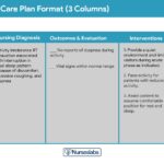
Once nursing diagnoses are identified, care plans provide a framework for prioritizing and delivering patient-centered care. Here are examples of nursing care plans for pleural effusion, focusing on common nursing diagnoses.
Nursing Diagnosis: Acute Pain
Related to: Pleural inflammation and irritation
As evidenced by:
- Reports of sharp or burning chest pain
- Guarding behavior
- Pain worsened by inhalation or coughing
- Shallow breathing
Expected Outcomes:
- Patient will report reduced pain (pain scale ≤ 2/10) within 24-48 hours.
- Patient will demonstrate relaxed breathing pattern without guarding.
- Patient will participate in activities of daily living without significant pain interference.
Assessments:
- Pain Assessment: Assess pain level using a pain scale, location, quality, aggravating and relieving factors. Differentiate pleuritic pain from other types of chest pain.
- Nonverbal Pain Cues: Observe for facial grimacing, guarding, reluctance to move, and shallow breathing.
Interventions:
- Administer Analgesics: Administer prescribed pain medications, typically NSAIDs initially for pleuritic pain. Opioids may be necessary for severe pain.
- Non-Pharmacologic Pain Relief:
- Repositioning: Help patient find a comfortable position; often, lying on the affected side can splint the chest and reduce pain.
- Guided Imagery and Relaxation Techniques: To reduce pain perception and anxiety.
- Splinting the Chest: Instruct patient to splint the chest with a pillow during coughing to minimize pain.
- Rest and Activity Management: Encourage rest periods and simplify activities to reduce exertion and pain exacerbation.
- Deep Breathing and Coughing Exercises: Teach and encourage deep breathing exercises to prevent atelectasis and improve oxygenation, despite pain. Medicate prior to exercises if needed to manage pain.
Nursing Diagnosis: Impaired Gas Exchange
Related to: Alveolar-capillary membrane changes secondary to pleural fluid, decreased lung expansion.
As evidenced by:
- Dyspnea, shortness of breath
- Abnormal arterial blood gases (ABGs) – hypoxemia, hypercapnia
- Restlessness, anxiety
- Changes in mental status (confusion, lethargy)
- Tachycardia, tachypnea
- Decreased oxygen saturation (SpO2 < 95%)
- Diminished breath sounds on auscultation
Expected Outcomes:
- Patient will demonstrate improved gas exchange evidenced by ABGs within patient’s baseline normal limits.
- Patient will maintain SpO2 ≥ 95% on room air or prescribed oxygen.
- Patient will exhibit reduced dyspnea and improved breathing pattern.
Assessments:
- Auscultate Lung Sounds: Assess for diminished or absent breath sounds, adventitious sounds. Note areas of decreased air entry.
- Monitor ABGs and Oxygen Saturation: Evaluate oxygenation and ventilation status.
- Assess Respiratory Rate, Rhythm, and Effort: Note for tachypnea, labored breathing, use of accessory muscles.
- Monitor for Signs of Hypoxia: Restlessness, confusion, cyanosis, altered mental status.
Interventions:
- Positioning: Elevate head of bed to semi-Fowler’s or high-Fowler’s position to maximize lung expansion. Lateral positioning (good lung down) may improve ventilation-perfusion matching in unilateral effusions.
- Supplemental Oxygen: Administer oxygen therapy as prescribed to maintain adequate oxygenation. Monitor response to oxygen therapy.
- Encourage Deep Breathing and Coughing: Promote lung expansion and secretion mobilization.
- Anxiety Reduction: Provide calm environment, reassurance, and emotional support to reduce anxiety associated with dyspnea.
- Prepare for Procedures: Prepare patient for thoracentesis, chest tube insertion, or other procedures to remove pleural fluid and improve gas exchange. Explain procedure and provide pre- and post-procedure care.
Nursing Diagnosis: Fluid Volume Excess
While not explicitly listed as a nursing diagnosis in the original article, fluid volume excess is intrinsically linked to pleural effusion, especially transudative types.
Related to: Compromised regulatory mechanisms (e.g., heart failure, renal failure, liver cirrhosis), excess fluid shift into pleural space.
As evidenced by:
- Pleural effusion on chest X-ray
- Dyspnea, orthopnea
- Peripheral edema, weight gain
- Jugular venous distension (JVD)
- Crackles on lung auscultation (may be present if underlying CHF)
- Decreased urine output (in some cases of renal dysfunction)
- Electrolyte imbalances (depending on underlying cause)
Expected Outcomes:
- Patient will demonstrate reduced pleural effusion size on follow-up imaging.
- Patient will exhibit balanced fluid volume evidenced by stable weight, reduced edema, and clear breath sounds (or improved from baseline).
- Patient will maintain stable vital signs and electrolyte balance.
Assessments:
- Monitor Fluid Balance: Strict intake and output, daily weights, assess for edema (peripheral, sacral), JVD.
- Assess Respiratory Status: Dyspnea, orthopnea, breath sounds (crackles may indicate pulmonary edema in CHF).
- Monitor Cardiovascular Status: Vital signs, heart sounds (S3 gallop in CHF).
- Review Laboratory Data: Electrolytes, renal function tests, liver function tests, serum protein and albumin levels to assess underlying causes and impact of fluid excess.
Interventions:
- Fluid Restriction: Implement prescribed fluid restriction based on patient’s condition and underlying cause.
- Sodium Restriction: Dietary sodium restriction to reduce fluid retention, especially in CHF or liver cirrhosis.
- Administer Diuretics: Administer prescribed diuretics to promote fluid excretion. Monitor electrolyte levels (especially potassium) and renal function.
- Monitor Response to Therapy: Assess for improvement in respiratory symptoms, edema reduction, weight loss, and electrolyte balance.
- Positioning: Elevate legs when sitting or lying to promote venous return and reduce peripheral edema. Semi-Fowler’s or Fowler’s position to ease breathing.
- Patient Education: Educate patient about fluid and sodium restrictions, importance of medication adherence, and signs and symptoms to report.
Other Nursing Diagnoses (Briefly Addressed in Original Article)
- Impaired Spontaneous Ventilation: Related to ventilatory compromise secondary to pleural effusion. Interventions focus on drainage, positioning, and respiratory support.
- Ineffective Airway Clearance: Related to fluid accumulation and cough. Interventions include assisting with drainage, oxygen therapy, medications, and preparing for thoracostomy.
- Ineffective Breathing Pattern: Related to compromised lung expansion. Interventions include medications, oxygen, elevating HOB, and preparing for procedures.
References
While the original article does not list specific references, for a comprehensive and evidence-based approach, incorporating references would strengthen the article. Examples of relevant references could include:
- UpToDate – for clinical information on pleural effusion diagnosis and management.
- Nursing textbooks focused on respiratory and medical-surgical nursing.
- PubMed Central – for research articles on pleural effusion and nursing care.
- Professional guidelines from organizations like the American Thoracic Society (ATS) or European Respiratory Society (ERS).
By expanding on the original article, providing detailed nursing care plans, and focusing on the keywords, this revised article aims to be a more comprehensive and SEO-optimized resource for nurses seeking information on pleural effusion and fluid excess management.

