Oral potentially malignant disorders (OPMDs) represent a significant area of concern in dental and medical fields. These conditions, which include leukoplakia, erythroplakia, oral submucous fibrosis, actinic cheilitis, and oral lichen planus (OLP), are defined as lesions or disorders within the oral cavity that carry a potential risk of transforming into oral squamous cell carcinoma. Accurate and timely Oral Lesions Differential Diagnosis is paramount for effective management and prevention of malignant progression. This article will delve into these pre-malignant disorders, emphasizing the importance of differential diagnosis in identifying and managing them effectively.
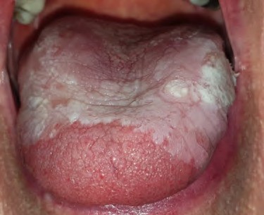
Oral leukoplakia is characterized by white plaques on the oral mucosa of unknown etiology. It’s crucial in oral lesions differential diagnosis to distinguish leukoplakia from benign conditions such as candidiasis, hairy leukoplakia, lupus erythematosus, and morsicatio buccarum (cheek biting). Smoking significantly increases the risk of leukoplakia development, making smokers six times more susceptible. Leukoplakia carries an estimated 1% annual risk of malignant transformation. Two primary types of leukoplakia are recognized: homogenous and non-homogenous. Homogenous leukoplakia presents as a uniformly white, flat, and thick lesion, while non-homogenous leukoplakia, also known as erythroleukoplakia or speckled leukoplakia, exhibits a mixed red and white appearance. Non-homogenous leukoplakia, appearing speckled or nodular, is particularly concerning due to its higher risk of malignant transformation and may necessitate surgical excision.
Erythroplakia, another significant OPMD, manifests as red patches on the oral mucosa. In oral lesions differential diagnosis, erythroplakia is defined by exclusion, meaning it is diagnosed when red patches cannot be clinically attributed to any other identifiable condition. The bright red appearance is due to underlying vascularity and reduced keratinization. Erythroplakia carries a higher malignant transformation rate compared to leukoplakia, making accurate differential diagnosis and prompt management critical.
Oral submucous fibrosis (OSF) is a chronic, progressive condition affecting the oral cavity, characterized by inflammation and fibrosis of the submucosal layer. Patients with OSF often report limitations in mouth opening (trismus), intolerance to spicy foods, and a burning sensation in the mouth. Clinical examination reveals mucosal stiffening and blanching due to fibrosis. In the oral lesions differential diagnosis of conditions causing restricted mouth opening and mucosal changes, OSF must be considered, especially in regions where betel quid chewing is prevalent. OSF has a substantial 10-year malignant transformation risk, highlighting the importance of early detection and intervention.
Actinic cheilitis is a pre-malignant condition specifically affecting the lip vermilion, primarily caused by chronic exposure to solar ultraviolet (UV) radiation. Histopathological changes include solar elastosis, chronic inflammatory infiltrate, vasodilation, and hyperkeratosis. Actinic cheilitis can progress to squamous cell carcinoma if left untreated. Clinically, it presents with lip dryness, atrophy, scaly lesions, ulcerations, and a blurring of the vermilion border. When performing oral lesions differential diagnosis for lip lesions, actinic cheilitis should be considered in individuals with a history of sun exposure, particularly fair-skinned individuals over 40.
In conclusion, accurate oral lesions differential diagnosis is critical in managing oral potentially malignant disorders. A biopsy is often indispensable to establish a definitive diagnosis, assess the degree of dysplasia, and differentiate OPMDs from other benign inflammatory and atrophic lesions. Early identification and appropriate management strategies based on accurate differential diagnosis are essential for preventing the progression of these lesions to oral squamous cell carcinoma and improving patient outcomes.